. 2023 Aug;620(7972):128-136.
doi: 10.1038/s41586-023-06331-x. Epub 2023 Jul 19.

A common allele of HLA is associated with asymptomatic SARS-CoV-2 infection

Affiliations

A common allele of HLA is associated with asymptomatic SARS-CoV-2 infection

Danillo G Augusto et al. Nature. 2023 Aug.

Abstract

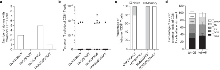
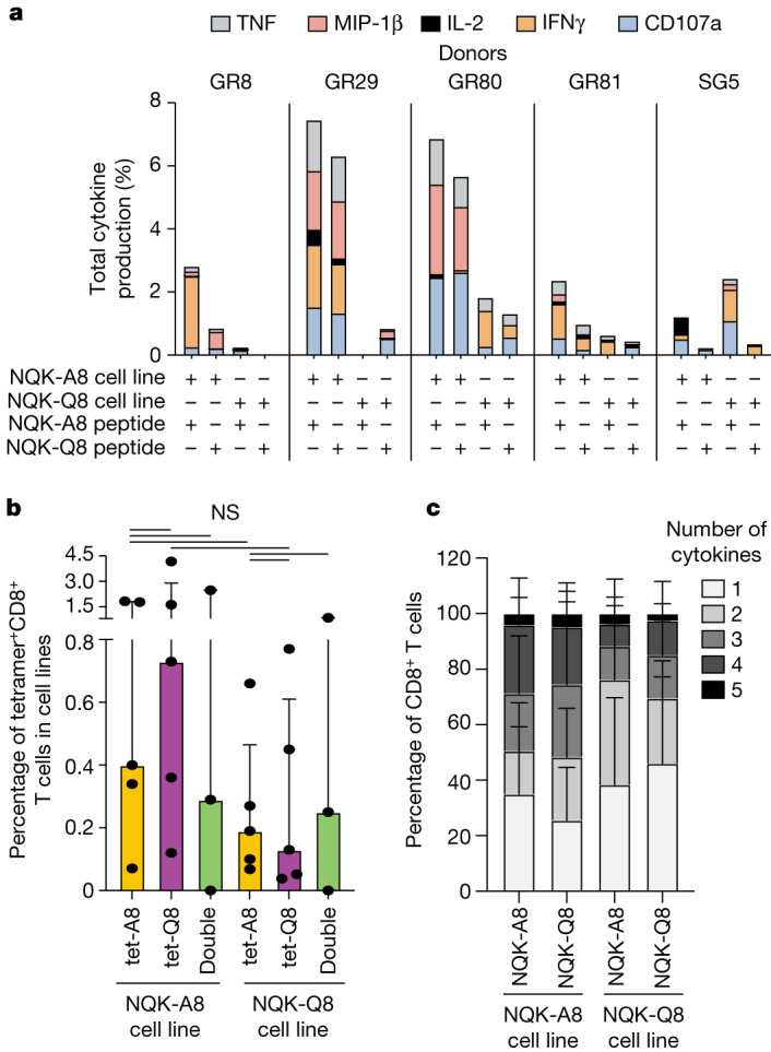
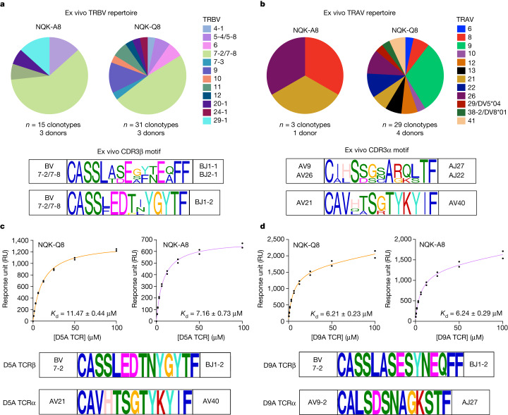
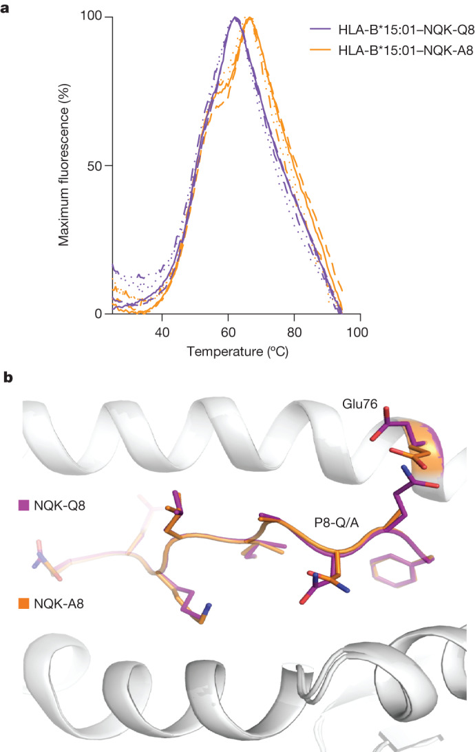
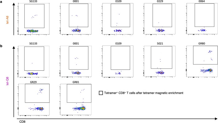
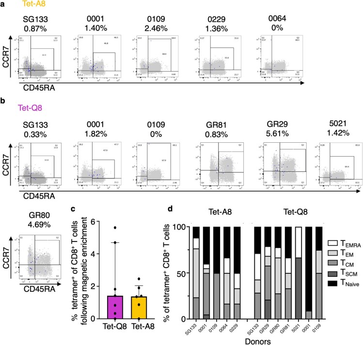
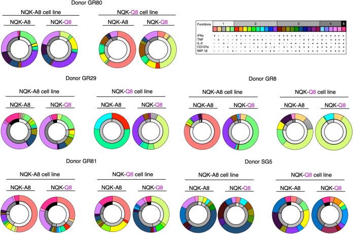
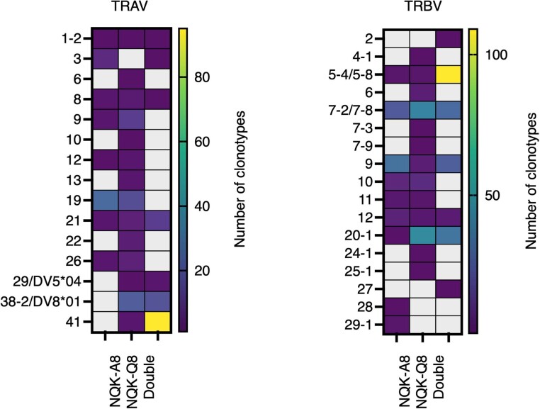
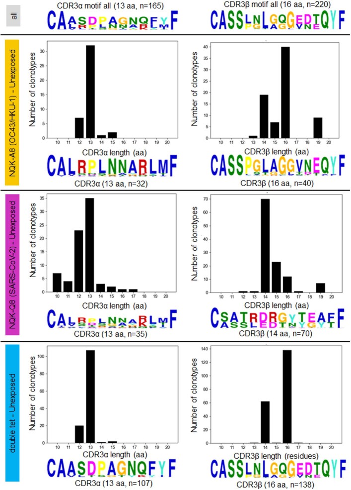
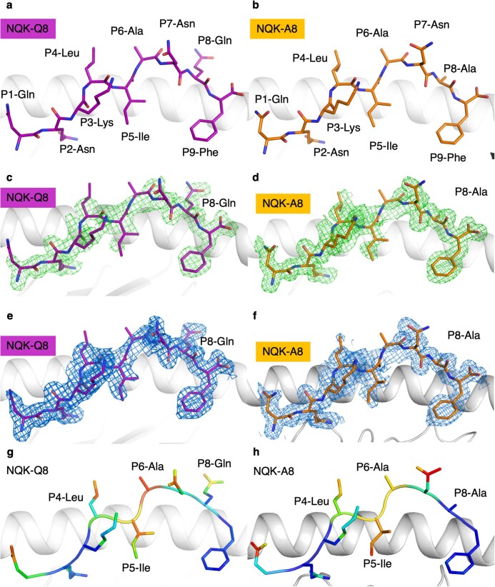
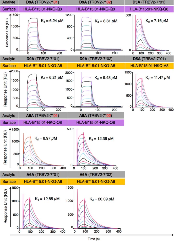
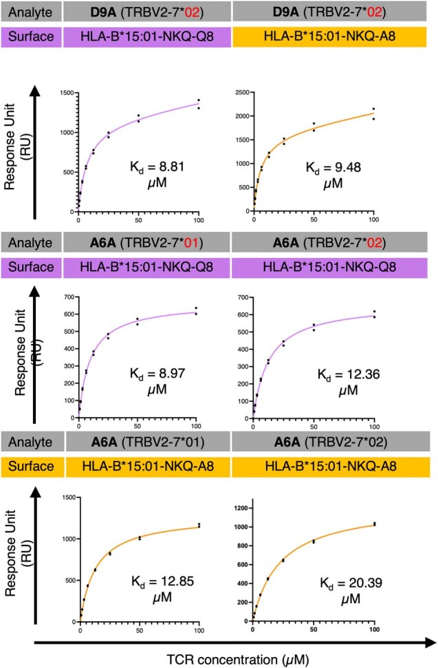
Studies have demonstrated that at least 20% of individuals infected with SARS-CoV-2 remain asymptomatic1-4. Although most global efforts have focused on severe illness in COVID-19, examining asymptomatic infection provides a unique opportunity to consider early immunological features that promote rapid viral clearance. Here, postulating that variation in the human leukocyte antigen (HLA) loci may underly processes mediating asymptomatic infection, we enrolled 29,947 individuals, for whom high-resolution HLA genotyping data were available, in a smartphone-based study designed to track COVID-19 symptoms and outcomes. Our discovery cohort (n = 1,428) comprised unvaccinated individuals who reported a positive test result for SARS-CoV-2. We tested for association of five HLA loci with disease course and identified a strong association between HLA-B*15:01 and asymptomatic infection, observed in two independent cohorts. Suggesting that this genetic association is due to pre-existing T cell immunity, we show that T cells from pre-pandemic samples from individuals carrying HLA-B*15:01 were reactive to the immunodominant SARS-CoV-2 S-derived peptide NQKLIANQF. The majority of the reactive T cells displayed a memory phenotype, were highly polyfunctional and were cross-reactive to a peptide derived from seasonal coronaviruses. The crystal structure of HLA-B*15:01-peptide complexes demonstrates that the peptides NQKLIANQF and NQKLIANAF (from OC43-CoV and HKU1-CoV) share a similar ability to be stabilized and presented by HLA-B*15:01. Finally, we show that the structural similarity of the peptides underpins T cell cross-reactivity of high-affinity public T cell receptors, providing the molecular basis for HLA-B*15:01-mediated pre-existing immunity.


Conflict of interest statement

The authors declare no competing interests.

Figures

Fig. 1
Fig. 1. T cell reactivity in pre-pandemic samples from individuals carrying HLA-B*15:01.
ac, Ex vivo combinatorial tetramer analysis for the four indicated peptides was performed in nine pre-pandemic donor samples. a,b, The number of donors with detectable tetramer+CD8+ T cells (a) and their frequencies (b) are shown. c, The proportion of naive (CD45RA+CCR7+) and memory (combination of CD45RACCR7, CD45RACCR7+, CD45RA+CCR7) cells among tetramer+CD8+ T cells (based on n = 9 samples). d, Phenotypic analysis after TAME of ex vivo tetramer+ NQK-Q8-specific (tet-Q8) and NQK-A8-specific (tet-A8) T cells in 7 and 6 donors, respectively. Cell types were defined as follows: Tnaive (CD45RA+CCR7+CD95); TSCM (stem cell memory, CD45RA+CCR7+CD95+); TCM (central memory, CD45RACCR7+); TEM (effector memory, CD45RACCR7); TEMRA (terminally differentiated, CD45RA+CCR7). Data are mean ± s.e.m.
Fig. 2
Fig. 2. NQK-specific T cells are cross-reactive.
a, Total cytokine production by CD8+ T cells in NQK-A8- and NQK-Q8-specific T cell lines. Each peptide-specific T cell line was restimulated individually with its cognate peptide or the homologous peptide as indicated, and the cytokine response was measured by intracellular cytokine staining (n = 5 donors). Percentages of effector functions (IFNγ, TNF, IL-2, MIP-1β, CD107a) minus the no peptide control are reported. b, In vitro tetramer analysis for the NQK-Q8- and NQK-A8-specific T cell lines (n = 5 donors). The cell lines were tetramer stained with a single tet-A8 (orange bar) or tet-Q8 (purple bar) tetramer or both tetramers (green bar). The frequency of tetramer+CD8+ T cells is shown. Data are median ± interquartile range. Differences between two groups were compared using two-tailed unpaired t-tests. P < 0.05 was considered to be significant. NS, not significant. c, Polyfunctionality analysis of CD8+ NQK-peptide-specific T cells from five unexposed donors. The number of functions is shown on a scale from 5 (black) to 1 (white). Data are the relative frequency (%) of total cytokine+CD8+ T cells. Data are mean ± s.e.m. Differences between two groups were determined using two-tailed unpaired t-tests. P < 0.05 was considered to be significant, and the result was not significant.
Fig. 3
Fig. 3. NQK-specific T cells are characterized by the presence of high-affinity public TCRs in unexposed donors.
a,b, Analysis of the ex vivo TCR repertoire for NQK-A8- and NQK-Q8-specific T cells after TAME on the basis of TRBV (a) and TRAV (b) use. The pie charts show the percentage of each TRBV or TRAV used in the single-cell sorted clonotypes. The CDR3β and CDR3α sequence motifs are shown below the pie charts, respectively, for the TCRs with the biased expression of TRBV7-2/7-8 paired with TRAV9/26 or TRAV21, including the public TCRs. c,d, The binding response (response units (RU)) of D5A (c) and D9A (d) TCRs (analyte) against HLA-B*15:01–NQK complexes (A8 in orange and Q8 in purple). The public TCR D5A expresses TRBV7-2 paired with TRAV21, and the public D9A TCR expresses TRBV7-2 paired with TRAV9-2; the CDR3β and CDR3α sequences for each public TCRs are shown below the binding curves. The SPR steady-state binding curves represent binding under a TCR concentration range of 0.39–100 μM. n = 2 biologically independent experiments performed in duplicate; the graph shows the results from one experiment performed in duplicate, represented by the black dots.
Fig. 4
Fig. 4. NQK peptides are stable and adopt the same conformation bound to the HLA-B*15:01 molecule.
a, DSF plots showing the normalized fluorescence intensity versus temperature for HLA-B*15:01 in a complex with the NQK-Q8 (purple) or NQK-A8 (orange) peptide measured at concentrations of 5 μM and 10 μM. n = 2 biologically independent experiments performed in duplicate, represented by the different lines. b, Superimposition of the crystal structures of HLA-B*15:01 (white cartoon) in a complex with either the NQK-Q8 (purple stick) or the NQK-A8 (orange stick) peptide.
Extended Data Fig. 1
Extended Data Fig. 1. Ex vivo raw data from pre-pandemic samples from individuals carrying HLA-B*15:01.
(a–b) Individual FACS plots for each pre-pandemic donor samples used for ex vivo tetramer staining following tetramer magnetic enrichment with tet-A8 tetramer (a) and with tet-Q8 tetramer (b), from 5 and 7 donors, respectively. The enriched tetramer+ CD8+ T cells observed are indicated within the inner square.
Extended Data Fig. 2
Extended Data Fig. 2. Ex vivo tetramer and phenotype analysis of NQK-Q8- and NQK-A8-specific T cells, related to Fig. 1d.
(a,b) FACS plots of NQK-A8-tetramer+ CD8+ T cells following tetramer magnetic enrichment (a) and NQK-Q8-tetramer+ CD8+ T cells (b). The NQK-A8- and NQK-Q8- tetramer+ CD8+ T cells shown as blue dots were plotted versus CD3+ T cells shown as grey dots and gated on CCR7 and CD45RA to determine memory status, following tetramer magnetic enrichment for each donor. (c) The percentage of tetramer+ of CD8+ T cells after tetramer magnetic enrichment is reported for each donor with the purple bar for the Tet-Q8 tetramer and the orange bar for the Tet-A8 tetramer (n = 7 and n = 6 biologically independent samples, respectively). Data are presented as median values with IQR (interquartile range). The individual percentage for each donor is indicated on panels a and b. (d) The proportion of Tnaïve (CD45RA+CCR7+CD95-); TSCM (stem cell memory, CD45RA+CCR7+CD95+); TCM (central memory, CD45RA-CCR7+); TEM (effector memory, CD45RA-CCR7-); TEMRA (terminally differentiated, CD45RA+CCR7-) cells in different donors.
Extended Data Fig. 3
Extended Data Fig. 3. Functionality pie charts of T cell lines.
Frequency of CD8+ T cells with different effector functions (IFNγ, TNF, IL-2, MIP-1β, CD107a) was determined, along with the number of different functions, minus the no peptide control. The outer ring of the double ring pie shows the function profile in different colours according to the table, and the inner ring represents the number of functions with black for 5 and white for 1.
Extended Data Fig. 4
Extended Data Fig. 4. TRAV and TRBV usage specific for each tetramer used in unexposed individuals.
Heatmaps displaying preferred TRAV (left) and TRBV (right) usage of NQK-Q8-, NQK-A8-, or both (double) peptide-specific TCRs in all unexposed donors collectively.
Extended Data Fig. 5
Extended Data Fig. 5. NQK-specific TCR repertoire CDR3 analysis in unexposed donors.
Summary of CDR3α (left) and CDR3β (right) lengths and motif analysis for the NQK-A8 (top), NQK-Q8 (middle), and both peptides (bottom)-specific TCR clonotypes in unexposed donors. The MEME motif discovery program was used to identify aa motifs enriched; the relative size of each residue symbol is proportional to its frequency, while the total height of aa symbols indicates the information content of the position in bits. The motif on top left panel is the CDR3α 13 amino acid (aa) long sequence motif derived from all CDR3α sequences obtained (n = 165); on the right top panel is the CDR3β 16 aa long sequence motif derived from all CDR3β sequences obtained (n = 220).
Extended Data Fig. 6
Extended Data Fig. 6. HLA-B*15:01 interaction with peptides derived from SARS-CoV-2 and seasonal coronaviruses.
(a–b) Structures of HLA-B*15:01-NQK-Q8 (peptide as purple sick) (a) and HLA-B*15:01-NQK-A8 (peptide as orange stick) (b), with the HLA chain in white cartoon. (c–d) Electron density map around the NQK-Q8 (purple stick) and NQK-A8 (orange stick) peptides, respectively, in green at 3s for the Fo-Fc map. (e–f) Electron density map around the NQK-Q8 (purple stick) and NQK-A8 (orange stick) peptides, respectively, in blue at 1s for the 2Fo-Fc map. (g–h) Bfactor analysis of the atoms of the NQK-Q8 and NQK-A8 peptides, respectively. The atoms are coloured according to their Bfactor that indicates the mobility of each atom from low (blue) to high (red).
Extended Data Fig. 7
Extended Data Fig. 7. Surface plasmon resonance sensorgrams.
SPR sensograms for the 5 tested TCRs with each curve showing different concentration of the TCR.
Extended Data Fig. 8
Extended Data Fig. 8. Steady-state binding curves.
Steady-state binding curve for the five tested TCRs towards HLA*15:01-NKQ-Q8 or -A8 complex. Analytes (TCRs) flowed over immobilized HLA*15:01-NKQ-Q8 or -A8 with a concentration range of 0 to 100 μM. n = 2 biologically independent experiments performed in duplicate, with the graph showing results from one experiment.

Update of

  • A common allele of HLA mediates asymptomatic SARS-CoV-2 infection.
    Augusto DG, Yusufali T, Sabatino JJ Jr, Peyser ND, Murdolo LD, Butcher X, Murray V, Pae V, Sarvadhavabhatla S, Beltran F, Gill G, Lynch K, Yun C, Maguire C, Peluso MJ, Hoh R, Henrich TJ, Deeks SG, Davidson M, Lu S, Goldberg SA, Kelly JD, Martin JN, Viera-Green CA, Spellman SR, Langton DJ, Lee S, Marcus GM, Olgin JE, Pletcher MJ, Gras S, Maiers M, Hollenbach JA. Augusto DG, et al. medRxiv [Preprint]. 2022 Oct 12:2021.05.13.21257065. doi: 10.1101/2021.05.13.21257065. medRxiv. 2022. Update in: Nature. 2023 Aug;620(7972):128-136. doi: 10.1038/s41586-023-06331-x. PMID: 34031661 Free PMC article. Updated. Preprint.

Similar articles

  • A common allele of HLA mediates asymptomatic SARS-CoV-2 infection.
    Augusto DG, Yusufali T, Sabatino JJ Jr, Peyser ND, Murdolo LD, Butcher X, Murray V, Pae V, Sarvadhavabhatla S, Beltran F, Gill G, Lynch K, Yun C, Maguire C, Peluso MJ, Hoh R, Henrich TJ, Deeks SG, Davidson M, Lu S, Goldberg SA, Kelly JD, Martin JN, Viera-Green CA, Spellman SR, Langton DJ, Lee S, Marcus GM, Olgin JE, Pletcher MJ, Gras S, Maiers M, Hollenbach JA. Augusto DG, et al. medRxiv [Preprint]. 2022 Oct 12:2021.05.13.21257065. doi: 10.1101/2021.05.13.21257065. medRxiv. 2022. Update in: Nature. 2023 Aug;620(7972):128-136. doi: 10.1038/s41586-023-06331-x. PMID: 34031661 Free PMC article. Updated. Preprint.
  • Rapid, point-of-care antigen tests for diagnosis of SARS-CoV-2 infection.
    Dinnes J, Sharma P, Berhane S, van Wyk SS, Nyaaba N, Domen J, Taylor M, Cunningham J, Davenport C, Dittrich S, Emperador D, Hooft L, Leeflang MM, McInnes MD, Spijker R, Verbakel JY, Takwoingi Y, Taylor-Phillips S, Van den Bruel A, Deeks JJ; Cochrane COVID-19 Diagnostic Test Accuracy Group. Dinnes J, et al. Cochrane Database Syst Rev. 2022 Jul 22;7(7):CD013705. doi: 10.1002/14651858.CD013705.pub3. Cochrane Database Syst Rev. 2022. PMID: 35866452 Free PMC article.
  • Antibody tests for identification of current and past infection with SARS-CoV-2.
    Fox T, Geppert J, Dinnes J, Scandrett K, Bigio J, Sulis G, Hettiarachchi D, Mathangasinghe Y, Weeratunga P, Wickramasinghe D, Bergman H, Buckley BS, Probyn K, Sguassero Y, Davenport C, Cunningham J, Dittrich S, Emperador D, Hooft L, Leeflang MM, McInnes MD, Spijker R, Struyf T, Van den Bruel A, Verbakel JY, Takwoingi Y, Taylor-Phillips S, Deeks JJ; Cochrane COVID-19 Diagnostic Test Accuracy Group. Fox T, et al. Cochrane Database Syst Rev. 2022 Nov 17;11(11):CD013652. doi: 10.1002/14651858.CD013652.pub2. Cochrane Database Syst Rev. 2022. PMID: 36394900 Free PMC article.
  • A pan-beta-coronavirus vaccine bearing conserved and asymptomatic B- and T-cell epitopes protects against highly pathogenic Delta and highly transmissible Omicron SARS-CoV-2 variants.
    Vahed H, Prakash S, Quadiri A, Ibraim IC, Omorogieva E, Patel S, Tadros J, Liao EJ, Lau L, Chentoufi AA, Nesburn AB, Kuppermann BD, Ulmer JB, Gil D, BenMohamed L. Vahed H, et al. Hum Vaccin Immunother. 2025 Dec;21(1):2527438. doi: 10.1080/21645515.2025.2527438. Epub 2025 Jul 4. Hum Vaccin Immunother. 2025. PMID: 40613499 Free PMC article.
  • The effect of sample site and collection procedure on identification of SARS-CoV-2 infection.
    Davenport C, Arevalo-Rodriguez I, Mateos-Haro M, Berhane S, Dinnes J, Spijker R, Buitrago-Garcia D, Ciapponi A, Takwoingi Y, Deeks JJ, Emperador D, Leeflang MMG, Van den Bruel A; Cochrane COVID-19 Diagnostic Test Accuracy Group. Davenport C, et al. Cochrane Database Syst Rev. 2024 Dec 16;12(12):CD014780. doi: 10.1002/14651858.CD014780. Cochrane Database Syst Rev. 2024. PMID: 39679851 Free PMC article.

Cited by

References

    1. Meyerowitz EA, Richterman A, Bogoch II, Low N, Cevik M. Towards an accurate and systematic characterisation of persistently asymptomatic infection with SARS-CoV-2. Lancet Infect. Dis. 2020;21:e163–e169. doi: 10.1016/S1473-3099(20)30837-9. - DOI - PMC - PubMed
    1. Oran DP, Topol EJ. Prevalence of asymptomatic SARS-CoV-2 infection. Ann. Intern. Med. 2020;173:M20-3012. doi: 10.7326/M20-3012. - DOI - PMC - PubMed
    1. Buitrago-Garcia D, et al. Occurrence and transmission potential of asymptomatic and presymptomatic SARS-CoV-2 infections: a living systematic review and meta-analysis. PLoS Med. 2020;17:e1003346. doi: 10.1371/journal.pmed.1003346. - DOI - PMC - PubMed
    1. Ra SH, et al. Upper respiratory viral load in asymptomatic individuals and mildly symptomatic patients with SARS-CoV-2 infection. Thorax. 2021;76:61–63. doi: 10.1136/thoraxjnl-2020-215042. - DOI - PubMed
    1. Docherty AB, et al. Features of 20 133 UK patients in hospital with covid-19 using the ISARIC WHO Clinical Characterisation Protocol: prospective observational cohort study. Brit. Med. J. 2020;369:m1985. doi: 10.1136/bmj.m1985. - DOI - PMC - PubMed