. 2023 Aug;620(7974):669-675.
doi: 10.1038/s41586-023-06349-1. Epub 2023 Jul 19.

Diverse modes of H3K36me3-guided nucleosomal deacetylation by Rpd3S

Affiliations

Diverse modes of H3K36me3-guided nucleosomal deacetylation by Rpd3S

Haipeng Guan et al. Nature. 2023 Aug.

Abstract

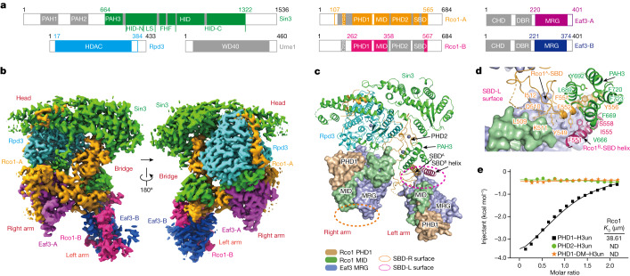
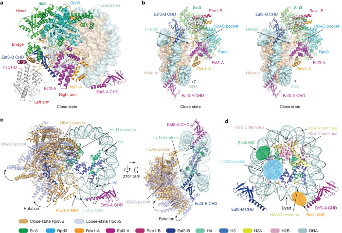
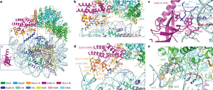
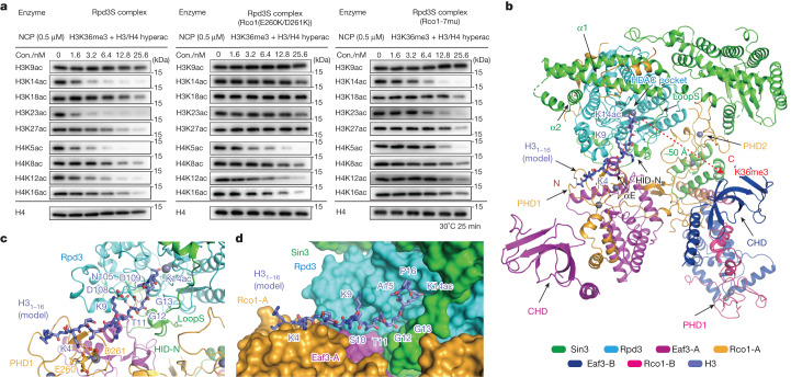
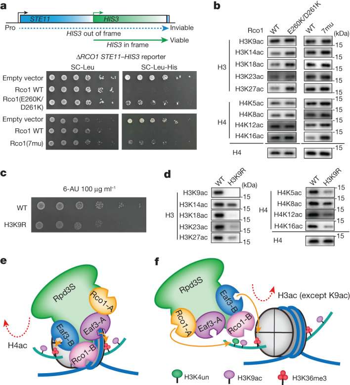
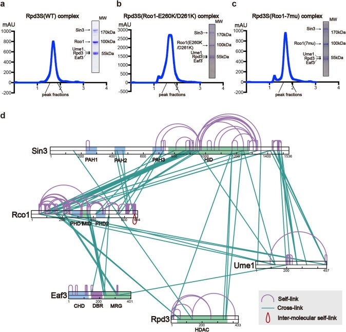
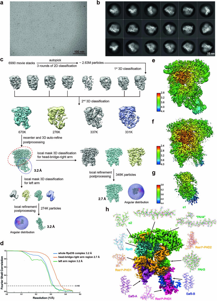
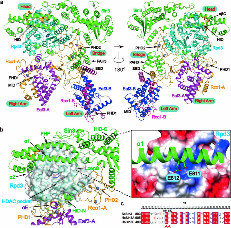
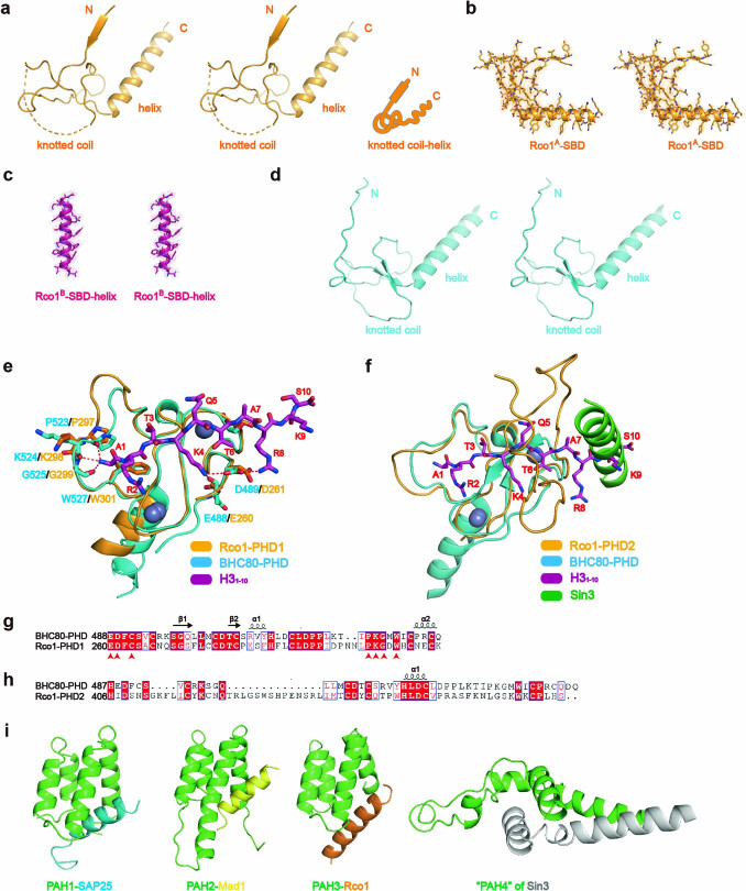
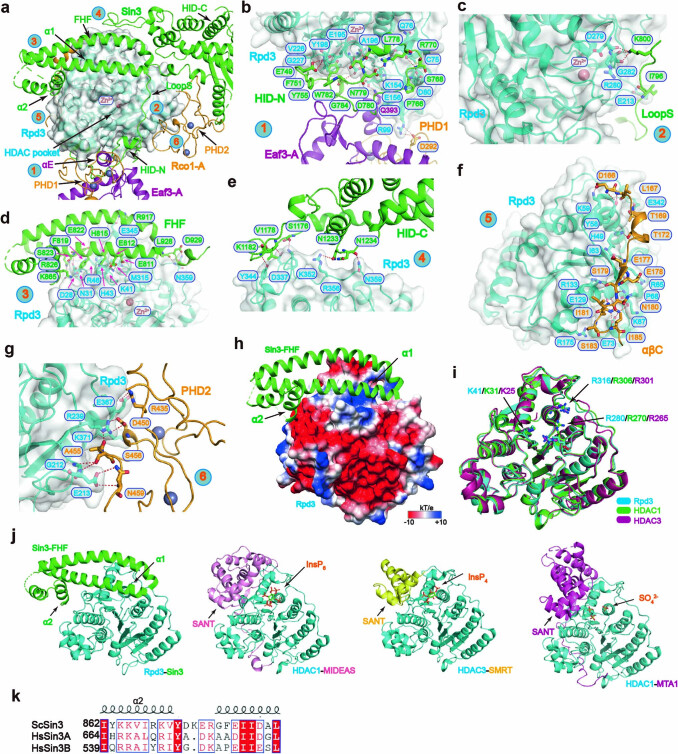
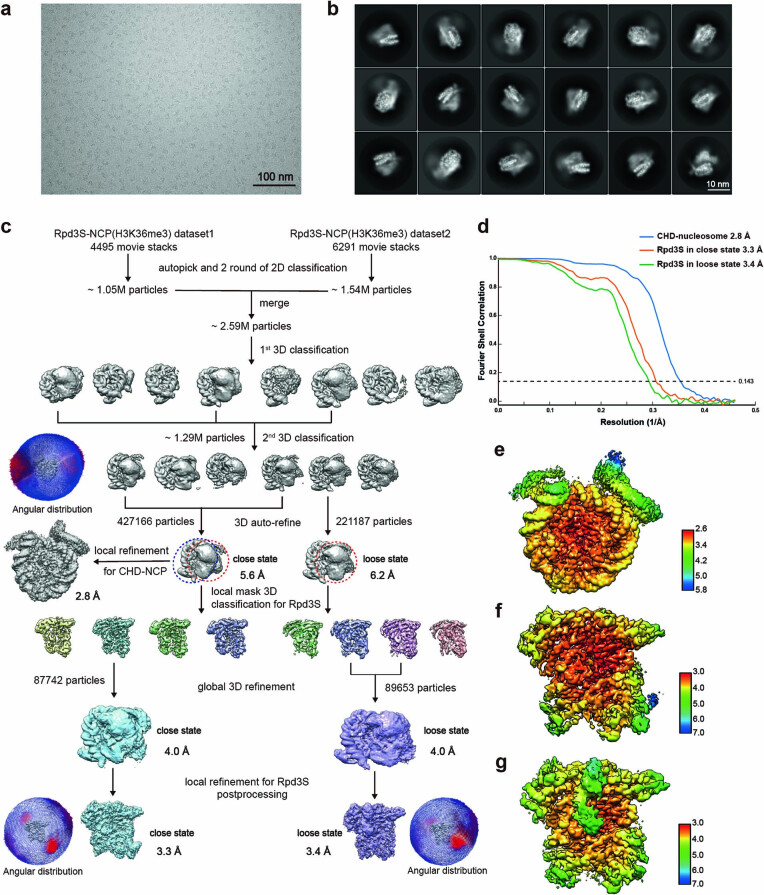
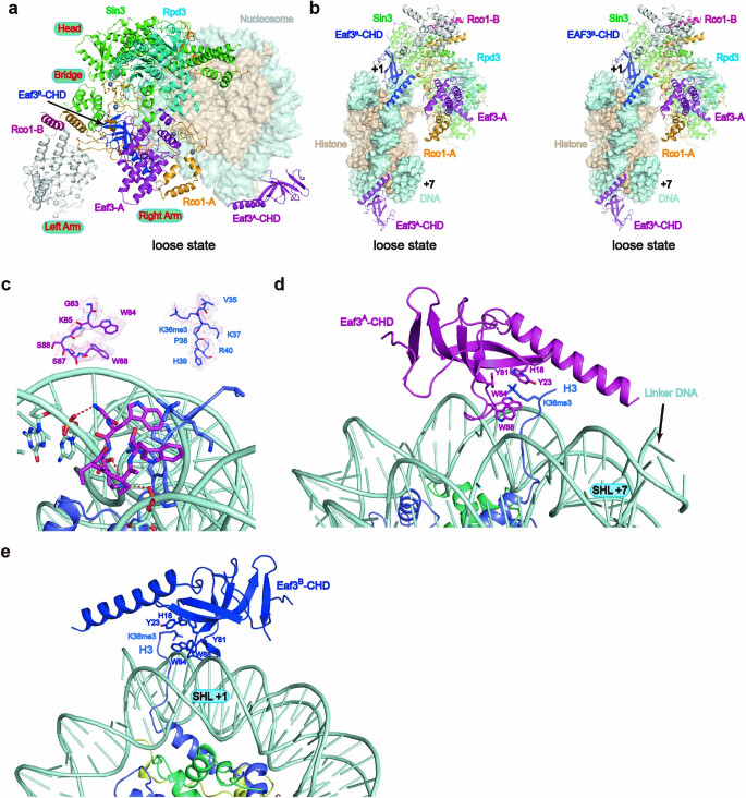
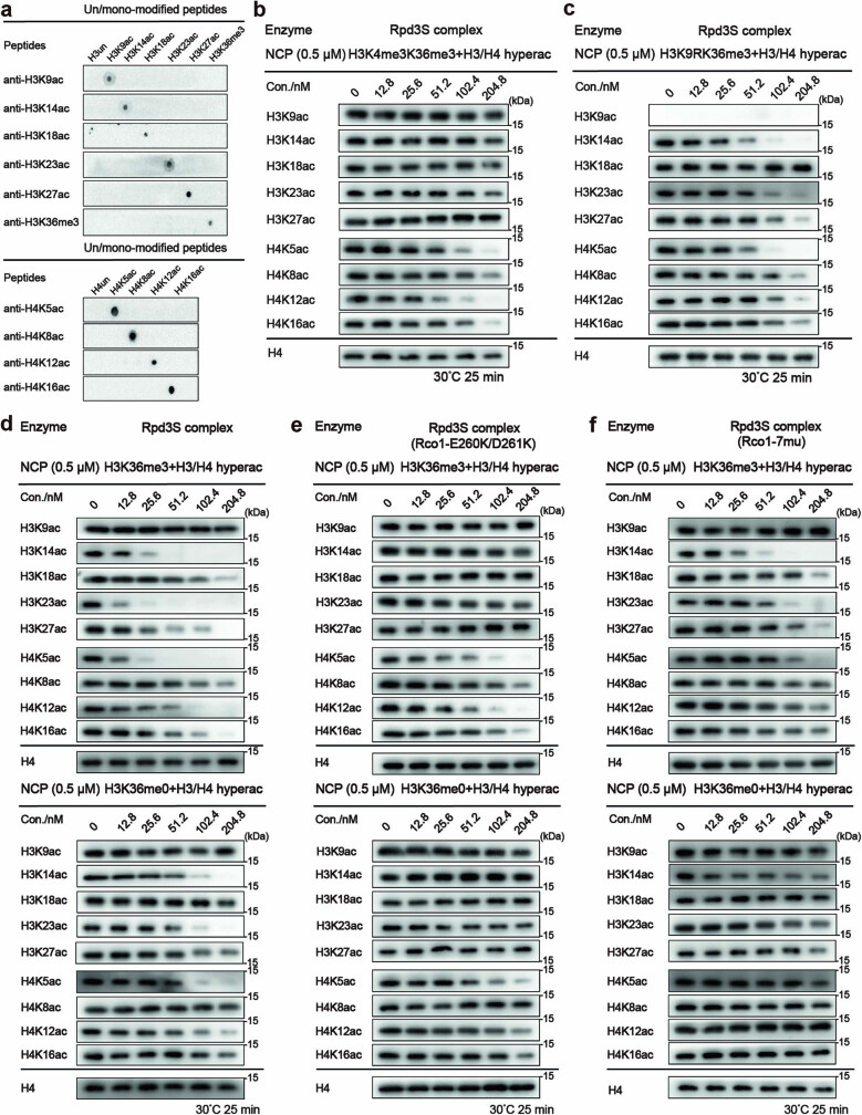
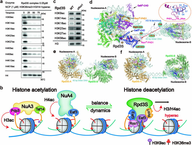
Context-dependent dynamic histone modifications constitute a key epigenetic mechanism in gene regulation1-4. The Rpd3 small (Rpd3S) complex recognizes histone H3 trimethylation on lysine 36 (H3K36me3) and deacetylates histones H3 and H4 at multiple sites across transcribed regions5-7. Here we solved the cryo-electron microscopy structures of Saccharomyces cerevisiae Rpd3S in its free and H3K36me3 nucleosome-bound states. We demonstrated a unique architecture of Rpd3S, in which two copies of Eaf3-Rco1 heterodimers are asymmetrically assembled with Rpd3 and Sin3 to form a catalytic core complex. Multivalent recognition of two H3K36me3 marks, nucleosomal DNA and linker DNAs by Eaf3, Sin3 and Rco1 positions the catalytic centre of Rpd3 next to the histone H4 N-terminal tail for deacetylation. In an alternative catalytic mode, combinatorial readout of unmethylated histone H3 lysine 4 and H3K36me3 by Rco1 and Eaf3 directs histone H3-specific deacetylation except for the registered histone H3 acetylated lysine 9. Collectively, our work illustrates dynamic and diverse modes of multivalent nucleosomal engagement and methylation-guided deacetylation by Rpd3S, highlighting the exquisite complexity of epigenetic regulation with delicately designed multi-subunit enzymatic machineries in transcription and beyond.


Conflict of interest statement

The authors declare no competing interests.

Figures

Fig. 1
Fig. 1. Cryo-EM structure of Rpd3S complex and interactions among subunits.
a, Schematic representation of the domain organizations of the Rpd3S complex. The colour scheme for Rpd3S complex subunits is indicated in b. Domains that are not structurally resolved are coloured grey. b, Cryo-EM map of the entire Rpd3S complex integrated with a 2.7 Å map of the head–bridge–right arm region and a 3.2 Å map of the left arm region. WD40, WD40 β-propeller domain. c, Global view of the Rpd3S complex, highlighting the Rco1–Eaf3 heterodimers. The PHD1 (light brown) and MID (reseda) domains of Rco1 and the MRG domain (blue violet) of Eaf3 are shown in surface representation; other domains are shown in cartoon form. The dotted lines represent the SBD surfaces (SBD-R and SBD-L) at different angles. d, Close-up view of interactions of the SBD domains of Rco1 and the PAH3 domain of Sin3 in the SBD-L surface and bridge region. e, Isothermal titration calorimetry fitting curves for indicated histone peptides with PHDs of Rco1. H3un, unmodified H31–10 peptide; PHD1-DM, PHD1 double mutant (E260K/D261K). ND, not determined.
Fig. 2
Fig. 2. Cryo-EM structure of Rpd3S complex bound to H3K36me3 nucleosome.
a, A model of the core Rpd3S complex bound to the H3K36me3 modified nucleosome in the close state. b, A stereo view of contact between the core Rpd3S complex and the H3K36me3 modified nucleosome in the close state. Sin3, Rpd3, Eaf3 and Rco1 are shown in cartoon form. The nucleosome is shown in surface representation. The invisible left arm region is coloured grey. c, Two views of the superimposition of close and loose state of core Rpd3S complex are shown in orange yellow and pale cyan, respectively. H3 and H4 of the histone octamer are highlighted in cartoon form. d, A model of the CHD domains with the H3K36me3 modified nucleosome. The tails of histone H2A and H2B are labelled with arrows. The positions of nucleosomal DNA are labelled as SHL positions.
Fig. 3
Fig. 3. Details of the interface between the Rpd3S complex and the H3K36me3 nucleosome.
a, A model of the core Rpd3S complex bound to the H3K36me3 modified nucleosome following histone H4 deacetylation. Interactions are outlined and shown in close up in the indicated panel. b, Detailed view of the location between the N terminus of H4 and Rpd3 in the close state as shown in a. c, Detailed view of the interactions between CHD and the H3K36me3 modified nucleosome as shown in a. The residues of CHD and the nucleotides of nucleosomal DNA involved in recognition and H3 tail residues are shown as sticks. Selected hydrogen bonds are shown as red dashed lines. d, Detailed view of interactions between the Rco1 MID and linker DNA of nucleosome as shown in a. Residues at the interface are depicted as sticks. e, Detailed view of interactions between Sin3 and nucleosomal DNA at SHL +2.5 as shown in a. The residues of Sin3 and the nucleotides of nucleosomal DNA involved in recognition are shown as sticks. Selected hydrogen bonds are shown as red dashed lines.
Fig. 4
Fig. 4. Combinatorial readout-guided histone deacetylation by Rpd3S complexes.
a, A representative HDAC assay measuring activity of Rpd3S complexes containing wild-type Rco1 (left), Rco1 PHD1 mutants (middle) and Rco1 left arm region mutants (right) on H3K36me3 and hyperacetylated (hyperac) nucleosome. The reaction products were identified by western blot. Data are representative of three independent experiments. b, Global view of the docking model of the Rpd3S complex with histone H31–16K14ac on histone H3 deacetylation. c,d, Detailed views of interactions between the Rpd3S complex and histone H3K14ac in cartoon (c) and surface (d) representation.
Fig. 5
Fig. 5. In vivo modification crosstalk studies.
a, The test cryptic transcription phenotype caused by Rco1(E260K/D261K) or Rco1(7mu) mutants in a STE11-HIS3 reporter strain (YBL853). WT, wild type. b, Western blot showing H3 and H4 acetylation levels at different sites in Rco1 wild type, E260K/D261K and 7mu mutants. c, Growth of H3 wild type and H3K9R mutant yeast. Fivefold serial dilutions of H3 wild type and H3K9R mutant cells were spotted onto plates with synthetic complete medium (with glucose) containing 100 μg ml−1 6-azauracil (6-AU) and cultivated at 30 °C for 3 days. d, Western blot showing global H3 and H4 acetylation defects in the H3K9R mutant yeast strain. ad, One representative example of three independent experiments. e,f, Models of the Rpd3S complex bound to the H3K36me3 modified nucleosome on histone H4 deacetylation (e) and on histone H3 deacetylation (f).
Extended Data Fig. 1
Extended Data Fig. 1. Protein purification and XL-MS analysis of Rpd3S complex.
a-c. Purification of Rpd3S and mutant complexes. Purification of the Rpd3S and mutant complexes. The complexes were purified using size-exclusion chromatography (Superose 6), and the peak fractions were subjected to SDS-PAGE for Coomassie blue staining. The complexes include: (a) wild-type of Rpd3S complex; (b) Rco1-E260A/D261A of the Rpd3S complex to disrupt the PHD1 of Rco1; (c) Rco1-7mutants of Rpd3S complex to disrupt the left arm region. d. Schematic representation of the intermolecular cross-links within the Rpd3S complex. The domains of Rpd3S are indicated, and the identified inter-subunit cross-links or subunit self-links are shown as cyan or modena solid lines, respectively. The special intermolecular self-links in the C-terminal of Rco1 are shown as red solid lines.
Extended Data Fig. 2
Extended Data Fig. 2. Data collection, image processing, Cryo-EM reconstructions and structural model of the Rpd3S complex.
Representative cryo-EM micrograph (a) and 2D class averages (b) of various projection views of the Rpd3S complex. c. Flowchart of the cryo-EM image processing, 3D reconstructions for the Rpd3S complex and angular distribution of EM maps. d. Resolution estimation of the EM maps. e. Estimated resolution of the cryo-EM reconstructions of the whole Rpd3S complex. Local resolution estimation of the cryo-EM reconstructions of the head-bridge-right arm region (f) and the bridge-left arm region (g) of the Rpd3S complex. h. The locally refined cryo-EM map of the Rpd3S complex. Close-up views of fragments of Rpd3S subunits with cryo-EM densities shown as meshes. The residues are shown as sticks representations.
Extended Data Fig. 3
Extended Data Fig. 3. Detailed structure of the Rpd3S complex.
a. A cartoon model of Rpd3S complex shown in two different views. PAH, paired amphipathic helice; HID, HDAC-interaction domain; αβC, “α-β-coil” motif; PHD, plant homeobox domain ; MID, MRG-interacting domain; SBD, Sin3-binding domain. b. The interactions between the core enzyme Rpd3 and its neighboring subunits are shown. Acidic residues of the α1 of Sin3 pack against the basic surface of the Rpd3. The negatively charged residues are shown as sticks. c. Sequence conservation analysis of the α1 of Sin3 from yeast to human.
Extended Data Fig. 4
Extended Data Fig. 4. Detailed structure of Rco1-SBD, Rco1-PHDs and Sin3-PAH4.
a. Stereo view of the SBD domain of Rco1-A shown in cartoon form. Detailed structures of visible SBD domain of Rco1-A (b) and Rco1-B (c). Residues are depicted as sticks. d. Stereo view of the SBD domain of Rco1-A predicted by AlphaFold. e and f. Comparison of the PHD1 and PHD2 of Rco1 with BHC80-PHD (PDB ID: 2PUY). g and h. Sequence conservation analysis of PHD1 and PHD2 of Rco1 with BHC80-PHD. i. Structural comparison of PAH domains. PAH1-SAP25 (PDB ID: 2RMS), PAH2-Mad1 (PDB ID: 1G1E), PAH3-Rco1 in Rpd3S complex, “PAH4” of Sin3 in the Rpd3S complex. The PAH domains are colored in green.
Extended Data Fig. 5
Extended Data Fig. 5. The interface details and regulatory modes between the core enzyme Rpd3 and its neighboring subunits.
a. A global view of the interactions around the core enzyme Rpd3. The positions of interaction are marked with numbers. Detailed views of the interactions between Rpd3 and HID-N of Sin3, PHD1 of Rco1-A, Eaf3-A (b); LoopS of Sin3 (c); FHF of Sin3 (d); HID-C of Sin3 (e); αβC of Rco1-A (f); PHD2 of Rco1-A (g). Residues at the interface are depicted as sticks. h. Close-up view of interactions between the HID domain of Sin3 shown in cartoon and Rpd3 shown in surface representation. i. Comparison of the overall structure and key amino acids in the basic pocket of HDACs. Rpd3S is colored in blue, HDAC1 is colored in green, and HDAC3 is colored in purple. j. Structural comparison of HDAC complexes in inositol phosphates regulation. Rpd3S complex: Rpd3-Sin3, MiDAC complex: HDAC1-MIDEAS (PDB ID: 6Z2J), SMRT complex: HDAC3-SMRT (PDB ID: 4A69), NuRD complex: HDAC1-MTA1 (PDB ID: 4BKX). k. Sequence conservation analysis of the α2 of Sin3 from yeast to human.
Extended Data Fig. 6
Extended Data Fig. 6. Data collection, image processing, Cryo-EM reconstructions and structural models of the Rpd3S-nucleosome.
Representative cryo-EM micrograph (a) and 2D class averages (b) of various projection views of Rpd3S-nucleosome. c. Flowcharts of the cryo-EM image processing, 3D reconstructions for the Rpd3S-nucleosome and angular distribution of EM maps. d. Resolution estimation of the EM maps. Local estimated resolution of the cryo-EM reconstructions of the CHD-nucleosome (e), Rpd3S in close state (f), and Rpd3S in loose state (g).
Extended Data Fig. 7
Extended Data Fig. 7. Cryo-EM structure of Rpd3S complex bound to H3K36me3 nucleosome in the loose state and the interface details between CHD domains and histone H3K36me3.
a. Core Rpd3S complex bound to the H3K36me3 modified nucleosome in the loose state. b. Stereo view of contact between core Rpd3S complex and H3K36me3 modified nucleosome in the loose state. Sin3, Rpd3, Eaf3, and Rco1 are shown in cartoon form. Nucleosome is shown in surface representation. The invisible left arm region is colored white. c. Detailed view of interactions between CHD and H3 tail. Close-up views of the loop of CHD and H3 tail for interactions with cryo-EM densities shown as meshes. d. Detailed view of the interactions between Eaf3A-CHD and the H3K36me3 modified nucleosome. e. Detailed view of the interactions between Eaf3B-CHD and the H3K36me3 modified nucleosome. The residues of CHDs and H3 tail residues are shown as sticks. The positions of nucleosomal DNA are labeled with SHL numbers.
Extended Data Fig. 8
Extended Data Fig. 8. The catalytic activity of Rpd3S on modified nucleosomes.
a. Validation of the site-specificity of antibodies used in HDAC assay. b. A representative HDAC assay measuring activity of Rpd3S complex on H3K4me3K36me3 nucleosome. c. A representative HDAC assay measuring activity of Rpd3S complex on H3K9RK36me3 nucleosome. A representative HDAC assay measuring the activity of Rpd3S complex containing wild-type (d), mutants of PHD1 (e), and left arm region (f) on H3K36me3 and H3K36me0 nucleosomes. The reaction products were identified using Western blot. One representative example of three (a-f) independent experiments is shown.
Extended Data Fig. 9
Extended Data Fig. 9. The regulatory models of Rpd3S complex.
a. A representative HDAC assay measuring the activity of Rpd3S complex in sufficient time, where only H3K9ac can be retained over time. b. A “seeding mark” model of “Rpd3S-NuA3/NuA4” enzymatic pairs in balancing chromatin acetylation levels during transcription. The complexes are shown in a cartoon model. The Rpd3S complex recognizes H3K36me3 and removes most N-terminal acetylation marks of H3 and H4, except for H3K9ac. The Rpd3S-resistent H3K9ac, along with H3K36me3, may serve as “seeding marks” that can recruit NuA3 and NuA4 for the reestablishment of hyperacetylated histones H3 and H4, respectively. c. The H3K56ac modification is not directly affected by the Rpd3S complex in vivo. Western blot shows H3 acetylation levels at different sites in Rpd3S wild-type and Rco1-deleted strains. The alterations observed in H3K9ac and H3K56ac are not significant in comparison to other H3 sites in Rco1-deleted strains. The black vertical lines in the figure indicate that the rearranged lanes are from non-adjacent lanes within the same gels. The catalytic models of Rpd3S with di-nucleosome (df). d. The two CHD domains of Eaf3-A and Eaf3-B with PHD1 of Rco1-A are involved in recognizing one nucleosome, and PHD1 of Rco1-B is involved in recognizing another nucleosome. e and f. Two Rpd3S can bind to two nucleosomes, respectively, at a suitable 40bp linker DNA length. The latter Rpd3S complexes may be assembled on two nucleosome discs respectively. One representative example of three (a,c) independent experiments is shown.

Similar articles

Cited by

References

    1. Cheung P, Allis CD, Sassone-Corsi P. Signaling to chromatin through histone modifications. Cell. 2000;103:263–271. doi: 10.1016/S0092-8674(00)00118-5. - DOI - PubMed
    1. Kouzarides T. Chromatin modifications and their function. Cell. 2007;128:693–705. doi: 10.1016/j.cell.2007.02.005. - DOI - PubMed
    1. Bannister AJ, Kouzarides T. Regulation of chromatin by histone modifications. Cell Res. 2011;21:381–395. doi: 10.1038/cr.2011.22. - DOI - PMC - PubMed
    1. Stillman B. Histone modifications: insights into their influence on gene expression. Cell. 2018;175:6–9. doi: 10.1016/j.cell.2018.08.032. - DOI - PubMed
    1. Li B, et al. Infrequently transcribed long genes depend on the Set2/Rpd3S pathway for accurate transcription. Genes Dev. 2007;21:1422–1430. doi: 10.1101/gad.1539307. - DOI - PMC - PubMed

MeSH terms

LinkOut - more resources