

Bone biopsies guided by augmented reality: a pilot study
- PMID: 37468652
- PMCID: PMC10356701
- DOI: 10.1186/s41747-023-00353-w
Bone biopsies guided by augmented reality: a pilot study
Abstract
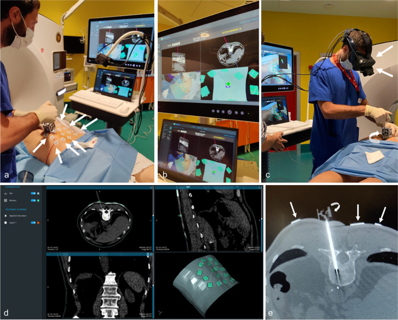
Purpose: To test the technical feasibility of an augmented reality (AR) navigation system to guide bone biopsies.
Methods: We enrolled patients subjected to percutaneous computed tomography (CT)-guided bone biopsy using a novel AR navigation system. Data from prospectively enrolled patients (AR group) were compared with data obtained retrospectively from previous standard CT-guided bone biopsies (control group). We evaluated the following: procedure duration, number of CT passes, patient's radiation dose (dose-length product), complications, and specimen adequacy. Technical success was defined as the ability to complete the procedure as planned, reaching the target center. Technical efficacy was assessed evaluating specimen adequacy.
Results: Eight patients (4 males) aged 58 ± 24 years (mean ± standard deviation) were enrolled in the AR group and compared with 8 controls (4 males) aged 60 ± 15 years. No complications were observed. Procedure duration, number of CT passes, and radiation dose were 22 ± 5 min, 4 (median) [4, 6 interquartile range] and 1,034 ± 672 mGy*cm for the AR group and 23 ± 5 min, 9 [7.75, 11.25], and 1,954 ± 993 mGy*cm for controls, respectively. No significant differences were observed for procedure duration (p = 0.878). Conversely, number of CT passes and radiation doses were significantly lower for the AR group (p < 0.001 and p = 0.021, respectively). Technical success and technical efficacy were 100% for both groups.
Conclusions: This AR navigation system is safe, feasible, and effective; it can decrease radiation exposure and number of CT passes during bone biopsies without increasing duration time.
Relevance statement: This augmented reality (AR) navigation system is a safe and feasible guidance for bone biopsies; it may ensure a decrease in the number of CT passes and patient's radiation dose.
Key points: • This AR navigation system is a safe guidance for bone biopsies. • It ensures decrease of number of CT passes and patient's radiation exposure. • Procedure duration was similar to that of standard CT-guided biopsy. • Technical success was 100% as in all patients the target was reached. • Technical efficacy was 100% as the specimen was adequate in all patients.
Trial registration: ClinicalTrials.gov NCT05732558.
Keywords: Augmented reality; Bone; Image-guided biopsy; Radiology (interventional); Tomography (x-ray computed).
© 2023. The Author(s).
Conflict of interest statement
L. M. Sconfienza is a member of the European Radiology Experimental Editorial Board. He has not taken part in the review or selection process of this article. The other authors declare that they have no competing interests.
Figures
References
Publication types
MeSH terms
Associated data
LinkOut - more resources
Full Text Sources
Medical
Research Materials