

Sulphate-reducing bacteria-mediated pyrite formation in the Dachang Tongkeng tin polymetallic deposit, Guangxi, China
- PMID: 37468706
- PMCID: PMC10356812
- DOI: 10.1038/s41598-023-38827-x
Sulphate-reducing bacteria-mediated pyrite formation in the Dachang Tongkeng tin polymetallic deposit, Guangxi, China
Abstract
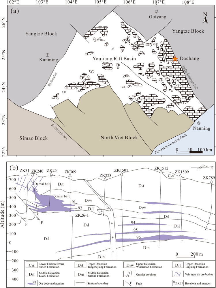
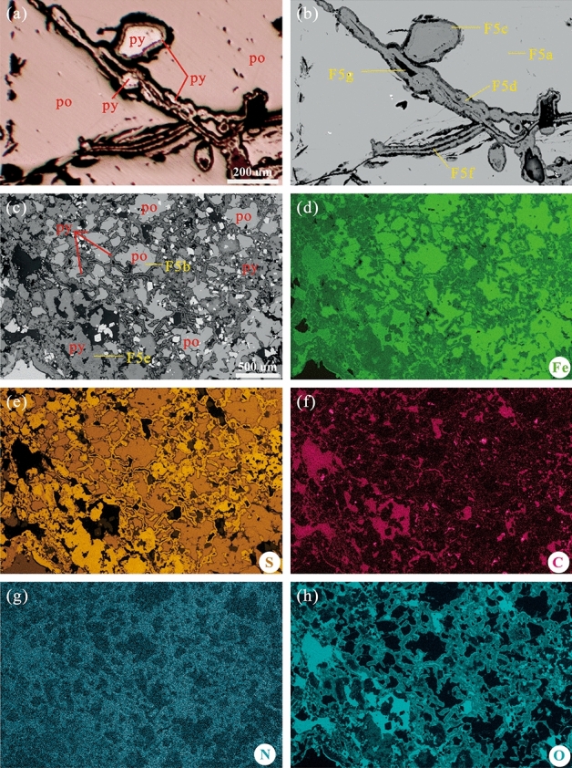
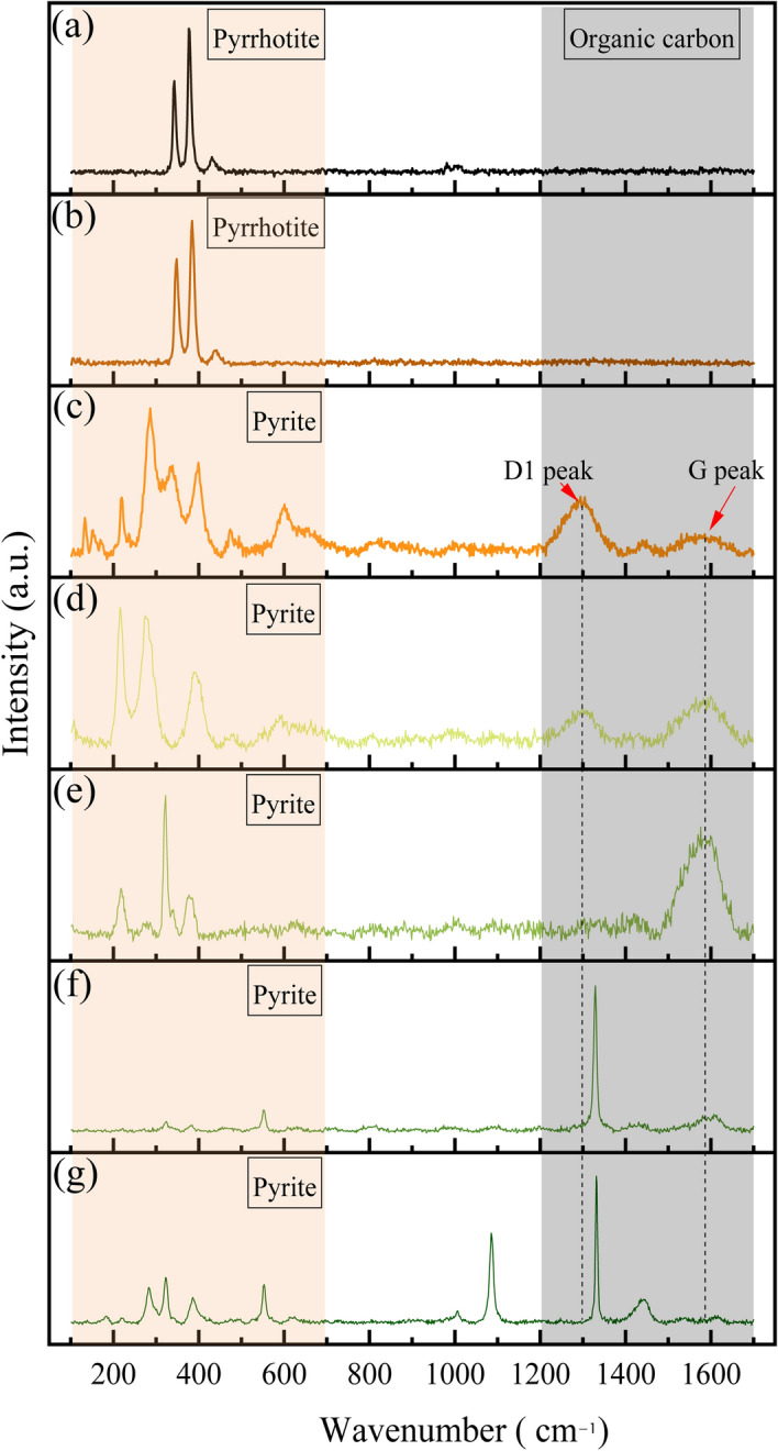
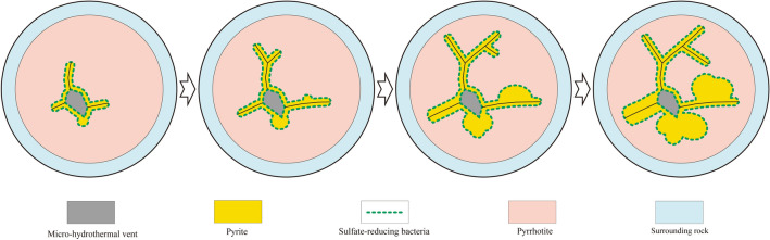
Mediation by sulphate-reducing bacteria (SRB) is responsible for pyrite (FeS2) formation. The origin of the Dachang tin polymetallic ore field is related to the mineralisation of submarine hydrothermal vent sediments. Here, we investigated SRB in these ores via morphological, chemical, and isotopic analyses. Polarised and scanning electron microscopy indicated that trace SRB fossils in the metal sulphide ore were present in the form of tubular, beaded, and coccoidal bodies comprising FeS2 and were enclosed within a pyrrhotite (FeS) matrix in the vicinity of micro-hydrothermal vents. The carbon (C), nitrogen (N), and oxygen (O) contents in the FeS2 synthesised by SRB were high, and a clear biological Raman signal was detected. No such signals were discerned in the peripheral FeS. This co-occurrence of FeS, FeS2, and the remains of bacteria (probably chemoautotrophic bacteria) was interpreted as the coprecipitation process of SRB-mediated FeS2 formation, which has, to the best of our knowledge, not been reported before. Our study also illustrates that combined energy-dispersive X-ray spectroscopy, Raman spectroscopy, and isotopic analysis can be used as a novel methodology to document microbial-mediated processes of mineral deposition in submarine hydrothermal vent ecology on geological time scales.
© 2023. The Author(s).
Conflict of interest statement
The authors declare no competing interests.
Figures


Similar articles
-
Phylogenetic diversity of sulphate-reducing Desulfovibrio associated with three South China Sea sponges.Lett Appl Microbiol. 2015 May;60(5):504-12. doi: 10.1111/lam.12400. Epub 2015 Mar 12. Lett Appl Microbiol. 2015. PMID: 25661682
-
Rapid pyritization in the presence of a sulfur/sulfate-reducing bacterial consortium.Sci Rep. 2020 May 19;10(1):8264. doi: 10.1038/s41598-020-64990-6. Sci Rep. 2020. PMID: 32427954 Free PMC article.
-
Pyrite formation from FeS and H2S is mediated through microbial redox activity.Proc Natl Acad Sci U S A. 2019 Apr 2;116(14):6897-6902. doi: 10.1073/pnas.1814412116. Epub 2019 Mar 18. Proc Natl Acad Sci U S A. 2019. PMID: 30886102 Free PMC article.
-
Reductive biomining of pyrite by methanogens.Trends Microbiol. 2022 Nov;30(11):1072-1083. doi: 10.1016/j.tim.2022.05.005. Epub 2022 May 24. Trends Microbiol. 2022. PMID: 35624031 Review.
-
Iron sulphides mediated autotrophic denitrification: An emerging bioprocess for nitrate pollution mitigation and sustainable wastewater treatment.Water Res. 2020 Jul 15;179:115914. doi: 10.1016/j.watres.2020.115914. Epub 2020 May 6. Water Res. 2020. PMID: 32413614 Review.
References
-
- Fisher CR, Takai K, Le Bris N. Hydrothermal vent ecosystems. Oceanography. 2007;20:14–23. doi: 10.5670/oceanog.2007.75. - DOI
Publication types
MeSH terms
Substances
LinkOut - more resources
Full Text Sources
Miscellaneous
