. 2023 Jul 19;13(1):11623.
doi: 10.1038/s41598-023-38834-y.

Microfluidic-based prostate cancer model for investigating the secretion of prostate-specific antigen and microRNAs in vitro

Affiliations

Microfluidic-based prostate cancer model for investigating the secretion of prostate-specific antigen and microRNAs in vitro

Adventina Padmyastuti et al. Sci Rep. .

Abstract

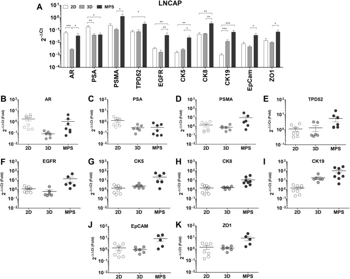
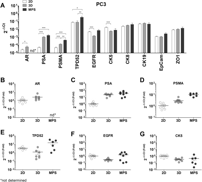
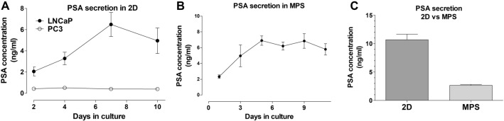
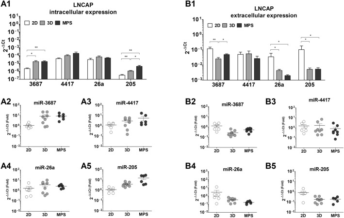
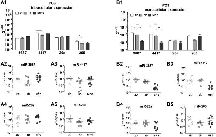
The study of prostate cancer in vitro relies on established cell lines that lack important physiological characteristics, such as proper polarization and expression of relevant biomarkers. Microphysiological systems (MPS) can replicate cancer microenvironments and lead to cellular phenotypic changes that better represent organ physiology in vitro. In this study, we developed an MPS model comprising conventional prostate cancer cells to evaluate their activity under dynamic culture conditions. Androgen-sensitive (LNCaP) and androgen-insensitive (PC3) cells were grown in conventional and 3D cultures, both static and dynamic. Cell morphology, the secretion of prostate-specific antigen, and the expression of key prostate markers and microRNAs were analyzed. LNCaP formed spheroids in 3D and MPS cultures, with morphological changes supported by the upregulation of cytokeratins and adhesion proteins. LNCaP also maintained a constant prostate-specific antigen secretion in MPS. PC3 cells did not develop complex structures in 3D and MPS cultures. PSA expression at the gene level was downregulated in LNCaP-MPS and considerably upregulated in PC3-MPS. MicroRNA expression was altered by the 3D static and dynamic culture, both intra- and extracellularly. MicroRNAs associated with prostate cancer progression were mostly upregulated in LNCaP-MPS. Overall dynamic cell culture substantially altered the morphology and expression of LNCaP cells, arguably augmenting their prostate cancer phenotype. This novel approach demonstrates that microRNA expression in prostate cancer cells is sensitive to external stimuli and that MPS can effectively promote important physiological changes in conventional prostate cancer models.


Conflict of interest statement

The authors declare no competing interests.

Figures

Figure 1
Figure 1
HUMIMIC chip 2 microphysiological system. (A) HUMIMIC chip 2 with the chambers of the right perfusion circuit open. An agar/collagen I hydrogel is shown next to the chip (highlighted circle). (B) Transmitted light image (2x) of the hydrogel used, where the conical shape of the gel is evident, as well as the embedded cellular 3D structure. (C) Detail of a hydrogel (10x) showing the density of LNCaP spheroids. (D) Representation of the microfluidic circuits of the HUMIMIC chip 2, which comprises 2 independent circulations with two culture chambers each, encased in a Polydimethylsiloxane (PDMS) body. The circuits are located at the bottom of the chip, with a glass body on top, evident in panel A. (E) Representation of a cross-section of a culture chamber with the hydrogel inside. The perfusion is driven from the bottom of the chamber (blue arrows), and flow disperses through the gel and supernatant in cycles. E: Representation of the bottom of the chip, which is comprised of a clear microscopy glass that enables direct visualization of the chambers.
Figure 2
Figure 2
Immunofluorescent characterization of LNCaP and PC3 cells. (A1) Schematic representation of the morphology of LNCaP cell in 2D (top) and dynamic culture (bottom). In all images Hoechst33342 (nuclei, blue)) and Phalloidin-488 (f-actin, green) were used as co-stain. The markers of interest were detected using Alexa-555 antibodies (red) (A2) Epidermal growth factor receptor (EGFR) is seemingly dispersed in the cytoplasm of LNCaP cells in 2D. (A3) Zonula ocludens-1 (ZO1) is present in the cellular boundaries of LNCaP, although to a lesser extent. (A4) LNCaP cells in MPS develop into non-hollow spheroids with EGFR predominantly expressed in the outer boundaries of the structures. (A5) In MPS, the expression of ZO1 in LNCaP cells is pronounced and evident between cells. F-actin filaments also present a more consistent organization in MPS and are predominantly concentrated in the periphery of the cells. (A6) EGFR and nuclear stain alone, in LNCaP-MPS. (A7) ZO1 and nuclear stain alone, in LNCaP-MPS. (B1) Schematic representation of the morphology of PC3 cells in 2D (top) and dynamic culture (bottom). (B2) EGFR is concentrated in endosomes dispersed in the cytoplasm of PC3 cells in 2D. (B3) ZO1 expression is pronounced in the cellular boundaries of PC3 cells. (B4) In MPS, PC3 do not develop into 3D structures and remain as individual cells, suspended in the gel. Nonetheless, EGFR and f-actin are predominately found in the periphery of the cells. (B5) ZO1 is dispersed in the cytoplasm of PC3 in MPS, lacking proper membrane localization.
Figure 3
Figure 3
Differences in gene expression between LNCaP cells in 2D, 3D, and MPS: (A) Absolute expression values (2-ΔCt), presented in a log10 scale. There is evident deregulation of several key prostate cancer markers when cells are cultured under 3D -static or -dynamic conditions, relative to conventional culture. Significant statistical differences were found in the expression of all genes analyzed across the three culture conditions tested. This data represents a minimum of six independent samples analyzed using three technical replicates. Each data point represents the average value of the technical replicates. Statistical significances were determined using a two-tailed unpaired t-test (*p < 0.05; **p < 0.01; ***p < 0.001).
Figure 4
Figure 4
Differences in gene expression between PC3 cells in 2D, 3D, and MPS: (A) Absolute expression values (2-ΔCt), presented in a log10 scale. There is evident deregulation of certain key prostate cancer markers when cells are cultured under 3D -static or -dynamic conditions, relative to conventional culture. Significant statistical differences were found in the expression of AR (B), PSA (C), prostate-specific membrane antigen (PSMA) (D), TPD52 (E), EGFR (F), and CK5 (G). 3D culture under static seems to exert the most differences in gene expression of PC3 cells. This data represents a minimum of six independent samples analyzed using three technical replicates. Each data point represents the average value of the technical replicates. Statistical significances were determined using a two-tailed unpaired t-test (*p < 0.05; **p < 0.01; ***p < 0.001).
Figure 5
Figure 5
PSA secretion. (A) In 2D conventional culture PSA levels in PC3 are marginal and unaltered over time, in culture. In contrast, PSA secretion in LNCaP cells is seen increasing over the culture period. Cells were culture in a 24-well format with 500µL of culture media (B) Secretion of PSA over 11 days in LNCaP-MPS, with samples collected in 2–3 days intervals. PSA levels increase up to day 5 in culture and are maintained constant afterward. Cells were culture at a density of 100 000 cells per hydrogel in a volume of 250µL. (C) PSA levels in LNCaP-MPS, after 4 days in culture, are substantially lower than the levels determined for 3D static and 2D culture when the cells were cultured for the same period, cell density and media volume.
Figure 6
Figure 6
MicroRNA expression in LNCaP cells. (A1) Absolute expression values (2-ΔCt), presented in a log10 scale for microRNA levels determined in cell samples (intracellular). There is evident deregulation of prostate cancer-associated microRNAs when LNCaP cells are cultured under 3D -static or -dynamic conditions, relative to conventional culture. The levels of miR-3667 (A2), miR-4417, and miR-205 (A5) are seemingly upregulated while the expression of miR-26a is unaltered (A4). In supernatant samples (B1), only the levels of miR-3687 are seemingly deregulated (B2). The expressions of miR-4417 (B3), miR-26a (B4), and miR-205 (B5) were determined to be considerably variable in the LNCaP supernatant. This data represents a minimum of six independent samples analyzed using three technical replicates. Each data point represents the average value of the technical replicates. Statistical significances were determined using a two-tailed unpaired t-test   (*p < 0.05; ***p < 0.001).
Figure 7
Figure 7
MicroRNA expression in PC3 cells. (A1) Absolute expression values (2-ΔCt), presented in a log10 scale for microRNA levels determined in cell samples (intracellular). Evident deregulation of intracellular prostate cancer-associated microRNAs—miR-3687 (A2), miR-4417 (A3), and miR-205 (A5) was observed in PC3 cells after 3D -static or -dynamic culture condition. The expressions of miR-26a (B4) and miR-205 (B5) were determined to be considerably variable in PC3 supernatant samples. This data represents a minimum of six independent samples analyzed using three technical replicates. Each data point represents the average value of the technical replicates. Statistical significances were determined using a two-tailed unpaired t-test (*p < 0.05; ***p < 0.001).

Similar articles

Cited by

References

    1. Ferlay J, et al. Estimates of worldwide burden of cancer in 2008: GLOBOCAN 2008. Int. J. Cancer. 2010;127:2893–2917. doi: 10.1002/ijc.25516. - DOI - PubMed
    1. Padala SA, et al. Epidemiology of renal cell carcinoma. World J. Oncol. 2020;11:79–87. doi: 10.14740/wjon1279. - DOI - PMC - PubMed
    1. Kirby M, Hirst C, Crawford ED. Characterising the castration-resistant prostate cancer population: A systematic review. Int. J. Clin. Pract. 2011;65:1180–1192. doi: 10.1111/j.1742-1241.2011.02799.x. - DOI - PubMed
    1. Aly A, Mullins CD, Hussain A. Understanding heterogeneity of treatment effect in prostate cancer. Curr. Opin. Oncol. 2015;27:209–216. doi: 10.1097/CCO.0000000000000172. - DOI - PubMed
    1. Bedke J, et al. The 2021 updated European association of urology guidelines on renal cell carcinoma: Immune checkpoint inhibitor–based combination therapies for treatment-naive metastatic clear-cell renal cell carcinoma are standard of care. Eur. Urol. 2021;80:393–397. doi: 10.1016/j.eururo.2021.04.042. - DOI - PubMed

Publication types