Review
. 2023 Aug;25(8):1089-1100.
doi: 10.1038/s41556-023-01194-w. Epub 2023 Jul 19.

Advances and prospects for the Human BioMolecular Atlas Program (HuBMAP)

Collaborators, Affiliations
Review

Advances and prospects for the Human BioMolecular Atlas Program (HuBMAP)

Sanjay Jain et al. Nat Cell Biol. 2023 Aug.

Erratum in

  • Author Correction: Advances and prospects for the Human BioMolecular Atlas Program (HuBMAP).
    Jain S, Pei L, Spraggins JM, Angelo M, Carson JP, Gehlenborg N, Ginty F, Gonçalves JP, Hagood JS, Hickey JW, Kelleher NL, Laurent LC, Lin S, Lin Y, Liu H, Naba A, Nakayasu ES, Qian WJ, Radtke A, Robson P, Stockwell BR, Van de Plas R, Vlachos IS, Zhou M; HuBMAP Consortium; Börner K, Snyder MP. Jain S, et al. Nat Cell Biol. 2024 May;26(5):839. doi: 10.1038/s41556-024-01384-0. Nat Cell Biol. 2024. PMID: 38429479 Free PMC article. No abstract available.

Abstract

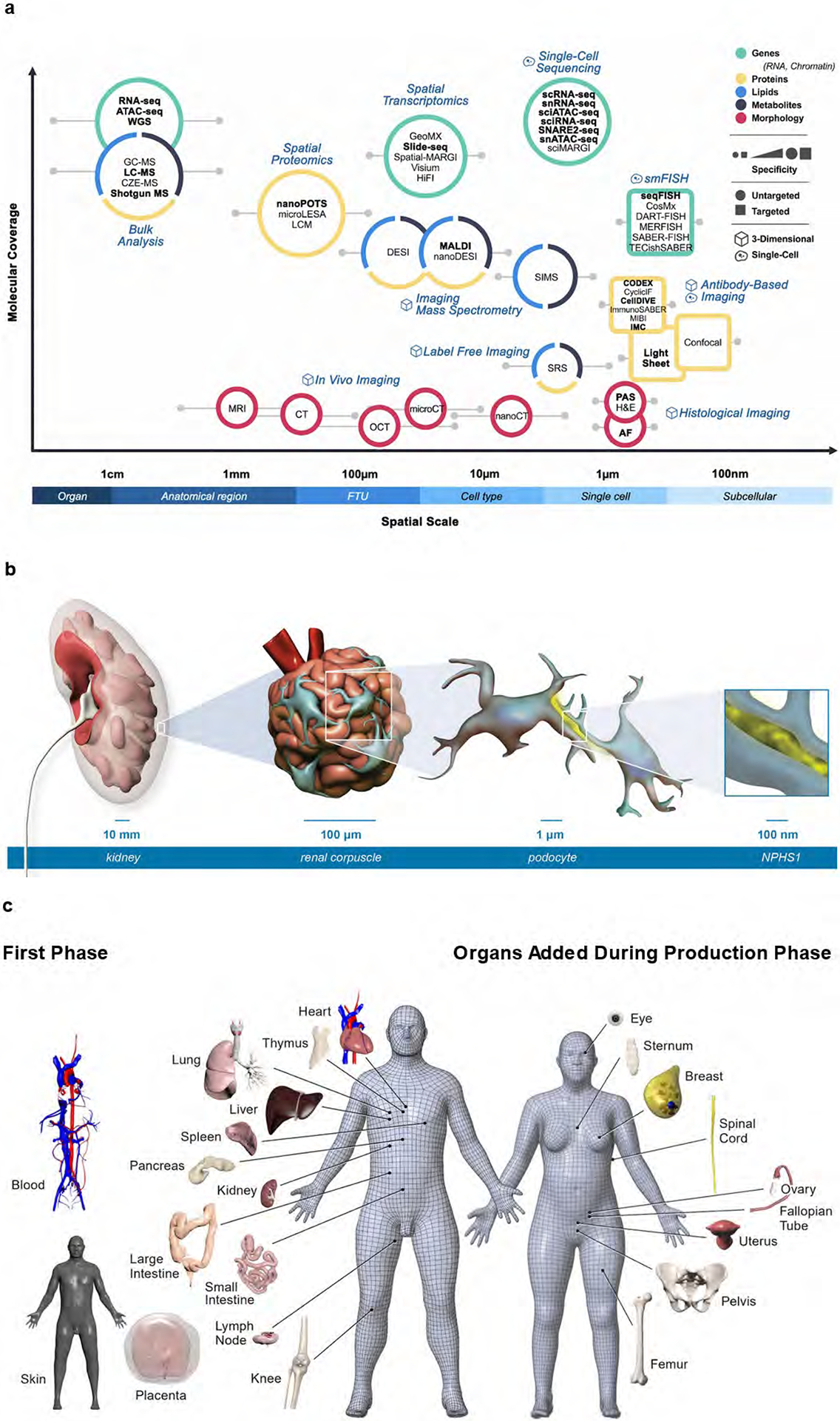
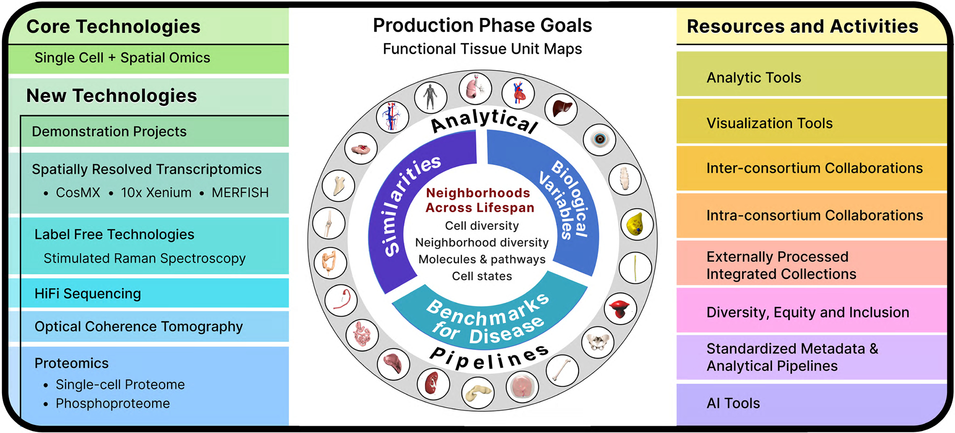
The Human BioMolecular Atlas Program (HuBMAP) aims to create a multi-scale spatial atlas of the healthy human body at single-cell resolution by applying advanced technologies and disseminating resources to the community. As the HuBMAP moves past its first phase, creating ontologies, protocols and pipelines, this Perspective introduces the production phase: the generation of reference spatial maps of functional tissue units across many organs from diverse populations and the creation of mapping tools and infrastructure to advance biomedical research.


Conflict of interest statement

Competing Interests

The authors declare the following competing interests.

F.G. are employees of GE Research. B.R.S. is an inventor on patents and patent applications involving small molecule drug discovery, and the 3F3-FMA antibody, co-founded and serves as a consultant to Inzen Therapeutics, Nevrox Limited, Exarta Therapeutics, and ProJenX Inc.; and serves as a consultant to Weatherwax Biotechnologies Corporation and Akin Gump Strauss Hauer & Feld LLP. M.P.S. is cofounder and advisory board member of Personalis, Qbio, January AI, Mirvie, Filtricine, Fodsel, Lollo, and Protos. I.S.V. consults for Guidepoint Global, Cowen, Mosaic, and NextRNA. N.G. is a co-founder and equity owner of Datavisyn. H.L. is a co-founder and equity owner of ExoMira Medicine, Inc. The remaining authors declare no competing interests.

Figures

Figure 1.
Figure 1.. Molecular coverage and spatial scale of different assay types.
About 40 different analytical technologies are used in HuBMAP. a. Molecular coverage versus spatial scale. Data publicly available via the HuBMAP portal is rendered in bold. b. The multi-scale HRA covers more than 1,500 anatomical structures in the male and female body. A zoom into the kidney (10 mm level) reveals a representative view of a renal corpuscle (200 μm level), a subsegment of one of the ca. 1 million FTUs (nephron) of the kidney that is important in filtration. Podocytes, one of the cells important in filtration (μm level) with nucleus (in blue) and protein NPHS1 that maintains the structural integrity of the filtration barrier (yellow) is illustrated. c. Three-dimensional reference objects exist for 53 organs (counting left/right and female/male organs). Shown on left of the 3D reference bodies are female/male organs for which HRA data exists on the HuBMAP Portal, note that placenta is full-term; on right of the reference bodies are female/male organs that will be added during the production phase. The 3D reference organs are used during tissue registration to automatically assign anatomical structure tags and they serve as landmarks during spatial search for tissue datasets with specific anatomical structures, FTUs, cell types, or biomarkers.
Figure 2.
Figure 2.. Organs and assay types publicly available via HuBMAP portal (as of May 2023).
Organs are sorted alphabetically. Assay types are grouped into bulk, single-cell, and imaging. LC-MS includes Imaging Mass Cytometry (2D), Imaging Mass Cytometry (3D), LC-MS, LC-MS Bottom Up, and LC-MS Top Down. Abbreviations: Assay for Transposase-Accessible Chromatin using sequencing (ATAC-seq), Mass Spectrometry (MS), Whole-genome sequencing (WGS).
Figure 3.
Figure 3.. Unique functional tissue unit neighborhoods in different biological contexts in the human body.
Illustrated are three themes demonstrating functional and neighborhood relationships revealed through HuBMAP atlas data. a) Remarkable differences in interstitial neighborhood of kidney tubules in homeostasis and altered states. Single cell analysis of healthy and diseased kidneys uncovered healthy and several altered cell states. Spatial analysis (2D and 3D) led to the discovery of differential enrichment of immune and stromal cells in adaptive (successful or failed repair of tubules) and degenerative (severely injured cells with degenerative changes) states. The failed repair tubules show enrichment of macrophages, adaptive fibroblasts and myofibroblasts with fibrosis due to collagen deposition, while degenerative tubule cells associate more with CD3+ lymphocytes. b) Distinct immune cell neighborhood of immature and remodeled spiral arteries at the maternal-fetal interface at different gestational ages coincides with maturing placental villi. As gestation progresses, the immune cell composition changes from high-NK/low-macrophages to high-macrophages/ow-NK cells, a pattern that can ascertain gestational age and is associated with paracrine interactions between the placental villous trophoblasts and spiral artery endothelial cell mediated remodeling. c) The healthy human intestine was analyzed at the single-cell level with spatial resolution using a multi-hierarchical approach to define cellular neighborhoods and multi-neighborhood communities. Biological insights were obtained at three scales, for example: (i) at the cell type level, M1 macrophages positively correlate with body mass index (BMI); (ii) at the neighborhood level, the “CD8+ T cell IEL” multicellular neighborhood decreased in the colon compared to the small intestine and for patients with a history of hypertension; (iii) at the cellular community level, distinct cellular communities were identified and found to be layered from the submucosa to the lumen, driven by distinct compositions of epithelial, immune, and mesenchymal cells in these communities. These illustrations relay concepts and are not intended to be truly reflective of actual biological scale and cellular composition. Some of the icons were created with BioRender.com.
Figure 4.
Figure 4.. New technologies, resources, integrated knowledge base and mapping in the production phase.
Left. All organs generate molecular data from core technologies that include single-cell/single-nucleus RNA-seq, morphology, antibody-based protein expression and metabolomics. Several new technologies will be applied in addition. Right. Resources for the community and atlasing efforts. Analytical tools include Azimuth for ATAC-seq and RNA-seq, cell segmentation and neighborhood mapping. Visualization tools include enhancement of Vitessce for 2D and 3D viewing of single cell and spatial data and tracking of specimens with associated data and ASCT+B on the Human Reference Atlas portal. Inter-consortium efforts include harmonizing nomenclature via ASCT+B tables and 2D/3D anatomical structure references, tissue exchange and community tools for mapping and visualization. Intra-consortium efforts include collaborations among the various components of HuBMAP, such as common antibody or targeted transcriptome panels, cross organ comparative analysis such as vasculature or extracellular matrix. External processed integrated collections will provide a mechanism to import external QC data into HuBMAP portal and create opportunities for synergies in organ mapping projects and discovery. Diversity, equity and inclusion efforts are dedicated to establishing infrastructure to attract individuals from underrepresented and disadvantaged backgrounds and provide opportunities for cutting-edge research and to foster their career goals. Standardized data and analytical pipelines will enhance quality control by harmonizing metadata standards across tissue mapping centers and technologies with detailed documentation of metadata, assays, and analytical parameters. Artificial intelligence (AI) tools are resources where users will be able to visualize omics data integrated with histology, quantify tissue components at the level of cell state and predict gene or protein expression or cell identity from histology slides. Center. The information from technologies and resources will be leveraged to create spatial maps of Functional Tissue Units (FTUs) and 2D/3D neighborhoods across HuBMAP organs. The knowledge base created will enable comparative analysis of cell types and neighborhoods across organs to identify similarities, understand how diversity (age, sex, and race) affects FTU maps and create a benchmark reference atlas of FTUs for studying changes in disease and targets for therapies. Some of the icons were created with BioRender.com.

References

    1. Snyder MP et al. The human body at cellular resolution: the NIH Human Biomolecular Atlas Program. Nature 574, 187–192 (2019). - PMC - PubMed
    1. HuBMAP Data Portal. https://portal.hubmapconsortium.org/.
    1. Hao Y et al. Integrated analysis of multimodal single-cell data. Cell 184, 3573–3587.e29 (2021). - PMC - PubMed
    1. Börner K et al. Anatomical structures, cell types and biomarkers of the Human Reference Atlas. Nat. Cell Biol 23, 1117–1128 (2021). - PMC - PubMed
    1. Hickey JW et al. Spatial mapping of protein composition and tissue organization: a primer for multiplexed antibody-based imaging. Nat. Methods 19, 284–295 (2022). - PMC - PubMed

Grants and funding