

Neurotoxin-Derived Optical Probes for Biological and Medical Imaging
- PMID: 37468801
- PMCID: PMC10598172
- DOI: 10.1007/s11307-023-01838-1
Neurotoxin-Derived Optical Probes for Biological and Medical Imaging
Abstract
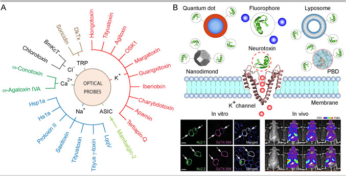
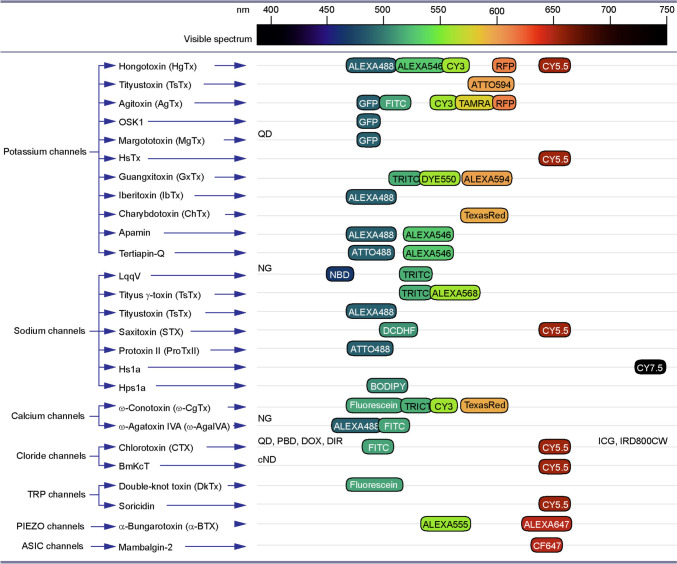
The superb specificity and potency of biological toxins targeting various ion channels and receptors are of major interest for the delivery of therapeutics to distinct cell types and subcellular compartments. Fused with reporter proteins or labelled with fluorophores and nanocomposites, animal toxins and their detoxified variants also offer expanding opportunities for visualisation of a range of molecular processes and functions in preclinical models, as well as clinical studies. This article presents state-of-the-art optical probes derived from neurotoxins targeting ion channels, with discussions of their applications in basic and translational biomedical research. It describes the design and production of probes and reviews their applications with advantages and limitations, with prospects for future improvements. Given the advances in imaging tools and expanding research areas benefiting from the use of optical probes, described here resources should assist the discovery process and facilitate high-precision interrogation and therapeutic interventions.
Keywords: Advanced biomaterials; Animal toxins; Fluorescent probes; ICG; Ion channels; Optical imaging; Visualisation.
© 2023. The Author(s).
Conflict of interest statement
The authors declare no competing interests.
Figures

Similar articles
-
Neurotoxin-Derived Optical Probes for Elucidating Molecular and Developmental Biology of Neurons and Synaptic Connections : Toxin-Derived Optical Probes for Neuroimaging.Mol Imaging Biol. 2024 Dec;26(6):912-925. doi: 10.1007/s11307-024-01954-6. Epub 2024 Sep 30. Mol Imaging Biol. 2024. PMID: 39348040 Free PMC article. Review.
-
Labelled animal toxins as selective molecular markers of ion channels: Applications in neurobiology and beyond.Neurosci Lett. 2018 Jul 13;679:15-23. doi: 10.1016/j.neulet.2017.10.050. Epub 2017 Nov 22. Neurosci Lett. 2018. PMID: 29107087 Review.
-
Inorganic ion-sensitive imaging probes for biomedical applications.Chem Commun (Camb). 2023 May 11;59(39):5807-5822. doi: 10.1039/d3cc00418j. Chem Commun (Camb). 2023. PMID: 37078643 Review.
-
Recent Progress of Hybrid Optical Probes for Neural Membrane Potential Imaging.Biotechnol J. 2020 Dec;15(12):e2000086. doi: 10.1002/biot.202000086. Epub 2020 Jul 30. Biotechnol J. 2020. PMID: 32662937 Review.
-
Optical Probes for Neurobiological Sensing and Imaging.Acc Chem Res. 2018 May 15;51(5):1023-1032. doi: 10.1021/acs.accounts.7b00564. Epub 2018 Apr 13. Acc Chem Res. 2018. PMID: 29652127 Free PMC article. Review.
Cited by
-
Neurotoxin-Derived Optical Probes for Elucidating Molecular and Developmental Biology of Neurons and Synaptic Connections : Toxin-Derived Optical Probes for Neuroimaging.Mol Imaging Biol. 2024 Dec;26(6):912-925. doi: 10.1007/s11307-024-01954-6. Epub 2024 Sep 30. Mol Imaging Biol. 2024. PMID: 39348040 Free PMC article. Review.
-
Optical imaging guidance in oncologic surgery and interventional oncology.Pharmacol Res. 2025 Feb;212:107612. doi: 10.1016/j.phrs.2025.107612. Epub 2025 Jan 17. Pharmacol Res. 2025. PMID: 39826822 Free PMC article. Review.
References
Publication types
MeSH terms
Substances
LinkOut - more resources
Full Text Sources
Medical
