. 2023 Jul 19;20(1):29.
doi: 10.1186/s12989-023-00543-9.

Single-cell transcriptome sequencing-based analysis: probing the mechanisms of glycoprotein NMB regulation of epithelial cells involved in silicosis

Affiliations

Single-cell transcriptome sequencing-based analysis: probing the mechanisms of glycoprotein NMB regulation of epithelial cells involved in silicosis

Shaoqi Yang et al. Part Fibre Toxicol. .

Abstract

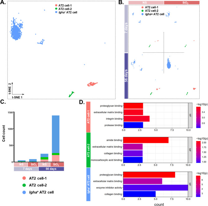
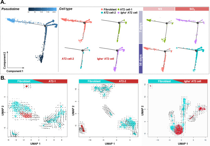
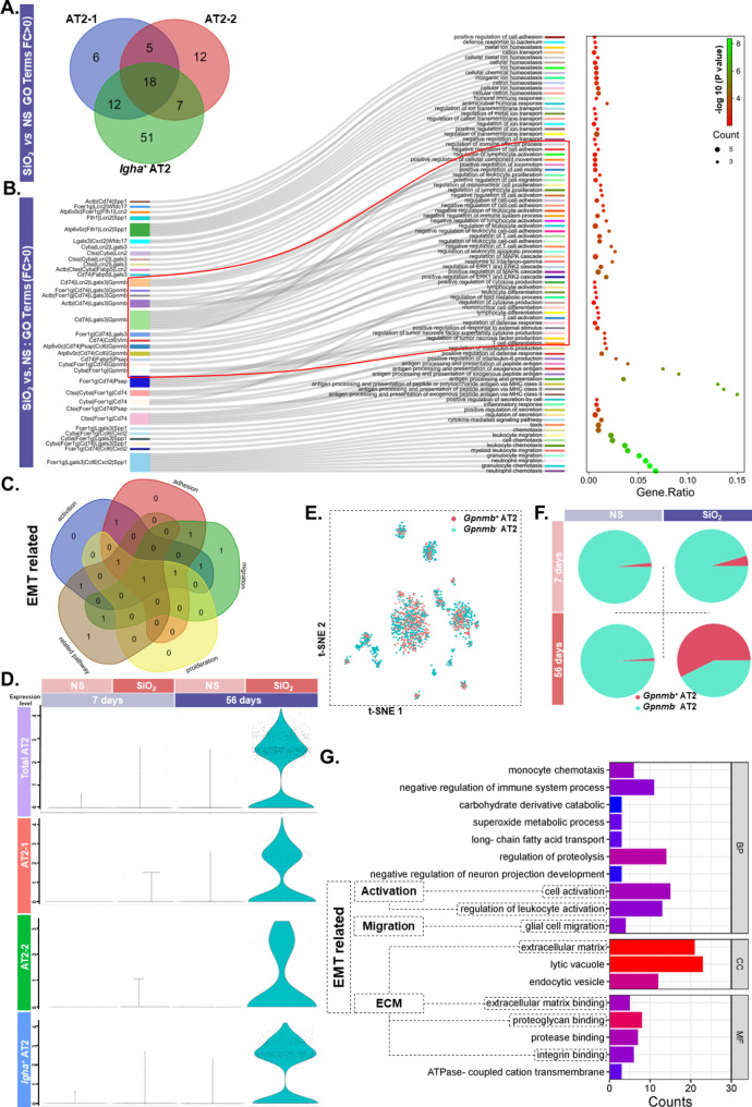
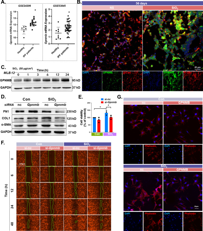
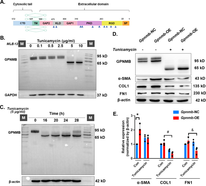
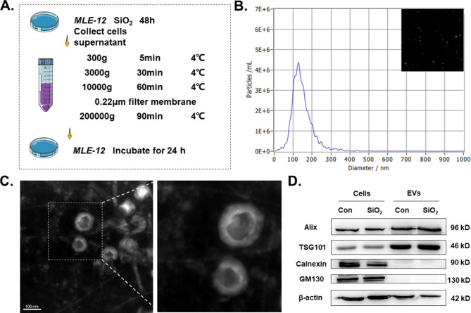
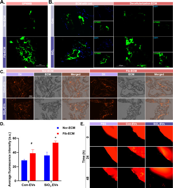
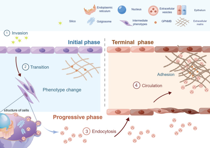
Chronic exposure to silica can lead to silicosis, one of the most serious occupational lung diseases worldwide, for which there is a lack of effective therapeutic drugs and tools. Epithelial mesenchymal transition plays an important role in several diseases; however, data on the specific mechanisms in silicosis models are scarce. We elucidated the pathogenesis of pulmonary fibrosis via single-cell transcriptome sequencing and constructed an experimental silicosis mouse model to explore the specific molecular mechanisms affecting epithelial mesenchymal transition at the single-cell level. Notably, as silicosis progressed, glycoprotein non-metastatic melanoma protein B (GPNMB) exerted a sustained amplification effect on alveolar type II epithelial cells, inducing epithelial-to-mesenchymal transition by accelerating cell proliferation and migration and increasing mesenchymal markers, ultimately leading to persistent pulmonary pathological changes. GPNMB participates in the epithelial-mesenchymal transition in distant lung epithelial cells by releasing extracellular vesicles to accelerate silicosis. These vesicles are involved in abnormal changes in the composition of the extracellular matrix and collagen structure. Our results suggest that GPNMB is a potential target for fibrosis prevention.

Keywords: Epithelial mesenchymal transformation; Extracellular matrix; Extracellular vesicles; Pulmonary fibrosis; Silica.


Conflict of interest statement

The authors declare no competing interests.

Figures

Fig. 1
Fig. 1
Probing alveolar type II epithelial cell fate changes via single-cell transcriptome sequencing. A. t-SNE demonstrating three subtypes of AT2. B. t-SNE demonstrating each subtype of AT2 in the saline and silica groups at 7 and 56 days. C. Changes in the number of alveolar type II epithelial cells. D. GO enrichment analysis of the differential genes (*P < 0.05, avg_logFC > 0) for each subtype of AT2 in the saline and silica groups at 7 and 56 days
Fig. 2
Fig. 2
Probing alveolar type II epithelial cell transition via bioinformatics analysis. A. Pseudo-time-series analysis showing the differentiation trajectory of AT2 and fibroblasts. B. RNA velocity analysis of fibroblasts and three cell types of AT2.
Fig. 3
Fig. 3
Screening of key molecules involved in the occurrence of EMT phenomenon in alveolar II epithelial cells. A. Venn diagram showing the differential genes for the three subtypes of AT2 (*P < 0.05, avg_logFC > 1.0). B. Sankey diagram showing the function of 18 differential genes. C. Venn diagram showing the differential genes for the three subtypes of AT2. D. Violin plot showing the expression of Gpnmb in AT2. E. t-SNE plot showing the distribution of the two subtypes of AT2, Gpnmb+AT2, and GpnmbAT2.F. Pie chart showing the percentage of each AT2 subtype occupying the total number of AT2 cells. G. GO enrichment analysis of the top 20 differential genes
Fig. 4
Fig. 4
GPNMB participation in the EMT process. A. Expression of GPNMB in the lung tissue of patients with IPF was significantly higher than that of the healthy group in the GEO database (*P < 0.05). B. Immunohistochemical staining showing that GPNMB was expressed on epithelial cells. Expression was higher in the experimental group than in the control group, scale bar: 20 μm. C. Representative western blot results showing that the expression of GPNMB was elevated over time in MLE-12 cells. D. Representative western blot results showing that the downregulation of GPNMB partially reversed the SiO2-induced upregulation of FN1, COL1, and α-SMA. E. CCK-8 assay results: #P < 0.05 indicates that the si-Gpnmb group had lower cell viability levels after SiO2 treatment than the si-nc group. *P < 0.05 indicates a significant increase in the viability of MLE-12 cells after 24 h of SiO2 treatment compared to the Con-nc group. F. Wound-healing experiments showing that downregulation of Gpnmb expression attenuated SiO2-induced cell migration. G. Phalloidin assay showing that the cytoskeleton distribution of microfilaments was coarser in the experimental group than in the control group, and the cytoskeleton level in the si-Gpnmb group was lower than that in the si-nc group, scale bar: 20 μm
Fig. 5
Fig. 5
Glycosylation level affectsGpnmb function. A. Structural domain of GPNMB protein. B. Molecular weight of GPNMB changed from 95 kD to 65 kD following treatment with 5 μg/ml of tunicamycin. C. Representative western blot results showing an action time of 16 h following treatment with 5 μg/ml of tunicamycin. D. Representative western blot results showing that the Gpnmb-OE group causes a reversal of the tunicamycin-induced elevation in α-SMA, COL1, and FN1 proteins compared to the Gpnmb-NC group. E. *P < 0.05 indicates that the α-SMA protein levels were higher in the Gpnmb-OE group than in the control group; #P < 0.05 indicates that the COL1 expression levels were lower in the Gpnmb-OE group than in the control group after tunicamycin treatment; and &P < 0.05 indicates that FN1 expression levels were lower in the Gpnmb-OE group than in the control group after tunicamycin treatment
Fig. 6
Fig. 6
Stimulation by SiO2 induces elevated GPNMB expression in EVs released from epithelial cells. A. Schematic diagram of the acquisition of EVs from MLE-12 cells. B. Measurement of EVs with NTA. C. Transmission electron microscopy of EVs isolated from MLE-12 cell cultures. D. Western blot analysis of Alix, TSG101, Calnexin, and GM130 in MLE-12 cells and MLE-12-EVs.
Fig. 7
Fig. 7
MLE-12 endocytosis of highly expressed Gpnmb EVs promotes the EMT process. A. Representative western blot results showing higher GPNMB expression in EVs in the SiO2 group than in the control group. B. Images of EVs incubated with MLE-12 for 0, 6, 12, 24, and 48 h. C. Representative western blot results showing elevated protein levels of COL1, FN1, and α-SMA in the SiO2-EVs group compared to the control group. D. *P < 0.05 indicates higher COL1 protein levels in the SiO2-EVs group compared to the control group, while #P < 0.05 indicates higher FN1 protein levels in the SiO2-EV group compared to the control group. E. Representative western blot results showing elevated expression of GPNMB in EVs secreted by cells in the Gpnmb-OE group compared to the Gpnmb-NC group. F. *P < 0.05 indicates that the GPNMB protein levels were higher in the Gpnmb-OE group than in the Gpnmb-NC group. G. Representative western blot results showing elevated protein levels of COL1, FN1, and α-SMA in the Gpnmb-OE group compared to the control group. H. *P < 0.05 indicates that the α-SMA protein level was higher in the Gpnmb-OE group compared to the control group; #P < 0.05 indicates that the COL1 protein level was higher in the Gpnmb-OE group compared to the control group; and &P < 0.05 indicates that the FN1 protein level was higher in the Gpnmb-OE group compared to the control group
Fig. 8
Fig. 8
Secreted MLE-12 EVs adhere to the extracellular matrix to exert their effects. A. Immunofluorescence staining to assess the GPNMB protein levels in the fibrotic ECM, scale bar: 200 μm. B. Elevated GPNMB protein levels in the fibrotic ECM originate from alveolar type II epithelial cells, scale bar: 275 μm. C. Adhesion of Dil-labeled EVs to the ECM, scale bar: 275 μm. D. Increased adhesion of EVs to Fib-ECM compared to Con-ECM. E. Three-dimensional cell migration assay showing that SiO2-EVs migrated from Fib-ECM at 24 h quicker than the control
Fig. 9
Fig. 9
Schematic representation of the mechanism of GPNMB regulation of silica-induced pulmonary fibrosis in alveolar type II epithelial cells. GPNMB expression in AT2 exposed to SiO2 was increased, promoting cell proliferation, accelerating migration, and regulating the EMT process. GPNMB was persistently highly expressed in AT2 and can be released extracellularly with extracellular vesicles to exert its effects, participating in the regulation of phenotypic transformation and functional changes in normal AT2. Fibrotic ECM showed abnormal accumulation of GPNMB, part of which originated from AT2, and GPNMB adhered to ECM in the form of extracellular vesicles to participate in the pulmonary fibrosis process and continued to exert a sustained amplifying effect on ECM accumulation

Similar articles

References

    1. Gan S, et al. Triiodothyronine attenuates Silica-Induced oxidative stress, inflammation, and apoptosis via thyroid hormone receptor alpha in differentiated THP-1 macrophages. Chem Res Toxicol. 2020;33(5):1256–65. doi: 10.1021/acs.chemrestox.0c00018. - DOI - PubMed
    1. Shi P, et al. Trends in global, regional and national incidence of pneumoconiosis caused by different aetiologies: an analysis from the global burden of Disease Study 2017. Occup Environ Med. 2020;77(6):407–14. doi: 10.1136/oemed-2019-106321. - DOI - PubMed
    1. Barnes H, et al. Silica-associated lung disease: an old-world exposure in modern industries. Respirol (Carlton Vic) 2019;24(12):1165–75. doi: 10.1111/resp.13695. - DOI - PubMed
    1. Wynn TA. Cellular and molecular mechanisms of fibrosis. J Pathol. 2008;214(2):199–210. doi: 10.1002/path.2277. - DOI - PMC - PubMed
    1. Parimon T et al. Alveolar epithelial type II cells as drivers of lung fibrosis in idiopathic pulmonary fibrosis. Int J Mol Sci, 2020. 21(7). - PMC - PubMed

Publication types