. 2023 Jul 19;12(1):61.
doi: 10.1186/s40164-023-00419-w.

Fibronectin promotes tumor angiogenesis and progression of non-small-cell lung cancer by elevating WISP3 expression via FAK/MAPK/ HIF-1α axis and activating wnt signaling pathway

Affiliations

Fibronectin promotes tumor angiogenesis and progression of non-small-cell lung cancer by elevating WISP3 expression via FAK/MAPK/ HIF-1α axis and activating wnt signaling pathway

Fei Zhou et al. Exp Hematol Oncol. .

Abstract

Background: Fibronectin, an extracellular matrix protein, has been reported to be associated with heterogeneous cancer stemness, angiogenesis and progression in multiple cancer types. However, the roles and the underlying mechanism of fibronectin on the progression NSCLC need to be further elucidated.

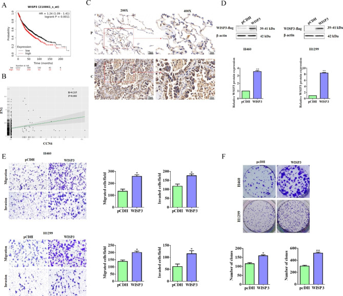
Methods: Public dataset such as Kaplan-Meier Plotter was used to determine the prognostic significance of genes. The correlation of different protein expression in clinical and xenograft tissues was tested by immunohistochemistry experiment. Both in vitro and in vivo experiments were performed to determine the role of fibronectin on the tumor growth, metastasis, and angiogenesis in NSCLC. The activation of key signaling pathway under fibronectin was examined by WB assay. RNA-seq was applicated to screening the target gene of fibronectin. Rescue experiment was performed to confirm the role of target gene in fibronectin-mediated function in NSCLC. Finally, luciferase and CHIP assays were used to elucidate the mechanism by which fibronectin regulated the target gene.

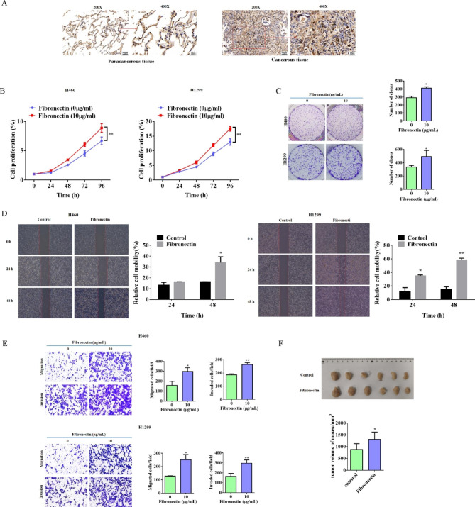
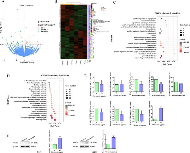
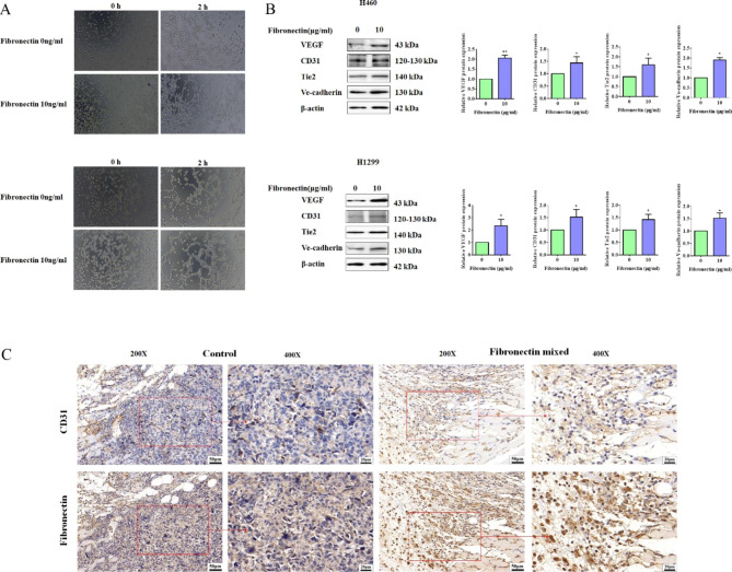
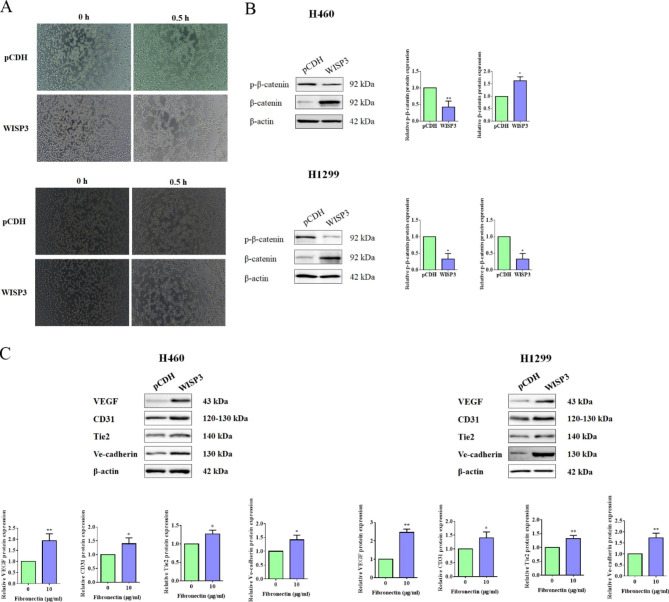
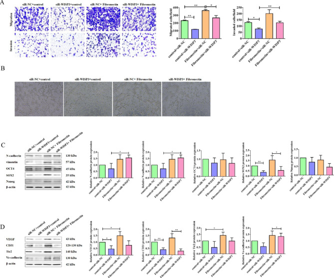
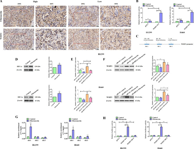
Results: Our results revealed that fibronectin was up-regulated in cancer tissues compared with the normal ones in NSCLC patients. Dish- coated fibronectin enhanced the tumor growth, metastasis, and angiogenesis of NSCLC in vitro and in vivo by promoting EMT and maintaining stemness of NSCLC cells. As expected, fibronectin activated FAK and its downstream MAPK/ERK signaling pathway. WISP3 was screened as a potential target gene of fibronectin. Interestingly, WISP3 effectively activated Wnt signaling pathway, and knockdown of WISP3 effectively blocked the influence of fibronectin on the migration, invasion and vascular structure formation potential of NSCLC cells. Our data also manifested that fibronectin elevated the transcription of WISP3 gene by promoting the binding of HIF-1α to the promoter region of WISP3 in NSCLC cells.

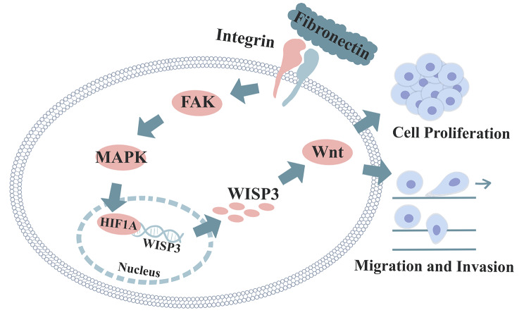
Conclusions: Our findings sketched the outline of the route for fibronectin exert its role in NSCLC, in which fibronectin activated downstream FAK and MAPK/ERK signaling pathways, and mediated the accumulation of HIF-1α. Then, HIF-1α enabled the transcription of WISP3, and subsequently promoted the activation of Wnt signaling pathway, and finally enhanced the tumor growth, metastasis, and angiogenesis in NSCLC.

Keywords: Angiogenesis; Fibronectin; MAPK/ERK; Metastasis; NSCLC; WISP3; Wnt.


Conflict of interest statement

The authors declare no competing interests.

Figures

Fig. 1
Fig. 1
Fibronectin significantly enhanced metastasis and slightly promoted tumor growth of NSCLC. (A) Representative IHC staining images for Fibronectin in paracancerous tissue (P) and cancer tissues from NSCLC patients. Brown color displays Fibronectin protein levels, with counterstaining by hematoxylin in blue. (B) The proliferation abilities of H460 and H1299 cells treated with or without dish-coated fibronectin (10 µg/mL) for 24 h were measured by MTT assay at the indicated time points. (C) Representative photographs of colony formation in H460 and H1299 cells that treated with or without dish-coated fibronectin (10 µg/mL) for 24 h. The number of colonies was photographed and analyzed in the histogram. (D) The migration abilities of H460 and H1299 cells treated with or without dish-coated fibronectin (10 µg/mL) for 24 h were detected by wound healing assay at 24 and 48 h. (E) The migration and invasion abilities of H460 and H1299 cells treated with or without dish-coated fibronectin (10 µg/mL) for 24 h were detected by Transwell assay. Quantitative analysis of migrated and invaded cells was shown in the histogram. (F) 5 × 106 H1299 cells were suspended in 0.2 ml saline with or without fibronectin (10 µg/mL), and subcutaneously injected into nude mice to generate xenograft model. Three weeks later, all tumors were removed from mice and listed for the photo. Data were presented by mean ± SD for cell experiment, or mean ± SEM animal experiments from three independent experiments. *P < 0.05; **P < 0.01 vs. control
Fig. 2
Fig. 2
WISP3 was identified as the candidate target of Fibronectin. (A) Volcano plots, showed genes with partial expression in the control and the dish-coated fibronectin-treated groups in H1299 cells. Red dots showed genes with dramatically higher abundances while the green dots represented genes with significantly lower levels of expression. Values on the x-axis was log2 fold change (log2FC), and the y-axis was log10 p value (log10pvalue). (B) Heat-map of differentially expressed genes from RNA-seq of the control and the fibronectin treated groups. (C and D) Functional analyzing by GO (C) and KEGG (D) of differentially expressed genes in control groups and fibronectin-treated groups. (E) The top 9 differentially expressed genes were subject to qPCR for validation. (F) Western blotting analysis of WISP3 in H460 and H1299 cells treated with or without dish-coated fibronectin (10 µg/mL). Data were presented by mean ± SD from three independent experiments. *P < 0.05; **P < 0.01 vs. control
Fig. 3
Fig. 3
Fibronectin activated FAK, WNT/β-catenin, MAPK/ERK signaling pathways, and promoted EMT and stem cell properties in NSCLC cells. (A) H460 and H1299 cells were treated with or without dish-coated fibronectin (10 µg/mL) for 24 h. Then, Western blot was performed for the detection the proteins of p-FAK, FAK, p-MEK, MEK, p-ERK and ERK. β-actin was used as a loading control. Quantitative analysis of phosphorylated proteins expression were shown in the histograms. (B) H460 and H1299 cells were treated with or without dish-coated fibronectin (10 µg/mL) for 24 h. Then, t Western blot was performed for the detection the proteins of p-β-catenin and β-catenin. β-actin was used as a loading control. Quantitative analysis of p-β-catenin and β-catenin expression were shown in the histogram. (C) H460 and H1299 cells were treated with or without dish-coated fibronectin (10 µg/mL) for 24 h. Then, Western blot was performed for the detection the proteins of N-cadherin, vimentin, OCT4 and Nanog. β-actin was used as a loading control. Quantitative analysis of proteins expression were shown in the histogram. Data were presented by mean ± SD from three independent experiments. *P < 0.05; **P < 0.01 vs. control
Fig. 4
Fig. 4
Fibronectin positively regulated the angiogenesis in NSCLC. (A) In vitro Matrigel assay was performed to detect the vascular structure formation in H460 and H1299 cells treated with or without dish-coated fibronectin (10 µg/mL) for 24 h. (B) H460 and H1299 cells were treated with or without dish-coated fibronectin (10 µg/mL) for 24 h. Then, Western blot was performed for the detection the proteins of VEGF, CD31, Tie2 and Ve-cadherin. β-actin was used as a loading control. Quantitative analysises of proteins expression were shown in the histogram. (C) IHC assay was used to determine the expression of fibronectin and CD31 in tumor tissues from xenograftsderived from H1299 cells mixed with or without fibronectin, and representative data were shown. Data were presented by mean ± SD from three independent experiments. *P < 0.05; **P < 0.01 vs. control
Fig. 5
Fig. 5
WISP3 strengthened cell migration, invasion and colony formation in NSCLC. (A) Correlation between expression of WISP3 and overall survival of lung cancer patients. Data were obtained from the Kaplan-Meier Plotter database. (B) The correlation of WISP3 (CCN6)and fibronectin (FN1) was analyzed using sequencing data from TCGA. (C) Representative IHC staining images for WISP3 in paracancerous tissues (P) and cancer tissues from NSCLC patients. Brown color displays Fibronectin protein levels, with counterstaining by hematoxylin in blue. (D) Western blot was performed to detect the overexpression of WISP3 in H460 and H1299 cells transfected with pCDH or pCDH-WISP3 for 24 h. Quantitative analysis of proteins expression were shown in the histogram. (E) The migration and invasion ability of H460 and H1299 cells transfected with pCDH or pCDH-WISP3 were detected by Transwell assay. Quantitative analysis of migrated and invaded cells was shown in the histogram. (F) Representative photographs of colony formation in H460 and H1299 cells that transfected with pCDH or pCDH-WISP3. The number of colonies was photographed and analyzed in the histogram. Data were presented by mean ± SD from three independent experiments. *P < 0.05; **P < 0.01 vs. control
Fig. 6
Fig. 6
WISP3 promoted tube forming potential of NSCLC cells in vitro. (A) In vitro Matrigel assay was performed to detect the vascular structure formation in H460 and H1299 cells transfected WISP3 overexpression plasmid (pCDH-WISP3) or empty vector (pCDH) for 24 h. (B) H460 and H1299 cells were transfected with pCDH or pCDH-WISP3 for 24 h. Then, Western blot was performed for the detection the proteins of p-β-catenin and β-catenin. β-actin was used as a loading control. Quantitative analysis of p-β-catenin and β-catenin expression were shown in the histogram. (C) H460 and H1299 cells were transfected with pCDH or pCDH-WISP3 for 24 h. Then, Western blot was performed for the detection the proteins of VEGF, CD31, Tie2 and Ve-cadherin. β-actin was used as a loading control. Quantitative analysis of proteins expression were shown in the histogram. Data were presented by mean ± SD from three independent experiments. *P < 0.05; **P < 0.01 vs. control
Fig. 7
Fig. 7
Knockdown of WISP3 blocked Fibronectin-promoted cell migration, invasion, angiogenesis and cancer stemness in NSCLC cells. (A) H1299 cells were transfected with siNC or siWISP3 for 24 h. Then, the migration and invasion ability of the transfected cells treated with or without dish-coated fibronectin were detected by transwell assay. Quantitative analysis of migrated and invaded cells was shown in the histogram. (B) In vitro Matrigel assay was performed to detect the vascular structure formation in H1299 cells treated as (A). (C) Western blot assay was used to detect the proteins of N-catenin, vimentin, OCT4, SOX2 and Nanog in H1299 cells as treated in (A). β-actin was used as a loading control. Quantitative analysis of proteins expression were shown in the histogram. (D) Western blot assay was used to detect the proteins of VEGF, CD31, Tie2 and Ve-cadherin in H1299 cells treated as (A). β-actin was used as a loading control. Quantitative analysis of proteins expression were shown in the histogram. Data were presented by mean ± SD from three independent experiments. *P < 0.05; **P < 0.01 vs. control
Fig. 8
Fig. 8
Fibronectin up-regulated WISP3 expression by activating HIF-1α in NSCLC cells. (A) IHC assay was performed to detect the WISP3 expression in NSCLC cancer tissues with high or low fibronectin expression, and representative data were shown. (B) Luciferase experiments were used to determine the role of fibronectin on the transcriptional activity of WISP3 promoter. (C) The potential binding site for HIF-1α to WISP3 promoter was predicted by JASPAR dataset and details were shown in the schematic diagram. (D) Western blot assay was used to detect the proteins of HIF-1α in H1299 cells treated with or without dish-coated fibronectin (10 µg/mL) for 24 h. β-actin was used as a loading control. Quantitative analysis of proteins expression were shown in the histogram. (E) H1299 and H460 cells were transfected with siRNA against HIF-1α or negative control (NC) for 24 h, followed by treating with or without dish-coated fibronectin (10 µg/mL) for 24 h. Then, qPCR was performed to examine the expression of WISP3 mRNA. (F) Western blot assay was used to detect the WISP3 proteins levels expressed in H1299 and H460 cells treated as (E). β-actin was used as a loading control. Quantitative analysis of proteins expression were shown in the histogram. (G) H1299 and H460 cells were treated with or without dish-coated fibronectin (10 µg/mL) for 24 h, CHIP assay was performed using HIF-1α antibody to determine the binding site of HIF-1α on WISP3 promoter. (H) Luciferase experiment was used to identify the role of fibronectin on the wildtype WISP3 promoter or the mutant one, in which the binding site1 to HIF-1α was mutant. Data were presented by mean ± SD from three independent experiments. *P < 0.05; **P < 0.01 vs. control
Fig. 9
Fig. 9
The schematic diagram for how fibronectin functions in NSCLC cells

Similar articles

Cited by

References

    1. Sung H, Ferlay J, Siegel RL, Laversanne M, Soerjomataram I, Jemal A, et al. Global Cancer Statistics 2020: GLOBOCAN estimates of incidence and Mortality Worldwide for 36 cancers in 185 countries. Cancer J Clin. 2021;71(3):209–49. doi: 10.3322/caac.21660. - DOI - PubMed
    1. Bao Y, Yang X, Men Y, Kang J, Sun X, Zhao M, et al. Postoperative radiotherapy improves survival of patients with ypN2 non-small cell lung cancer after neoadjuvant chemotherapy followed by surgery - A propensity score matching study of the Surveillance, Epidemiology, and end results database. Thorac cancer. 2022;13(3):404–11. doi: 10.1111/1759-7714.14273. - DOI - PMC - PubMed
    1. Rodak O, Peris-Diaz MD, Olbromski M, Podhorska-Okolow M, Dziegiel P. Current Landscape of Non-Small Cell Lung Cancer: Epidemiology, histological classification, targeted Therapies, and Immunotherapy. Cancers. 2021;13:18. doi: 10.3390/cancers13184705. - DOI - PMC - PubMed
    1. Hayashi H, Nadal E, Gray JE, Ardizzoni A, Caria N, Puri T, et al. Overall treatment strategy for patients with metastatic NSCLC with activating EGFR mutations. Clin Lung Cancer. 2022;23(1):e69–e82. doi: 10.1016/j.cllc.2021.10.009. - DOI - PubMed
    1. Insa A, Martin-Martorell P, Di Liello R, Fasano M, Martini G, Napolitano S, et al. Which treatment after first line therapy in NSCLC patients without genetic alterations in the era of immunotherapy? Crit Rev Oncol/Hematol. 2022;169:103538. doi: 10.1016/j.critrevonc.2021.103538. - DOI - PubMed