. 2023 Oct;14(5):2126-2142.
doi: 10.1002/jcsm.13290. Epub 2023 Jul 19.

Skeletal muscle-specific DJ-1 ablation-induced atrogenes expression and mitochondrial dysfunction contributing to muscular atrophy

Affiliations

Skeletal muscle-specific DJ-1 ablation-induced atrogenes expression and mitochondrial dysfunction contributing to muscular atrophy

Shuang Zhang et al. J Cachexia Sarcopenia Muscle. 2023 Oct.

Abstract

Background: DJ-1 is a causative gene for Parkinson's disease. DJ-1-deficient mice develop gait-associated progressive behavioural abnormalities and hypoactive forearm grip strength. However, underlying activity mechanisms are not fully explored.

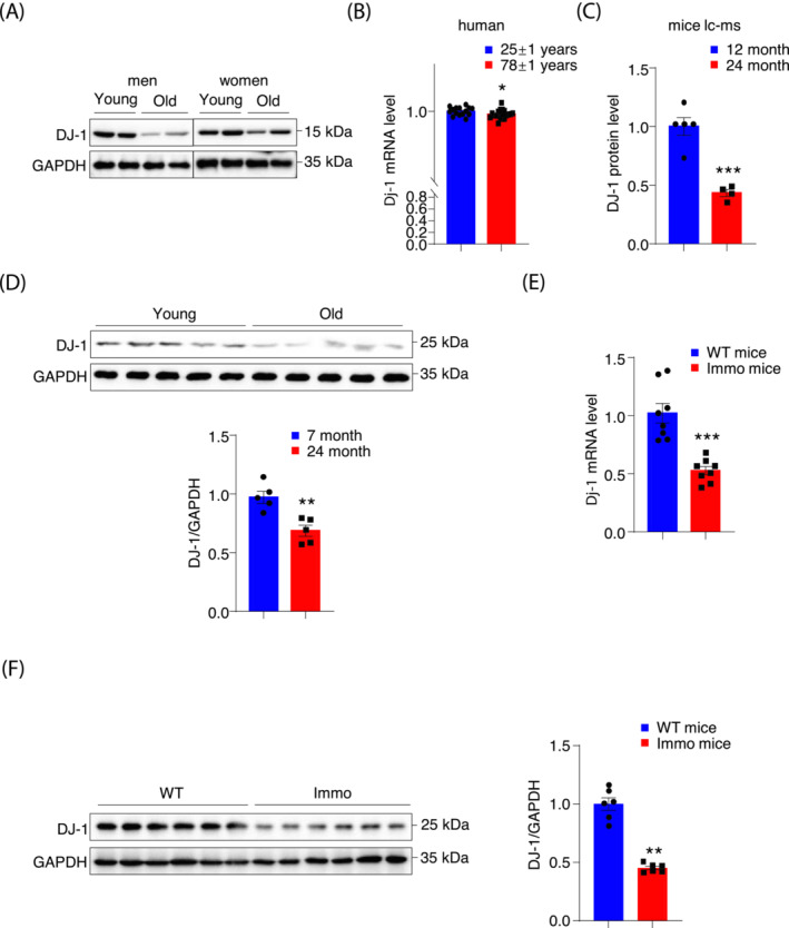
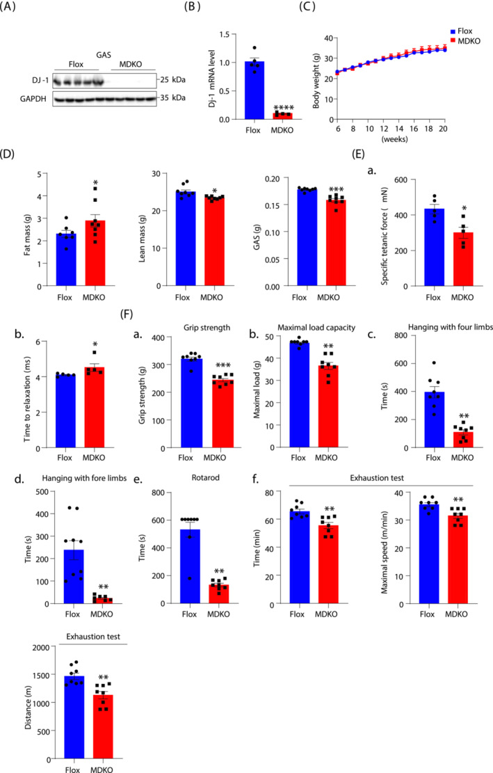
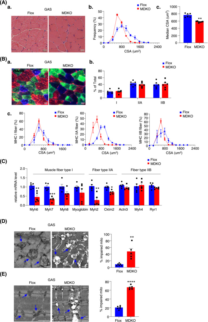
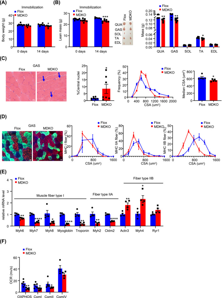
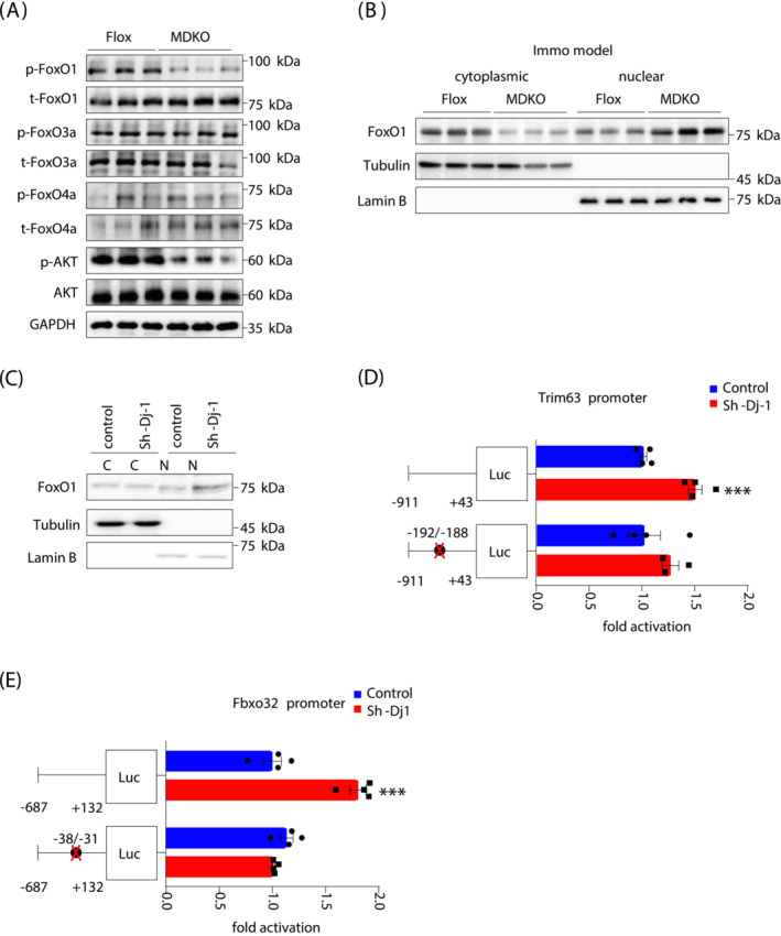
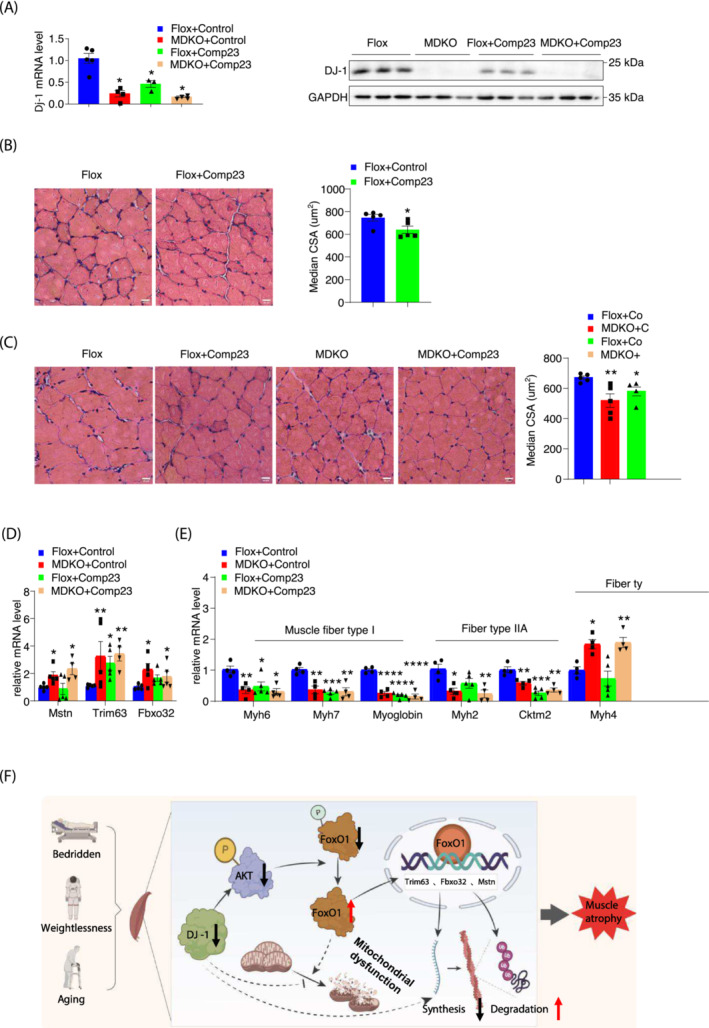
Methods: Western blotting and quantitative real-time polymerase chain reaction approaches were adopted to analyse DJ-1 expression in skeletal muscle from aged humans or mice and compared with young subjects. Skeletal muscle-specific-DJ-1 knockout (MDKO) mice were generated, followed by an assessment of the physical activity phenotypes (grip strength, maximal load capacity, and hanging, rotarod, and exercise capacity tests) of the MDKO and control mice on the chow diet. Muscular atrophy phenotypes (cross-sectional area and fibre types) were determined by imaging and quantitative real-time polymerase chain reaction. Mitochondrial function and skeletal muscle morphology were evaluated by oxygen consumption rate and electron microscopy, respectively. Tail suspension was applied to address disuse atrophy. RNA-seq analysis was performed to indicate molecular changes in muscles with DJ-1 ablation. Dual-luciferase reporter assays were employed to identify the promoter region of Trim63 and Fbxo32 genes, which were indirectly regulated by DJ-1 via the FoxO1 pathway. Cytoplasmic and nuclear fractions of DJ-1-deleted muscle cells were analysed by western blotting. Compound 23 was administered into the gastrocnemius muscle to mimic the of DJ-1 deletion effects.

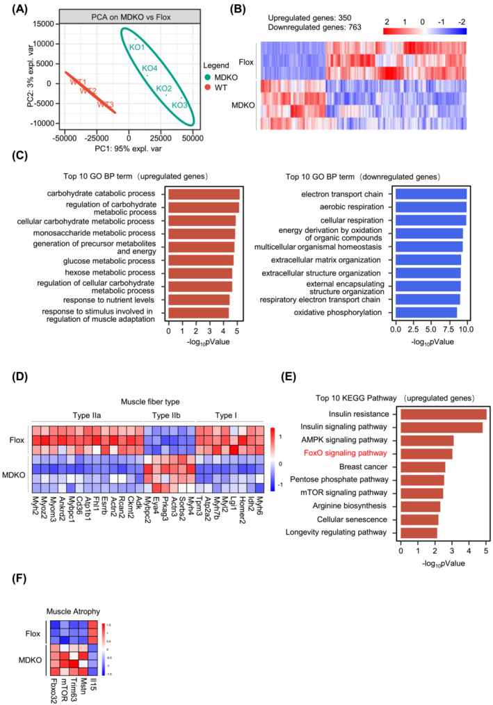
Results: DJ-1 expression decreased in atrophied muscles of aged human (young men, n = 2; old with aged men, n = 2; young women, n = 2; old with aged women, n = 2) and immobilization mice (n = 6, P < 0.01). MDKO mice exhibited no body weight difference compared with control mice on the chow diet (Flox, n = 8; MDKO, n = 9). DJ-1-deficient muscles were slightly dystrophic (Flox, n = 7; MDKO, n = 8; P < 0.05), with impaired physical activities and oxidative capacity (n = 8, P < 0.01). In disuse-atrophic conditions, MDKO mice showed smaller cross-sectional area (n = 5, P < 0.01) and more central nuclei than control mice (Flox, n = 7; MDKO, n = 6; P < 0.05), without alteration in muscle fibre types (Flox, n = 6; MDKO, n = 7). Biochemical analysis indicated that reduced mitochondrial function and upregulated of atrogenes induced these changes. Furthermore, RNA-seq analysis revealed enhanced activity of the FoxO1 signalling pathway in DJ-1-ablated muscles, which was responsible for the induction of atrogenes. Finally, compound 23 (an inhibitor of DJ-1) could mimic the effects of DJ-1 ablation in vivo.

Conclusions: Our results illuminate the crucial of skeletal muscle DJ-1 in the regulation of catabolic signals from mechanical stimulation, providing a therapeutic target for muscle wasting diseases.

Keywords: Atrogenes; Atrophy; DJ-1; Skeletal muscle.


Conflict of interest statement

The authors declare no competing interests.

Figures

Figure 1
Figure 1
DJ‐1 expression is decreased in skeletal muscle from human and mice with muscle atrophy. (A) DJ‐1 immunoblots of young and old aged men (young, 28 and 29 years; old, 65 and 88 years) and women (young, 15 and 26 years; old, 58 and 69 years) muscles lysates. (B) DJ‐1 RNA‐seq expression data from young (n = 15, 25 ± 1 years) and old (n = 21, 78 ± 1 years) human muscles in GSE25941 datasets. (C) The fold change of DJ‐1 protein expression in young (n = 5, 12 months) and old (n = 4, 24 months) male mice muscles. (D) DJ‐1 immunoblots of young (n = 5, 7 months) and old (n = 5, 24 months) male mice muscles lysates and the quantification of blots. (E) DJ‐1 expression in GAS muscle of Flox male mice and immobilized Flox male mice (n = 8). (F) DJ‐1 immunoblots of GAS from mice without/with immobilization and the quantification of blots. Data represented the mean ± SEM. *P < 0.05, **P < 0.01, ***P < 0.001, a two‐tailed Student's t‐test was used for statistical analysis.
Figure 2
Figure 2
Mice with DJ‐1‐specific knockout in skeletal muscle show decreased muscle mass and impaired exercise performance. (A) Western blot analysis of DJ‐1 in GAS of muscle‐specific DJ‐1 knockout (MDKO) and Flox mice. (B) DJ‐1 expression level in GAS by qPCR. (C) Body weight of male MDKO and Flox mice fed on chow diet (n = 8–9). (D) Body composition and muscle weight of male MDKO and Flox mice at the age of 18 weeks (n = 7–8). Fat mass, lean mass, GAS weight. (E) Tibialis anterior (TA) muscle force analysis of male mice at the age of 18 weeks (n = 5). Specific titanic force of TA muscles (A), relaxation time (B). (F) Exercise performance tests. Grip strength of mice at the age of 13 weeks (n = 8) (A). Maximum load capacity of mice in a resistance test at the age of 14 weeks (n = 8) (B). Maximum hanging time with four or fore limbs of mice at the age of 15 weeks (n = 8) (C, D). Rotarod performance test of mice at the age of 16 weeks (n = 8) (E). Total time, maximum speed, and total distance of mice in exhaustion test at the age of 17 weeks (n = 8) (F). Data represented the mean ± SEM. *P < 0.05, **P < 0.01, ***P < 0.001, a two‐tailed Student's t‐test was used for statistical analysis.
Figure 3
Figure 3
DJ‐1 ablation in skeletal muscle decreases fibre cross‐sectional area and impairs mitochondrial function. (A) Physiological consequences of GAS H&E staining in male mice at the age of 18 weeks (scale bars, 20 μm). Representative H&E staining of GAS (A). Fibre cross‐sectional area (CSA) distribution and median CSA in GAS of male mice (n = 6–7) (B, C). (B) Immunofluorescence analysis of fibre type composition in GAS. The different myosin heavy chain isoforms were stained in blue (MyHC‐I), green (MyHC‐IIa) or red (MyHC‐IIb) (scale bars, 20 μm). Representative immunofluorescence staining of GAS (A). The ratio of muscle fibre type (n = 6) (B). Fibre cross‐sectional area (CSA) distribution in different muscle fibres (n = 6) (C). (C) Expression of muscle fibre type related genes measured by qPCR (n = 5). (D) Representative electron micrographs of cross and the ratio of impaired mitochondria in GAS of male mice (scale bars, 0.5 μm). (E) Representative electron micrographs and quantification of abnormal mitochondria of longitudinal section. Blue arrows point to the healthy mitochondria in Flox mice and the impaired mitochondria in MDKO mice (scale bars, 0.5 μm). Data represented the mean ± SEM. *P < 0.05, **P < 0.01, ***P < 0.001, a two‐tailed Student's t‐test was used for statistical analysis.
Figure 4
Figure 4
DJ‐1 deletion in skeletal muscle aggravates atrophy during immobilization. (A) Body weight of male mice during immobilization at the age of 10‐week‐old (n = 6–7). (B) Lean mass, representative morphology and weight of muscles male mice during immobilization at the age of 10‐week‐old (n = 6–7). Lean mass. Representative morphology of quadriceps (QUA), GAS, soleus (SOL), TA, and extensor digitalis anterior (EDL) after immobilization. Weight of QUA, GAS, SOL, TA, and EDL after immobilization (n = 6–7). (C) Physiological consequences of GAS H&E staining in immobilized mice. Representative H&E staining of GAS (scale bars, 10 μm), the percentage of central nuclei after immobilization, CSA distribution, and median CSA in GAS of immobilized mice (n = 5). Blue arrows point to the nuclei inward migration in MDKO mice. (D) Physiological consequences of immunofluorescence staining in GAS. The different myosin heavy chain isoforms were stained in blue (MyHC‐I), green (MyHC‐IIa), and red (MyHC‐IIb) (scale bars, 20 μm). Representative immunofluorescence of muscle fibre type composition in GAS of immobilized mice and CSA distribution in GAS of immobilized mice (n = 6–7). (E) Expression of muscle fibre type related genes of GAS (n = 4). (F) Oroboros O2k respirometer oxygen flux analysis of permeabilized immobilized GAS (n = 5). Data represented the mean ± SEM. *P < 0.05, **P < 0.01, ***P < 0.001, ****P < 0.0001, a two‐tailed Student's t‐test was used for statistical analysis.
Figure 5
Figure 5
DJ‐1 knockout in skeletal muscle shows molecular signs of atrophy. (A) Principal component analysis (PCA) was analysed from RNA‐seq data of GAS in immobilized mice (n = 3–4). (B) Heatmap of 1113 differential genes of GAS in immobilized mice (n = 3–4). (C) GO enrichment analysis based on differential expression genes. Top 10 GO BP term of upregulated genes (left). Top 10 GO BP term down‐regulated genes (right). (D) Expression of muscle fibre type related genes from RNA‐seq data of immobilization models (n = 3–4). (E) KEGG enrichment analysis based on upregulated genes. (F) Atrogenes expression from RNA‐seq data (n = 3–4). Data represented the mean ± SEM. *P < 0.05, **P < 0.01, ***P < 0.001, a two‐tailed Student's t‐test was used for statistical analysis. UBI, ubiquitination; Immo model, immobilized model.
Figure 6
Figure 6
FoxO1 pathway is responsible for DJ‐1 ablation‐induced muscle atrophy. (A) Immunoblot analysis of FoxO family and AKT of GAS in immobilization models. (B) Protein level of FoxO1 in cytoplasmic and nuclear fractions of GAS in immobilization mice. (C) Protein level of FoxO1 in cytoplasmic and nuclear fractions of C2C12 myotubes with/without DJ‐1 knockdown. (D, E) Dual‐luciferase assays of Trim63 and Fbxo32 promoter. Luciferase activity was corrected for Renilla luciferase activity and normalized to control group. The putative FoxO site was noted by black circles. Binding site for FoxO1 in the Trim63 promoter was identified at −192/−188. Binding site for FoxO1 in the Fbxo32 promoter was identified at −38/−31. Data represented the mean ± SEM. *P < 0.05, ***P < 0.001, a two‐tailed Student's t‐test was used for statistical analysis. Immo model, immobilized model; Luc, luciferase.
Figure 7
Figure 7
Compound 23 mimics the effects of skeletal muscle DJ‐1 ablation in vivo. (A) DJ‐1 mRNA level and protein level expression of GAS from indicated mice after immobilization (n = 3–5). (B, C) Physiological consequences of GAS in comp23‐treated mice. Representative H&E staining of GAS in comp23‐treated mice (scale bars, 20 μm) (left). The median of fibre CSA in GAS from a (n = 5) (right). (D) Expression of GAS atrogenes (n = 4–5). (E) Expression of muscle fibre type related genes of GAS. (F) Working models. Data represented the mean ± SEM. *P < 0.05, **P < 0.01, ***P < 0.001, a two‐tailed Student's t‐test was used for statistical analysis. comp23, Compound 23.

Similar articles

Cited by

References

    1. Hasselgren PO, Fischer JE. Muscle cachexia: current concepts of intracellular mechanisms and molecular regulation. Ann Surg 2001;233:9–17. - PMC - PubMed
    1. Mitch WE, Goldberg AL. Mechanisms of muscle wasting. The role of the ubiquitin‐proteasome pathway. N Engl J Med 1996;335:1897–1905. - PubMed
    1. Tawa NE Jr, Odessey R, Goldberg AL. Inhibitors of the proteasome reduce the accelerated proteolysis in atrophying rat skeletal muscles. J Clin Invest 1997;100:197–203. - PMC - PubMed
    1. Clarke BA, Drujan D, Willis MS, Murphy LO, Corpina RA, Burova E, et al. The E3 Ligase MuRF1 degrades myosin heavy chain protein in dexamethasone‐treated skeletal muscle. Cell Metab 2007;6:376–385. - PubMed
    1. Attaix D, Baracos VE. MAFbx/Atrogin‐1 expression is a poor index of muscle proteolysis. Curr Opin Clin Nutr Metab Care 2010;13:223–224. - PubMed

Publication types