. 2023 Jul 4:14:1219359.
doi: 10.3389/fmicb.2023.1219359. eCollection 2023.

Divide and conquer: genetics, mechanism, and evolution of the ferrous iron transporter Feo in Helicobacter pylori

Affiliations

Divide and conquer: genetics, mechanism, and evolution of the ferrous iron transporter Feo in Helicobacter pylori

Camilo Gómez-Garzón et al. Front Microbiol. .

Abstract

Introduction: Feo is the most widespread and conserved system for ferrous iron uptake in bacteria, and it is important for virulence in several gastrointestinal pathogens. However, its mechanism remains poorly understood. Hitherto, most studies regarding the Feo system were focused on Gammaproteobacterial models, which possess three feo genes (feoA, B, and C) clustered in an operon. We found that the human pathogen Helicobacter pylori possesses a unique arrangement of the feo genes, in which only feoA and feoB are present and encoded in distant loci. In this study, we examined the functional significance of this arrangement.

Methods: Requirement and regulation of the individual H. pylori feo genes were assessed through in vivo assays and gene expression profiling. The evolutionary history of feo was inferred via phylogenetic reconstruction, and AlphaFold was used for predicting the FeoA-FeoB interaction.

Results and discussion: Both feoA and feoB are required for Feo function, and feoB is likely subjected to tight regulation in response to iron and nickel by Fur and NikR, respectively. Also, we established that feoA is encoded in an operon that emerged in the common ancestor of most, but not all, helicobacters, and this resulted in feoA transcription being controlled by two independent promoters. The H. pylori Feo system offers a new model to understand ferrous iron transport in bacterial pathogens.

Keywords: Feo; Fur; Helicobacter pylori; NikR; Vibrio cholerae; iron transport; nickel; operon.


Conflict of interest statement

The authors declare that the research was conducted in the absence of any commercial or financial relationships that could be construed as a potential conflict of interest.

Figures

Figure 1
Figure 1
Current understanding of the Feo system. (A) Feo works as a large (>250 kDa) complex embedded in the inner membrane (IM), likely consisting of trimers of FeoABC units. The N- and C-terminal domains of FeoB remain in the cytoplasm, and the N-terminal domain has ATPase and GTPase activity, which is essential for Fe2+ uptake. This model is largely based on observations made in V. cholerae (Gómez-Garzón and Payne, 2020). (B) In V. cholerae—as well as in most of the Gammaproteobacteria group—the feo genes form an operon controlled by Fur and FNR (shown as pale blue ovals). Strikingly, in H. pylori the operon architecture is not conserved. Instead, feoA is located between duf and nth, and feoB is separated from feoA by about 116 kbp. Diagrams are not to scale. The locus tags of feoA and feoB for three representative H. pylori strains are shown below the diagram. The RefSeq accession number of each genome is shown in brackets. Figure created with BioRender.com.
Figure 2
Figure 2
HpfeoA and HpfeoB are necessary and sufficient to support iron uptake. V. cholerae EPV6 requires heme supplementation to grow on LB agar in the absence of a functional iron transport system. EPV6 cells co-transformed with plasmids carrying HpfeoA and/or HpfeoB (pHpfeoA and pHpfeoB) were streaked on medium with (left panel) or without (right panel) heme. Expression of V. cholerae feo genes from the same backbones (pVcfeoA and pVcfeoBC) and the empty vectors in EPV6 served as positive and negative controls, respectively. The data are representative of multiple independent experiments with different transformants (biological replicates).
Figure 3
Figure 3
Deletion of feoA in H. pylori leads to increased sensitivity toward nickel. Similar to ΔfeoB (left panel), a ΔfeoA mutant (right panel) failed to grow on Ni2+ gradient HBA plates. Complementation of HpfeoA from a vector (pTMHpfeoA) restored the WT phenotype in the ΔfeoA mutant. All strains were able to grow on HBA plates without Ni2+ as shown in the bottom section of both panels. These plates correspond to a single representative experiment of multiple biological replicates.
Figure 4
Figure 4
Deletion of the duf-feoA-nth transcription unit. (A) Three regions mapping the junctions between duf, feoA, and nth (labeled as A, B, and C) were amplified by PCR from cDNA produced from RNA of H. pylori G27. The expected size for each product is shown above the approximate location (scheme not to scale). (B) Results of PCR amplifications shown in A. gDNA refers to the positive controls for the amplification conditions using genomic DNA instead of cDNA. RT(−) corresponds to the negative controls for DNA contamination, where the reverse transcription was carried out in the absence of reverse transcriptase. Numbers on the left show the approximated size in bp as estimated from a DNA ladder.
Figure 5
Figure 5
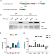
Activity of the putative feo promoters and gene transcription in H. pylori G27. (A) Schematic representation (not drawn to scale) of the transcriptional reporter used in these assays. Each putative promoter (depicted with red arrows) was amplified from H. pylori G27 genomic DNA and cloned upstream of a promoterless gfp gene in the pTM117 backbone. The junction between duf and feoA (Junct) is also shown. (B) Immunoblot analysis detecting GFP in H. pylori G27 strains, WT and Δfur, transformed with the promoter fusions shown in panel A. Ferritin (Pfr), shown in the bottom lane from a Coomassie-stained gel, was used as a control of a Fur-regulated gene (upregulated in the absence of Fur). Both gels were loaded with the same samples. (C) Relative fold changes in the expression levels of the HpfeoA-duf junction (Junct), HpfeoA, HpfeoB, duf, and pfr in the Δfur strain compared to the WT as determined by RT-qPCR. (D) Relative fold changes in gene expression in the Δfur and the WT strains upon iron depletion induced with 200 μM dpp. The p-values for C and D were determined by an unpaired, two-tailed Student’s t-test from the ΔCT values. Differences that were statistically significant are indicated (*p < 0.05, **p < 0.01, ***p < 0.001). The bars correspond to the relative means and standard deviations (error bars) from four biological replicates. Statistical analyses and bar graphs were generated with GraphPad Prism v9.5.0.
Figure 6
Figure 6
Schematic representation of the proposed HpfeoB promoter architecture. The gray boxes represent the identified operator sites for apo-Fur (afOP I and II), holo-Fur (hfOP), and NikR (nOP) with their relative positions indicated by the numbers around each box. All binding motifs were found in the same direction of HpfeoB, and their location in this scheme, either on the top or the bottom, is only for illustrative purposes. Proposed −10 and − 35 boxes are also shown. The transcription start site of HpfeoB (position +1) and the anti-sense RNAs (+260 and + 1,500) identified by Sharma et al. (2010) via RNA-Seq are depicted with the cyan (above the line) and magenta (below the line) arrows, respectively. The thick blue arrow represents the position and directionality of the HpfeoB coding sequence. This model is based on the H. pylori G27 genome and is not drawn to scale.
Figure 7
Figure 7
Phylogenetic reconstruction of the Campylobacterota group. This tree was constructed by the maximum likelihood method based on the alignment of concatenated RpoB-RpoC protein sequences. Bootstrap values were calculated with 300 replicates, and those values over 0.5 are shown on the corresponding nodes. Colors indicate clustering according to the currently proposed classification of the helicobacters. From top to bottom: H. pylori group (red), non-H. pylori gastric helicobacters (light blue); enterohepatic helicobacters groups A, B, C, and D (dark green, magenta, gold, and blue, respectively). The ones in black are not classified within these groups. Non-Helicobacter species are shown in the bottom and colored according to their genus affiliation: from Campylobacter spp. to Sulfurovum lithotrophicum (outgroup).
Figure 8
Figure 8
Genomic context of feoA loci among Helicobacter species. This cladogram shows the phylogenetic relationship as found in the organismal tree above for representative species of the gastric (Gast.) and non-H. pylori helicobacters (NHpH) as well as for the groups A, B, C, and D of the enterohepatic helicobacters (as indicated above the corresponding node). The AB node and the H. himalayensis branch, in which additional feoA rearrangements might have taken place, are indicated with an asterisk (*). The genomic context of the feoA and duf loci are shown after each species following the gene annotation deposited in GeneBank for each genome. Distances and gene sizes are not drawn to scale.
Figure 9
Figure 9
Formation of the transmembrane Feo complex by HpFeoA and HpFeoB in V. cholerae EPV6. (A) Immunoblot analysis of V. cholerae EPV6 co-transformed with plasmids carrying HpFeoAC-FLAG and HpFeoB. CH2O indicates whole cell pellets before (−) and after (+) formaldehyde crosslinking in vivo. (B) Samples obtained upon cell fractionation: cytoplasmic and membrane fractions (labeled as Cyto and Memb, respectively.) The numbers on the left indicate the estimated protein size in kDa. (C) Results for peptides matching HpFeoA and HpFeoB retrieved from mass spectroscopy analysis of those bands in the membrane fraction (labeled as 1 and 2) after immunoprecipitation with a monoclonal anti-FLAG antibody.
Figure 10
Figure 10
Structural model of the HpFeoA-HpFeoB interaction constructed with AlphaFold-Multimer. (A) 3D representation of the best model obtained for the interaction between HpFeoA (WP_000174130, in pink) and HpFeoB (WP_041201363, in green) by AlphaFold-Multimer. N′ and C′ correspond to the N- and C- termini of HpFeoB. The blue lines represent the predicted contacts between the two proteins, defined as residues at 3.00 Å or closer to each other. These contacts have a predicted aligned error value of 0. (B) Structural alignment of the HpFeoA-HpFeoB model shown in panel A and the 3D model for HpFeoB alone deposited in AlphaFold DB (ID: B5Z754_HELPG, shown in purple). N″ represent the N-terminus of the latter model as it does not align with that of the interaction model. (C) Predicted interacting peptides between HpFeoA and HpFeoB shown in (A), indicating the predicted distance between alpha carbon (Cα-Cα).

Similar articles

Cited by

References

    1. Agriesti F., Roncarati D., Musiani F., Del Campo C., Iurlaro M., Sparla F., et al. (2014). FeON-FeOFF: the Helicobacter pylori Fur regulator commutates iron-responsive transcription by discriminative readout of opposed DNA grooves. Nucleic Acids Res. 42, 3138–3151. doi: 10.1093/nar/gkt1258, PMID: - DOI - PMC - PubMed
    1. Arnold J., Mindiola D., Agapie T., Love J., Dincă M., Dauth A., et al. (2012). Dissecting the role of DNA sequence in Helicobacter pylori NikR/DNA recognition. Dalton Trans. 41, 7946–7951. doi: 10.1039/C2DT30504F, PMID: - DOI - PubMed
    1. Bailey T. L., Johnson J., Grant C. E., Noble W. S. (2015). The MEME suite. Nucleic Acids Res. 43, W39–W49. doi: 10.1093/nar/gkv416, PMID: - DOI - PMC - PubMed
    1. Barber M. F., Elde N. C. (2015). Buried treasure: evolutionary perspectives on microbial Iron piracy. Trends Genet. 31, 627–636. doi: 10.1016/j.tig.2015.09.001, PMID: - DOI - PMC - PubMed
    1. Bereswill S., Greiner S., Van Vliet A. H. M., Waidner B., Fassbinder F., Schiltz E., et al. (2000). Regulation of ferritin-mediated cytoplasmic iron storage by the ferric uptake regulator homolog (Fur) of Helicobacter pylori. J. Bacteriol. 182, 5948–5953. doi: 10.1128/JB.182.21.5948-5953.2000, PMID: - DOI - PMC - PubMed

LinkOut - more resources