Review
. 2023 Jul 4:11:1205792.
doi: 10.3389/fbioe.2023.1205792. eCollection 2023.

Engineered biomimetic micro/nano-materials for tissue regeneration

Affiliations
Review

Engineered biomimetic micro/nano-materials for tissue regeneration

Feng Han et al. Front Bioeng Biotechnol. .

Abstract

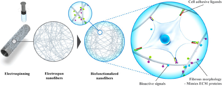
The incidence of tissue and organ damage caused by various diseases is increasing worldwide. Tissue engineering is a promising strategy of tackling this problem because of its potential to regenerate or replace damaged tissues and organs. The biochemical and biophysical cues of biomaterials can stimulate and induce biological activities such as cell adhesion, proliferation and differentiation, and ultimately achieve tissue repair and regeneration. Micro/nano materials are a special type of biomaterial that can mimic the microstructure of tissues on a microscopic scale due to its precise construction, further providing scaffolds with specific three-dimensional structures to guide the activities of cells. The study and application of biomimetic micro/nano-materials have greatly promoted the development of tissue engineering. This review aims to provide an overview of the different types of micro/nanomaterials, their preparation methods and their application in tissue regeneration.

Keywords: biomimetic microstructure; micro/nano-materials; regeneration; repair; tissue engineering.


Conflict of interest statement

The authors declare that the research was conducted in the absence of any commercial or financial relationships that could be construed as a potential conflict of interest.

Figures

FIGURE 1
FIGURE 1
Illustration of engineered biomimetic micro/nano-materials for tissue regeneration. The micro/nano structures including micro/nano fiber, nanoparticle, microsphere and 3D printing scaffold, and the biomedical applications referring to bone repair, AF repair and vascular grafts are shown as examples. Manufacturing methods for different micro/nano-materials are also shown.
FIGURE 2
FIGURE 2
Schematic of electrospinning setup and the fibers output. (A) mono-axial electrospinning; (B) side-by-side electrospinning; (C) coaxial electrospinning and (D) triaxial electrospinning (Luraghi et al., 2021).
FIGURE 3
FIGURE 3
Three main 3D bioprinting technologies. (A) Inkjet printing; (B) Extrusion or robotic dispensing bioprinters; (C) Laser-assisted bioprinting (Bartolo et al., 2022).
FIGURE 4
FIGURE 4
Microfluidic encapsulation platform using a novel custom design and device molding technique enables production of uniform hydrogel microspheres with a wide range of diameters. (A) Schematic of the microfluidic encapsulation platform. (B) Setup of the microfluidic encapsulation platform in a biosafety cabinet (Seeto et al., 2019).
FIGURE 5
FIGURE 5
Fabrication of bioactive electrospun fibers mimicking the native ECM (Taskin et al., 2021).
FIGURE 6
FIGURE 6
Three representative strategies for making high drug-loading nanoparticles. (A) Post-loading; (B) co-loading; (C) pre-loading (Liu et al., 2020).
FIGURE 7
FIGURE 7
Schematic illustration of advantages and design of LM and their applications in biomedicine (Wang et al., 2022).
FIGURE 8
FIGURE 8
Schematic illustration of the (SF/PCL)1:5/PVA-LBL20 coaxial fibers loaded with BMP2 and CTGF for bone tissue engineering (Cheng et al., 2019).
FIGURE 9
FIGURE 9
Schematic illustrations of the living GMs for cartilage regeneration. (A) The fabrication of GMs using microfluidic technology, and incorporation of PDGF-BB via the electrostatic force to engineer GMPs. (B) The BMSCs and PDGF-BB loaded GMs, or “the living GMs” were further developed through incubation with exogenous BMSCs. (C) The living GMs were injected into the joint cavity of the DMM rat. The enhanced paracrine activity was achieved by integrating the endogenous and exogenous regeneration mechanisms (Li et al., 2023).
FIGURE 10
FIGURE 10
Generation of helical hollow alginate hydrogel microfibers with different channels. (A) Schematic illustration of the fabrication of the helical hollow microfibers. (B, C-i, ii) Optical microscopy and iii, iv) SEM images of helical hollow microfibers with (B) straight and (C) helical channels. Scale bars, 200 μm (Jia et al., 2019).

Similar articles

Cited by

References

    1. Adeli-Sardou M., Yaghoobi M. M., Torkzadeh-Mahani M., Dodel M. (2019). Controlled release of lawsone from polycaprolactone/gelatin electrospun nano fibers for skin tissue regeneration. Int. J. Biol. Macromol. 124, 478–491. 10.1016/j.ijbiomac.2018.11.237 - DOI - PubMed
    1. Admane P., Gupta A. C., Jois P., Roy S., Chandrasekharan Lakshmanan C., Kalsi G., et al. (2019). Direct 3D bioprinted full-thickness skin constructs recapitulate regulatory signaling pathways and physiology of human skin. Bioprinting 15, 00051. 10.1016/j.bprint.2019.e00051 - DOI
    1. Agarwal P., Wang H., Sun M., Xu J., Zhao S., Liu Z., et al. (2017). Microfluidics enabled bottom-up engineering of 3D vascularized tumor for drug discovery. ACS Nano 11 (7), 6691–6702. 10.1021/acsnano.7b00824 - DOI - PMC - PubMed
    1. Ahmed T. A., Hincke M. T. (2010). Strategies for articular cartilage lesion repair and functional restoration. Tissue Eng. Part B Rev. 16 (3), 305–329. 10.1089/ten.TEB.2009.0590 - DOI - PubMed
    1. Arias L. S., Pessan J. P., Vieira A. P. M., Lima T. M. T., Delbem A. C. B., Monteiro D. R. (2018). Iron oxide nanoparticles for biomedical applications: A perspective on synthesis, drugs, antimicrobial activity, and toxicity. Antibiotics 7 (2), 46. 10.3390/antibiotics7020046 - DOI - PMC - PubMed

LinkOut - more resources