

Cerebral Sinus Vein Thrombosis following Sneezing: Case Report
- PMID: 37469522
- PMCID: PMC10353342
- DOI: 10.1159/000530812
Cerebral Sinus Vein Thrombosis following Sneezing: Case Report
Abstract
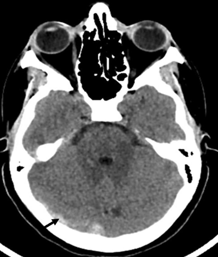
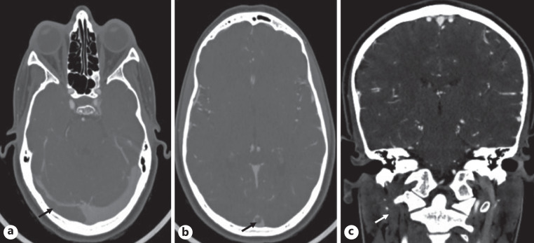
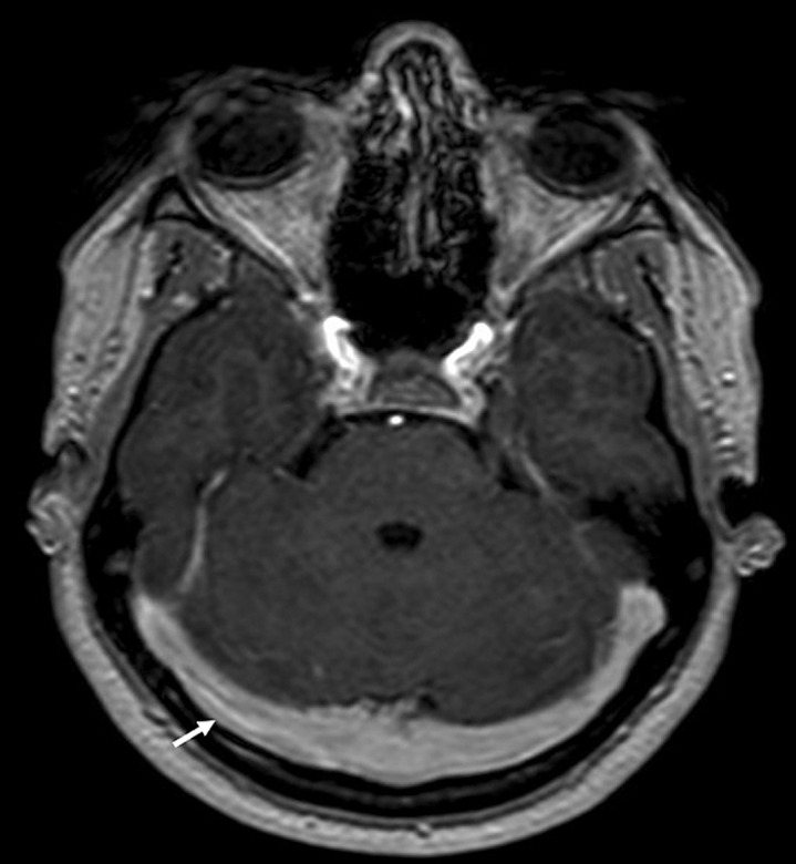
Cerebral sinus vein thrombosis (CVT) is a relatively rare neurovascular entity, usually associated with acquired or genetic hypercoagulable states, and in many cases it remains idiopathic. Trauma is also associated with CVT among patients with major head or neck trauma, including penetrating injuries. However, CVT associated with acceleration trauma has only been described in few cases so far. We present an unusual case of a 19-year-old woman with no past medical history, admitted with an extensive CVT following sneezing. A thorough investigation did not reveal any other potential etiology or risk factor other than estrogen-containing oral contraceptives. The patient was treated with anticoagulation and improved clinically with complete recanalization on follow-up imaging. This case suggests acceleration trauma may be a potential factor of risk for CVT.
Keywords: Acceleration trauma; Case report; Cerebral sinus vein thrombosis.
© 2023 The Author(s). Published by S. Karger AG, Basel.
Conflict of interest statement
None of the authors have any financial disclosures or conflicts of interests.
Figures
Similar articles
-
Sinus thrombosis after a jump from a small rock and a sneezing attack: minor endothelial trauma as a precipitating factor for cerebral venous thrombosis?Headache. 2004 Sep;44(8):812-5. doi: 10.1111/j.1526-4610.2004.04150.x. Headache. 2004. PMID: 15330829
-
Non-Vitamin K Antagonist Oral Anticoagulants for the Treatment of Cerebral Venous Sinus Thrombosis: a Retrospective, Matched Cohort Analysis.Neurocrit Care. 2021 Dec;35(3):783-788. doi: 10.1007/s12028-021-01244-x. Epub 2021 May 27. Neurocrit Care. 2021. PMID: 34046861 Clinical Trial.
-
Risk factors governing the development of cerebral vein and dural sinus thrombosis after craniotomy in patients with intracranial tumors.J Neurosurg. 2018 Feb;128(2):373-379. doi: 10.3171/2016.11.JNS161871. Epub 2017 Apr 7. J Neurosurg. 2018. PMID: 28387630
-
Recanalization in Cerebral Venous Thrombosis.Stroke. 2018 Aug;49(8):1828-1835. doi: 10.1161/STROKEAHA.118.022129. Stroke. 2018. PMID: 30021808
-
An Integrated Approach on the Diagnosis of Cerebral Veins and Dural Sinuses Thrombosis (a Narrative Review).Life (Basel). 2022 May 11;12(5):717. doi: 10.3390/life12050717. Life (Basel). 2022. PMID: 35629384 Free PMC article. Review.
References
-
- Ferro JM, Canhão P, Stam J, Bousser MG, Barinagarrementeria F; ISCVT Investigators . Prognosis of cerebral vein and dural sinus thrombosis: results of the international study on cerebral vein and dural sinus thrombosis (ISCVT). Stroke. 2004;35(3):664–70. 10.1161/01.STR.0000117571.76197.26. - DOI - PubMed
Publication types
LinkOut - more resources
Full Text Sources
Miscellaneous
