. 2023 Jul 3:17:1173086.
doi: 10.3389/fncel.2023.1173086. eCollection 2023.

Regulation of Schwann cell proliferation and migration via miR-195-5p-induced Crebl2 downregulation upon peripheral nerve damage

Affiliations

Regulation of Schwann cell proliferation and migration via miR-195-5p-induced Crebl2 downregulation upon peripheral nerve damage

Shiying Li et al. Front Cell Neurosci. .

Abstract

Background: Schwann cells acquire a repair phenotype upon peripheral nerve injury (PNI), generating an optimal microenvironment that drives nerve repair. Multiple microRNAs (miRNAs) show differential expression in the damaged peripheral nerve, with critical regulatory functions in Schwann cell features. This study examined the time-dependent expression of miR-195-5p following PNI and demonstrated a marked dysregulation of miR-195-5p in the damaged sciatic nerve.

Methods: CCK-8 and EdU assays were used to evaluate the effect of miR-195-5 on Schwann cell viability and proliferation. Schwann cell migration was tested using Transwell and wound healing assays. The miR-195-5p agomir injection experiment was used to evaluate the function of miR-195-5p in vivo. The potential regulators and effects of miR-195-5p were identified through bioinformatics evaluation. The relationship between miR-195-5p and its target was tested using double fluorescence reporter gene analysis.

Results: In Schwann cells, high levels of miR-195-5p decreased viability and proliferation, while suppressed levels had the opposite effects. However, elevated miR-195-5p promoted Schwann cell migration determined by the Transwell and wound healing assays. In vivo injection of miR-195-5p agomir into rat sciatic nerves promote axon elongation after peripheral nerve injury by affecting Schwann cell distribution and myelin preservation. Bioinformatic assessment further revealed potential regulators and effectors for miR-195-5p, which were utilized to build a miR-195-5p-centered competing endogenous RNA network. Furthermore, miR-195-5p directly targeted cAMP response element binding protein-like 2 (Crebl2) mRNA via its 3'-untranslated region (3'-UTR) and downregulated Crebl2. Mechanistically, miR-195-5p modulated Schwann cell functions by repressing Crebl2.

Conclusion: The above findings suggested a vital role for miR-195-5p/Crebl2 in the regulation of Schwann cell phenotype after sciatic nerve damage, which may contribute to peripheral nerve regeneration.

Keywords: Crebl2; Schwann cell; miR-195-5p; migration; peripheral nerve injury; proliferation.


Conflict of interest statement

The authors declare that the research was conducted in the absence of any commercial or financial relationships that could be construed as a potential conflict of interest.

Figures

FIGURE 1
FIGURE 1
Temporal expression patterns of miR-195-5p in damaged nerve stumps following PNI. Following sciatic nerve crush injury in rats, miR-195-5p expression levels in damaged sciatic nerve specimens were determined by qRT-PCR at 0, 1, 3, and 7 days. *P < 0.05 versus control group (day 0) (n = 3, mean ± SEM; one-way analysis of variance with post-hoc Dunnett’s test was carried out to analyze the data). d: day(s).
FIGURE 2
FIGURE 2
Effects of miR-195-5p on Schwann cell viability and proliferation. (A) Cells transfected with miR-195-5p mimic showed relatively enhanced miR-195-5p expression in comparison with MC transfection. *P < 0.05 versus MC group (mean ± SEM, n = 4; Student’s t-test). MC, mimic control. (B) Cells transfected with anti-miR-195-5p showed reduced miR-195-5p amounts in comparison with IC transfection. *P < 0.05 versus IC (mean ± SEM, n = 3; Student’s t-test). IC, inhibitor control. (C) Cell viability assessment in SCs after transfection with miR-195-5p mimic (miR-195-5p) or its control, and (D) with miR-195-5p inhibitor (anti-miR-195-5p) and its control by the CCK-8 assay. *P < 0.05 versus IC (mean ± SEM, n = 3; Student’s t-test). MC, mimic control; IC, inhibitor control. (E,F) Representative micrographs and proliferation rates of SCs transfected with miR-195-5p mimic (miR-195-5p) or its control. (G,H) Representative micrographs and proliferation rates of SCs transfected with miR-195-5p inhibitor (anti-miR-195-5p) or its control. Red, EdU-positive SCs; blue, Hoechst 33342 staining. Scale bar = 100 μm. *P < 0.05 versus MC or IC (mean ± SEM, n = 3; Student’s t-test). MC, mimic control; IC, inhibitor control.
FIGURE 3
FIGURE 3
Effect of miR-195-5p on Schwann cell migration. Micrographs showing SCs that traversed the membrane of the Transwell membrane after transfection with (A) miR-195-5p mimic (miR-195-5p) or its control, and (B) miR-195-5p inhibitor (anti-miR-195-5p) or its control. Violet, migratory SCs. Quantification used histograms. *P < 0.05 versus MC or IC (mean ± SEM, n = 3; Student’s t-test). MC, mimic control; IC, inhibitor control. Scale bar = 50 μm. (C) Representative micrographs depicting SC wound healing and quantification of SC migration following transfection with miR-195-5p mimic (miR-195-5p) or its control (MC), and (D) miR-195-5p inhibitor (anti-miR-195-5p) or its control (IC). *P < 0.05 versus MC or IC (mean ± SEM, n = 3; Student’s t-test). MC, mimic control; IC, inhibitor control. Scale bar = 100 μm.
FIGURE 4
FIGURE 4
miR-195-5p affects Schwann cell distribution, myelin preservation and axon elongation in vivo. (A,B) Representative immunostaining of sciatic nerves of miR-195-5p agomir injected rats at 3 days after crush injury. Green color represents MBP staining and red color represents S100β staining. Crush sites are labeled with dashed lines. White boxed areas are displayed at a higher magnification below. White arrows indicate normal myelin tissue. Scale bars represent 500 μm in the main image and 50 μm in the magnified image. (C,D) Representative immunostaining of sciatic nerves of agomir control injected rats at 3 days after crush injury. Agomir con represents agomir control. Green color represents MBP staining and red color represents S100β staining. Crush sites are labeled with dashed lines. White boxed areas are displayed at a higher magnification below. White arrows indicate normal myelin tissue. Scale bars represent 500 μm in the main image and 50 μm in the magnified image. (E) Representative immunostaining of sciatic nerves of agomir control or miR-195-5p agomir injected rats at 3 days after crush injury. Green color represents NF200 staining and blue color represents nucleus staining. Crush sites are labeled with dashed lines. Boxed areas are demonstrated at a higher magnification on the right side. Scale bars represent 500 μm in the main image and 50 μm in the magnified image. (F) Quantification of the length of regenerated nerve fibers at 3 days after crush injury. *P < 0.05 versus agomir control (mean ± SEM, n = 4; Student’s t-test).
FIGURE 5
FIGURE 5
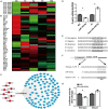
Identification of potential target mRNAs of miR-195-5p. (A) A heatmap showing the temporal expression patterns of lncRNAs in the miR-195-5p-centered ceRNA network for sciatic nerve stumps at 0, 1, 4, 7, and 14 days post-injury. Red, green, and gray indicate upregulation, downregulation, and no expression change, respectively. (B) A heatmap of the temporal expression patterns of mRNAs in the miR-195-5p-centered ceRNA network for sciatic nerve stumps at 0, 1, 4, 7, and 14 days post-nerve crush injury. ceRNA, competing endogenous RNA; lncRNA, long non-coding RNA; miRNA, microRNA. (C) Interactions among lncRNAs, miR-195-5p and target mRNAs in the miR-195-5p-centered ceRNA network. lncRNAs, the miRNA and mRNAs were labeled in red, yellow and blue, respectively. (D) Crebl2 mRNA amounts in SCs after transfection with miR-195-5p mimic (miR-195-5p) or its control (MC), and miR-195-5p inhibitor (anti-miR-195-5p) or its control (IC). *P < 0.05 versus MC or IC (mean ± SEM, n = 3; Student’s t-test). (E) Sequences of the predicted target site (1,250–1,257 bp) of miR-195-5p at the 3’-UTRs of Crebl2 in various species. (F) Constructions of wild-type and mutant pmiR-RB-UTR vectors. (G) Relative luciferase activities of 293T cells after transfection with WT or mutant pmiR-RB-UTR and miR-195-5p mimic (miR-195-5p) or mimic control (MC) *P < 0.05, mean ± SEM, n = 3; Student’s t-test.
FIGURE 6
FIGURE 6
Crebl2 affects Schwann cell phenotype. (A) Interference efficiencies of three Crebl2 siRNAs. Effects of Crebl2 siRNA on Schwann cell (B) proliferation, (C) migration, and (D) wound healing. In panels (B–D), scale bars are 100 μm, 50 μm and 100 μm, respectively. *P < 0.05 versus siRNA-con (mean ± SEM, n = 3; Student’s t-test). siRNA-con, control siRNA.
FIGURE 7
FIGURE 7
Crebl2 siRNA abrogates the effects of miR-195-5p inhibitor on Schwann cells. Rescue effect of Crebl2 siRNA in miR-195-5p inhibitor-regulated Schwann cell (A) proliferation and (B) migration. In panels (A,B), scale bars are 100 μm and 50 μm, respectively. siRNA-con, control siRNA; IC, inhibitor control; Anti-miR-195-5p, miR-195-5p inhibitor. *P < 0.05, mean ± SEM, n = 3; Student’s t-test.

Similar articles

Cited by

References

    1. Allodi I., Udina E., Navarro X. (2012). Specificity of peripheral nerve regeneration: interactions at the axon level. Prog. Neurobiol. 98 16–37. - PubMed
    1. Ambros V. (2004). The functions of animal microRNAs. Nature 431 350–355. - PubMed
    1. Bosch-Queralt M., Fledrich R., Stassart R. M. (2023). Schwann cell functions in peripheral nerve development and repair. Neurobiol. Dis. 176:105952. - PubMed
    1. Clements M. P., Byrne E., Camarillo Guerrero L. F., Cattin A. L., Zakka L., Ashraf A., et al. (2017). The wound microenvironment reprograms schwann cells to invasive mesenchymal-like cells to drive peripheral nerve regeneration. Neuron 96 98–114.e7. 10.1016/j.neuron.2017.09.008 - DOI - PMC - PubMed
    1. Diener C., Keller A., Meese E. (2022). Emerging concepts of miRNA therapeutics: from cells to clinic. Trends Genet. 38 613–626. - PubMed

LinkOut - more resources