

Extracellular vesicles in atherosclerosis and vascular calcification: the versatile non-coding RNAs from endothelial cells and vascular smooth muscle cells
- PMID: 37469665
- PMCID: PMC10352799
- DOI: 10.3389/fmed.2023.1193660
Extracellular vesicles in atherosclerosis and vascular calcification: the versatile non-coding RNAs from endothelial cells and vascular smooth muscle cells
Abstract
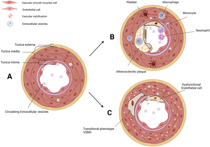
Atherosclerosis (AS) is characterized by the accumulation of lipids, fibrous elements, and calcification in the innermost layers of arteries. Vascular calcification (VC), the deposition of calcium and phosphate within the arterial wall, is an important characteristic of AS natural history. However, medial arterial calcification (MAC) differs from intimal calcification and cannot simply be explained as the consequence of AS. Endothelial cells (ECs) and vascular smooth muscle cells (VSMCs) are directly involved in AS and VC processes. Understanding the communication between ECs and VSMCs is critical in revealing mechanisms underlying AS and VC. Extracellular vesicles (EVs) are found as intercellular messengers in kinds of physiological processes and pathological progression. Non-coding RNAs (ncRNAs) encapsulated in EVs are involved in AS and VC, including microRNAs (miRNAs), long non-coding RNAs (lncRNAs), and circular RNAs (circRNAs). The effects of ncRNAs have not been comprehensively understood, especially encapsulated in EVs. Some ncRNAs have demonstrated significant roles in AS and VC, but it remains unclear the functions of the majority ncRNAs detected in EVs. In this review, we summarize ncRNAs encapsulated in EC-EVs and VSMC-EVs, and the signaling pathways that are involved in AS and VC.
Keywords: atherosclerosis (AS); extracellular vesicles (EVs); non-coding RNAs (ncRNAs); uremia; vascular calcification (VC).
Copyright © 2023 Yu, Duan, Liu, Huang, Xiao and He.
Conflict of interest statement
The authors declare that the research was conducted in the absence of any commercial or financial relationships that could be construed as a potential conflict of interest.
Figures


Similar articles
-
The roles of non-coding RNAs in vascular calcification and opportunities as therapeutic targets.Pharmacol Ther. 2021 Feb;218:107675. doi: 10.1016/j.pharmthera.2020.107675. Epub 2020 Sep 8. Pharmacol Ther. 2021. PMID: 32910935 Review.
-
RNA-seq analysis of extracellular vesicles from hyperphosphatemia-stimulated endothelial cells provides insight into the mechanism underlying vascular calcification.BMC Nephrol. 2022 May 21;23(1):192. doi: 10.1186/s12882-022-02823-6. BMC Nephrol. 2022. PMID: 35597927 Free PMC article.
-
Mechanisms of Action of MiRNAs and LncRNAs in Extracellular Vesicle in Atherosclerosis.Front Cardiovasc Med. 2021 Oct 8;8:733985. doi: 10.3389/fcvm.2021.733985. eCollection 2021. Front Cardiovasc Med. 2021. PMID: 34692785 Free PMC article. Review.
-
Vascular calcification: from the perspective of crosstalk.Mol Biomed. 2023 Oct 18;4(1):35. doi: 10.1186/s43556-023-00146-y. Mol Biomed. 2023. PMID: 37851172 Free PMC article. Review.
-
Extracellular Vesicles From LPS-Treated Macrophages Aggravate Smooth Muscle Cell Calcification by Propagating Inflammation and Oxidative Stress.Front Cell Dev Biol. 2022 Mar 9;10:823450. doi: 10.3389/fcell.2022.823450. eCollection 2022. Front Cell Dev Biol. 2022. PMID: 35356285 Free PMC article.
Cited by
-
Reactive Carbonyl Species and Protein Lipoxidation in Atherogenesis.Antioxidants (Basel). 2024 Feb 14;13(2):232. doi: 10.3390/antiox13020232. Antioxidants (Basel). 2024. PMID: 38397830 Free PMC article. Review.
-
Pathogenesis and Mechanism of Uremic Vascular Calcification.Cureus. 2024 Jul 17;16(7):e64771. doi: 10.7759/cureus.64771. eCollection 2024 Jul. Cureus. 2024. PMID: 39026575 Free PMC article. Review.
-
Extracellular Vesicles in Atherosclerosis: State of the Art.Int J Mol Sci. 2023 Dec 27;25(1):388. doi: 10.3390/ijms25010388. Int J Mol Sci. 2023. PMID: 38203558 Free PMC article. Review.
References
Publication types
LinkOut - more resources
Full Text Sources
Research Materials