. 2023 Jan:8:10-24.
doi: 10.1038/s41578-022-00488-z. Epub 2022 Oct 24.

The role of single protein elasticity in mechanobiology

Affiliations

The role of single protein elasticity in mechanobiology

Amy Em Beedle et al. Nat Rev Mater. 2023 Jan.

Abstract

In addition to biochemical signals and genetic considerations, mechanical forces are rapidly emerging as a master regulator of human physiology. Yet the molecular mechanisms that regulate force-induced functionalities across a wide range of scales, encompassing the cell, tissue or organ levels, are comparatively not so well understood. With the advent, development and refining of single molecule nanomechanical techniques, enabling to exquisitely probe the conformational dynamics of individual proteins under the effect of a calibrated force, we have begun to acquire a comprehensive knowledge on the rich plethora of physicochemical principles that regulate the elasticity of single proteins. Here we review the major advances underpinning our current understanding of how the elasticity of single proteins regulates mechanosensing and mechanotransduction. We discuss the present limitations and future challenges of such a prolific and burgeoning field.


Figures

Figure 1
Figure 1. Single molecule force spectroscopy techniques enable the mapping the (un)folding energy landscape of a protein under force.
The main force spectroscopy techniques rely on an individual molecule tethered between one fixed surface and a second surface, the position of which is precisely controlled. The adequacy of each technique depends on the mechanical properties of the studied protein and the desired sampling time. For example, magnetic tweezers are ideally suited to investigate the (un)folding dynamics of mechanically weak proteins over long times, up to hours or even days. At the other end of the spectrum, the AFM is ideally suited to examine high mechanical stability proteins, and experiments typically remain stable for a few seconds to minutes. (A) In a magnetic tweezers experiment, a molecule is attached at one end to a paramagnetic bead. The force applied to the bead is controlled by the localization of a pair of permanent magnets mounted above the surface (typically mounted on a voice coil or a piezoelectric actuator). Optical tweezers trap a bead in a laser beam which can both apply or sense movement. In a typical scenario, the protein is tethered between two long DNA handles that are, in turn, attached to the trapped (or fixed) beads. In the AFM, a molecule is tethered between a piezoelectric actuator and a flexible Hookean cantilever. Mechanical force is applied when the piezo retracts away from the cantilever, and the mechanical response of the molecule is measured by monitoring the deflection of a cantilever. (B) The application of mechanical force to the protein reduces the height of the energy barrier between the folded and the unfolded states. In the simplest approximation, the Bell/Arrhenius model predicts an exponential acceleration of the unfolding rate (αu) with the applied force (F), and indicates that the height of the energy barrier (ΔE) is effectively reduced by a factor -FΔx. The proportionality factor, Δx, is a direct indication of how sensitive (brittle) each protein is to force denaturation. If the applied force is high enough, the molecule will traverse the energy landscape and remain in the energetically-favorable unfolded state. However, if the force is low enough such that the unfolded and folded state are energetically similar, the protein can hop between the folded and unfolded state in equilibrium at that particular force.
Figure 2
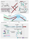
Figure 2. Nanomechanical regulation of the cell matrix and the focal adhesion hub.
The mechanical behaviour of the extracellular matrix (ECM) is governed by the mechanical behaviour of two types of molecular constituents. Molecules such as elastin and the polysaccharides readily extend under an applied force, hence behaving as entropic strings. They underpin the reversible elastic properties of the ECM. By contrast, molecules that exhibit significant resistance to mechanical unfolding, such as tenascin and fibronectin, tend to work as (often reversible) shock-absorbers. When streteched under constant velocity conditions, polyproteins that naturally work as shock absorbers display a saw-tooth like force extension profile, where each force peak corresponds to the unfolding and extension of one individual domains. The area under each peak is a direct measurement of the stored heat that is dissipated as the segmented protein is gradually elongated after mechanical unfolding. In particular, talin couples integrins at the cell surface with the actin cytoskeleton. All the talin rod domains are mechanically labile, unfolding between 5-25 pN. The R3 domain – one of the most mechanically vulnerable – functions as a bandpass filter, capable of filtering out mechanical noise and only sensing the average applied force, and is able to bind to vinculin once talin is mechanically unfolded and the previously cryptic binding sites become exposed to the solvent.
Figure 3
Figure 3. Mechanical force regulates the nature of interactions within the cytoskeleton and between cells.
(Upper panel) Single molecule force spectroscopy experiments reveal the plethora of mechanisms employed by actin binding proteins to regulate their mechanical properties. FilaminA exhibits hierarchy in the mechanical stability, ranging from 10-70 pN. The mechanical properties of the domain 20 switches between 5 pN and 20 pN depending on the conformation of a single proline. Furthermore, the α-actinin protein, which forms a mechanically stable connection with actin, regulates vinculin binding depending on the conformation of the 4th domain of the spectrin-like region. (Lower panel) The cadherin-catenin complex maintains the mechanical connection spanning from the extracellular interface to the actin cytoskeleton. The conformation of the cadherin dimer and the calcium concentration dictate the mechanical response of the interaction. The X-dimer exhibits both catch- and slip-bond behaviour depending on the magnitude of applied force. By contrast, the strand-swapped dimer only exhibits slip-bond behaviour.
Figure 4
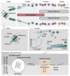
Figure 4. Lessons of muscle nanomechanics learnt with single molecule techniques.
The giant protein titin is responsible for the passive elasticity of muscle, and has arguably been the most studied protein at the nanomechanical level. (Upper panel) From a molecular perspective, titin is formed by a series of mechanically stiff Ig domains that work (that work as shock absorbers) intercalated by intrinsically disordered sequences (that behave like entropic springs). (Lower panel) Several molecular strategies, encompassing chaperone and ligand binding and a wide range of post-translational modifications, have been shown to affect titin nanomechanics.
Figure 5
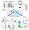
Figure 5. Integrating single molecule experiments into cells.
A number of emerging strategies aim to translate our single molecule understanding into the cellular context. A first general and promising approach modifies a single molecule either by the introduction of a point mutation or by the addition of a mechanical tag. These constructs can then be extensively characterized using single molecule approaches and then introduced into the cell. Subsequent changes in the cellular behaviour can then be directly attributed to changes in the mechanical properties of the individually modified protein. Alternatively, upon understanding the conformational changes undergone upon mechanical unfolding, it is possible to design probes that will specifically target cryptic protein residues that are exposed upon unfolding. The probe binding can be detected via western blots, mass spectrometry or fluorescence microscope. Therefore, these probes function as a direct readout of mechanical unfolding. Finally, FRET tension sensors combine the concepts of fluorescence resonance transfer with polymer physics. In this configuration, a donor and an acceptor fluorophore are separated by a mechanically characterized linker such that the FRET signal is a readout of the mechanical force applied to the system

Similar articles

Cited by

References

    1. Iskratsch T, Wolfenson H, Sheetz MP. Appreciating force and shape-the rise of mechanotransduction in cell biology. Nat Rev Mol Cell Biol. 2014;15:825–33. - PMC - PubMed
    1. Dumortier JG, et al. Hydraulic fracturing and active coarsening position the lumen of the mouse blastocyst. Science. 2019;365:465–468. - PubMed
    1. Engler AJ, Sen S, Sweeney HL, Discher DE. Matrix elasticity directs stem cell lineage specification. Cell. 2006;126:677–89. - PubMed
    1. Swift J, Discher DE. The nuclear lamina is mechano-responsive to ECM elasticity in mature tissue. J Cell Sci. 2014;127:3005–15. - PMC - PubMed
    1. Vining KH, Mooney DJ. Mechanical forces direct stem cell behaviour in development and regeneration. Nat Rev Mol Cell Biol. 2017;18:728–742. - PMC - PubMed