

Adenocarcinoma arising from widespread heterotopic gastric mucosa in the cervicothoracic esophagus: a case report
- PMID: 37470880
- PMCID: PMC10359231
- DOI: 10.1186/s40792-023-01707-7
Adenocarcinoma arising from widespread heterotopic gastric mucosa in the cervicothoracic esophagus: a case report
Abstract
Background: In Japan, about 6% of esophageal cancers are adenocarcinomas, although most of them arise from Barrett's epithelium. Adenocarcinoma arising from heterotopic gastric mucosa (HGM) is very rare. Due to its rarity, there is no unified view on its treatment strategy and prognosis.
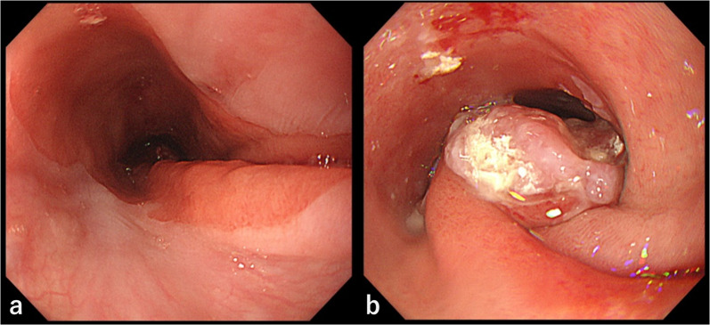
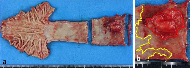
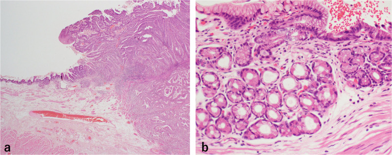
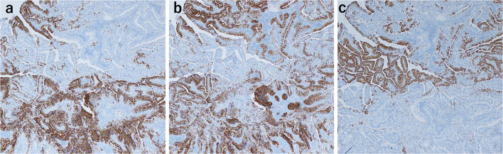
Case presentation: A 57-year-old man presented with a protruding lesion in the cervicothoracic esophagus that was detected by an upper gastrointestinal series at a medical checkup. Esophagoscopy revealed a 30 mm Type 1 tumor circumferentially surrounded by widespread HGM. Computed tomography (CT) and fluorodeoxyglucose (FDG) positron emission tomography (PET)/CT showed no metastasis or invasion of the surrounding organs. We diagnosed the lesion as cT2N0M0 cStageIIB [Union for International Cancer Control (UICC) 8th Ed] cancer and performed subtotal esophagectomy with three-field lymph node dissection. The tumor was determined to be a well-differentiated adenocarcinoma arising from HGM, with deep invasion of the submucosa. The patient underwent no adjuvant therapy and has currently survived without any evidence of recurrence for 15 months.
Conclusions: Although the treatment for adenocarcinoma arising from HGM is basically the same as that for squamous cell carcinoma (SCC) of the esophagus, it is important to determine the treatment strategy based on the characteristics of the adenocarcinoma arising from HGM.
Keywords: Adenocarcinoma; Esophagus; Heterotopic gastric mucosa.
© 2023. The Author(s).
Conflict of interest statement
The authors declare that they do not have any competing interests.
Figures


Similar articles
-
Adenocarcinoma arising from heterotopic gastric mucosa in the cervical esophagus and upper thoracic esophagus: two case reports and literature review.Expert Rev Gastroenterol Hepatol. 2016;10(3):405-14. doi: 10.1586/17474124.2016.1125780. Epub 2015 Dec 16. Expert Rev Gastroenterol Hepatol. 2016. PMID: 26610162 Review.
-
Primary esophageal adenocarcinoma arising from heterotopic gastric mucosa: report of a case.Surg Today. 2013 Apr;43(4):446-51. doi: 10.1007/s00595-012-0206-9. Epub 2012 Jun 17. Surg Today. 2013. PMID: 22706784 Review.
-
Primary adenocarcinoma of the cervical esophagus arising from heterotopic gastric mucosa.J Gastroenterol. 2001 Oct;36(10):704-9. doi: 10.1007/s005350170035. J Gastroenterol. 2001. PMID: 11686482 Review.
-
Adenocarcinoma of the upper esophagus arising in heterotopic gastric mucosa: common pathogenesis with Barrett's adenocarcinoma?Virchows Arch. 2002 Oct;441(4):406-11. doi: 10.1007/s00428-002-0697-7. Virchows Arch. 2002. PMID: 12516606 Review.
-
Primary adenocarcinoma of cervical esophagus.J Exp Clin Cancer Res. 2005 Jun;24(2):325-30. J Exp Clin Cancer Res. 2005. PMID: 16110768
Cited by
-
Transumbilical single-site laparoscopic treatment of primary splenic cyst in child: a rare case report and review of literature.Front Pediatr. 2024 Sep 25;12:1454487. doi: 10.3389/fped.2024.1454487. eCollection 2024. Front Pediatr. 2024. PMID: 39386018 Free PMC article.
-
A systematic review of fully circumferential inlet patches (heterotopic gastric mucosa): More complicated than regular inlet patches.Indian J Gastroenterol. 2025 Aug;44(4):443-456. doi: 10.1007/s12664-025-01738-y. Epub 2025 Mar 31. Indian J Gastroenterol. 2025. PMID: 40163316 Free PMC article.
References
-
- Tachimori Y, Ozawa S, Numasaki H, Ishihara R, Matsubara H, Muro K, et al. Comprehensive registry of esophageal cancer in Japan, 2012 the Registration Committee for Esophageal Cancer of the Japan Esophageal Society Preface 2012. Esophagus. 2019;16:221–245. doi: 10.1007/s10388-019-00674-z. - DOI - PMC - PubMed
-
- Weickert U, Wolf A, SchröDer C, Autschbach F, Vollmer H. Frequency, histopathological findings, and clinical significance of cervical heterotopic gastric mucosa (gastric inlet patch): a prospective study in 300 patients. Dis Esophagus. 2011;24:63–68. doi: 10.1111/j.1442-2050.2010.01091.x. - DOI - PubMed
LinkOut - more resources
Full Text Sources
Research Materials
