. 2023 Sep 15;133(18):e165370.
doi: 10.1172/JCI165370.

Cell-free, high-density lipoprotein-specific phospholipid efflux assay predicts incident cardiovascular disease

Affiliations

Cell-free, high-density lipoprotein-specific phospholipid efflux assay predicts incident cardiovascular disease

Masaki Sato et al. J Clin Invest. .

Abstract

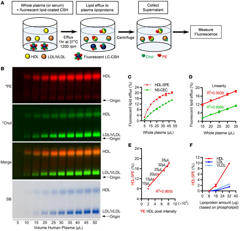
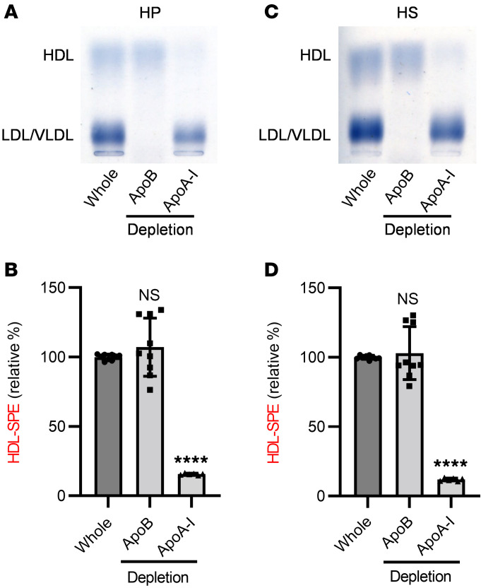
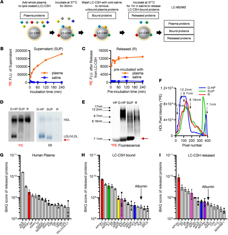
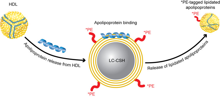
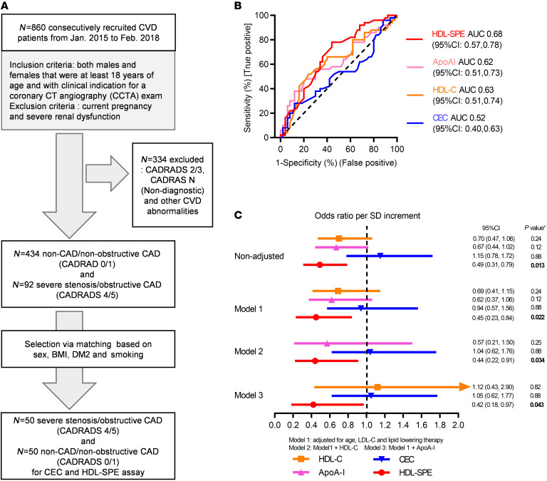
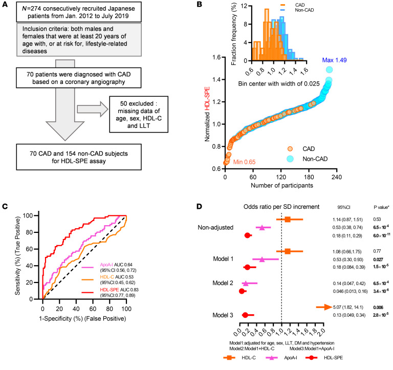
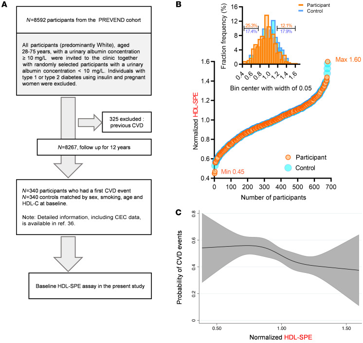
BACKGROUNDCellular cholesterol efflux capacity (CEC) is a better predictor of cardiovascular disease (CVD) events than HDL-cholesterol (HDL-C) but is not suitable as a routine clinical assay.METHODSWe developed an HDL-specific phospholipid efflux (HDL-SPE) assay to assess HDL functionality based on whole plasma HDL apolipoprotein-mediated solubilization of fluorescent phosphatidylethanolamine from artificial lipid donor particles. We first assessed the association of HDL-SPE with prevalent coronary artery disease (CAD): study I included NIH severe-CAD (n = 50) and non-CAD (n = 50) participants, who were frequency matched for sex, BMI, type 2 diabetes mellitus, and smoking; study II included Japanese CAD (n = 70) and non-CAD (n = 154) participants. We also examined the association of HDL-SPE with incident CVD events in the Prevention of Renal and Vascular End-stage Disease (PREVEND) study comparing 340 patients with 340 controls individually matched for age, sex, smoking, and HDL-C levels.RESULTSReceiver operating characteristic curves revealed stronger associations of HDL-SPE with prevalent CAD. The AUCs in study I were as follows: HDL-SPE, 0.68; apolipoprotein A-I (apoA-I), 0.62; HDL-C, 0.63; and CEC, 0.52. The AUCs in study II were as follows: HDL-SPE, 0.83; apoA-I, 0.64; and HDL-C, 0.53. Also longitudinally, HDL-SPE was significantly associated with incident CVD events independent of traditional risk factors with ORs below 0.2 per SD increment in the PREVEND study (P < 0.001).CONCLUSIONHDL-SPE could serve as a routine clinical assay for improving CVD risk assessment and drug discovery.TRIAL REGISTRATIONClinicalTrials.gov NCT01621594.FUNDINGNHLBI Intramural Research Program, NIH (HL006095-06).

Keywords: Cardiovascular disease; Lipoproteins; Molecular diagnosis; Vascular Biology.


Figures

Figure 1
Figure 1. Efflux of fluorescent PE from donor LC-CSH particles to plasma lipoproteins is HDL specific.
(A) Model of cell-free HDL-SPE and NS-CEC assays, as described in Methods. After centrifugation, lipoprotein-associated fluorescence in the supernatant is measured by agarose gel electrophoresis and fluorometry. HDL acquires both *PE and *Chol, whereas LDL/VLDL acquires only *Chol. (B) Effect of pooled normal HP volume on fluorescent lipid efflux. Agarose gel electrophoresis: *PE transfer from donor particles was HDL specific, whereas *Chol transfer to plasma lipoproteins was nonspecific. (C) Fluorometry of the reaction mixture supernatant revealed that the dependence of *PE and *Chol efflux on plasma volume differed. (D) Both HDL-SPE and NS-CEC were linear using 15–35 μL plasma. (E) Fluorometric and electrophoretic gel analyses of HDL-SPE were highly correlated. (F) LC-CSH *PE robustly effluxed to isolated HDL, whereas little to no *PE effluxed to isolated LDL or VLDL. All data are presented as the mean ± SD from triplicate assays unless otherwise stated. A P value of less than 0.05 was considered statistically significant. SUP, supernatant; R, released.
Figure 2
Figure 2. Efflux of fluorescent PE from donor LC-CSH particles to plasma HDL is apoA-I dependent.
HDL or LDL/VLDL was removed from (A and B) HP or (C and D) pooled normal HS by immunodepletion with anti–apoA-I or anti-apoB antibodies, respectively. SB staining demonstrated apoA-I immunodepletion of HDL and apoB immunodepletion of LDL/VLDL from both HP and HS with anti–apoA-I and anti-apoB antibodies, respectively. (B and D) HDL-SPE was markedly reduced in apoA-I– but not apoB-depleted samples. Dunnett’s multiple-comparison test was performed using whole plasma or serum samples as controls (n = 9). For apoB depletion, data represent 9 samples; triplicate assays were performed in 3 independent experiments. For apoA-I depletion data represent 6 samples; triplicate assays were performed in 2 independent experiments. ****P < 0.0001 versus control.
Figure 3
Figure 3. Exchangeable HDL apolipoproteins mediate HDL-SPE.
(A) Assay to identify plasma proteins that mediate HDL-SPE. (B) Time course of LC-CSH *PE efflux to HP versus saline after incubation at 37°C for the indicated durations. (C) Release of *PE from LC-CSH in A, after washing and subsequent incubation with saline at 37°C for 1 hour. (D) Agarose gel electrophoresis: D-HP, HP directly labeled with *PE; SUP, supernatant after incubation of HP with fluorescent *PE-labeled LC-SCH for 30 minutes; R, plasma proteins released to saline from LC-CSH preincubated with HP. (E) Fluorescent native gel lipoprotein electrophoresis and (F) Kymograph analysis of *PE fluorescence revealed that the supernatant and released plasma proteins contained mostly small *PE-tagged HDL-like particles. Red arrow indicates small HDL/albumin band. HP, unlabeled HP. (GI) iBAQ analyses revealed the relative distribution of the identified plasma proteins. (G) HP proteins, (H) LC-CSH–bound proteins, and (I) LC-CSH–released proteins. All data are the mean ± SD from triplicate assays.
Figure 4
Figure 4. Model of apoA-I/HDL–mediated *PE efflux from LC-CSH particles.
ApoA-I and other HDL-associated exchangeable apolipoproteins dissociate from HDL, bind to LC-CSH, solubilize *PE and other lipids, and are then released as small *PE-tagged lipidated apolipoprotein particles.
Figure 5
Figure 5. Clinical Study I: HDL-SPE, but not CEC, HDL-C, or apoA-I, associates with severe CAD in a CVD cohort, independent of traditional risk factors.
(A) Recruitment scheme for participants in Clinical Study I. (B) ROC curves for HDL-SPE, CEC, HDL-C, and apoA-I. (C) ORs are reported per 1 SD increment for HDL-SPE, CEC, HDL-C, and apoA-I based on univariate (nonadjusted) and multivariate regression analyses adjusted for risk factors and biomarkers as indicated. P values in C were calculated after adjusting the FDR, and the FDR-adjusted P value of less than 0.05 was considered statistically significant.
Figure 6
Figure 6. Clinical Study II: HDL-SPE highly and apoA-I significantly associate with CAD in a Japanese cohort.
(A) Recruitment scheme for participants in Clinical Study II. (B) Ranking and frequency distribution of HDL-SPE values among CAD and non-CAD participants. Max, maximum; Min, minimum. (C) ROC curves for HDL-SPE, apoA-I, and HDL-C. (D) ORs are reported per 1 SD increment for HDL-SPE and apoA-I and HDL-C levels based on univariate (nonadjusted) as well as multivariate logistic regression analyses adjusted for risk factors and biomarkers as indicated. The P value in D was obtained after adjusting the FDR, and the FDR-adjusted P value of less than 0.05 was considered statistically significant.
Figure 7
Figure 7. Clinical Study III: HDL-SPE is prospectively associated with CVD events independent of traditional risk factors.
(A) A prospective study among participants of the PREVEND cohort (36) was used to evaluate the prospective association of HDL-SPE with CVD events in the general population. (B) Ranking and frequency distribution of HDL-SPE values among participants who subsequently did (Particpant) or did not (Control) develop CVD events during follow-up. (C) Probabilities of CVD events associated with normalized HDL-SPE values were obtained by multivariate conditional logistic regression using restricted cubic splines with 4 knots, adjusted for BMI, DM, LDL-C and TG levels, hypertension, and hsCRP.

Similar articles

Cited by

References

    1. Grundy SM, et al. 2018 AHA/ACC/AACVPR/AAPA/ABC/ACPM/ADA/AGS/APhA/ASPC/NLA/PCNA Guideline on the management of blood cholesterol: a report of the American College of Cardiology/American Heart Association Task Force on Clinical Practice Guidelines. Circulation. 2019;73(24):e285–e1143. - PubMed
    1. Gordon DJ, et al. High-density lipoprotein cholesterol and cardiovascular disease. Four prospective American studies. Circulation. 1989;79(1):8–15. doi: 10.1161/01.CIR.79.1.8. - DOI - PubMed
    1. Castelli WP, et al. Incidence of coronary heart disease and lipoprotein cholesterol levels. The Framingham Study. JAMA. 1986;256(20):2835–2838. doi: 10.1001/jama.1986.03380200073024. - DOI - PubMed
    1. Zhong S, et al. Increased coronary heart disease in Japanese-American men with mutation in the cholesteryl ester transfer protein gene despite increased HDL levels. J Clin Invest. 1996;97(12):2917–2923. doi: 10.1172/JCI118751. - DOI - PMC - PubMed
    1. Madsen CM, et al. Extreme high high-density lipoprotein cholesterol is paradoxically associated with high mortality in men and women: two prospective cohort studies. Eur Heart J. 2017;38(32):2478–2486. doi: 10.1093/eurheartj/ehx163. - DOI - PubMed

Publication types

Associated data