. 2023 Sep 1;325(3):F328-F344.
doi: 10.1152/ajprenal.00267.2022. Epub 2023 Jul 20.

IL-6 mediates the hepatic acute phase response after prerenal azotemia in a clinically defined murine model

Affiliations

IL-6 mediates the hepatic acute phase response after prerenal azotemia in a clinically defined murine model

Kayo Okamura et al. Am J Physiol Renal Physiol. .

Abstract

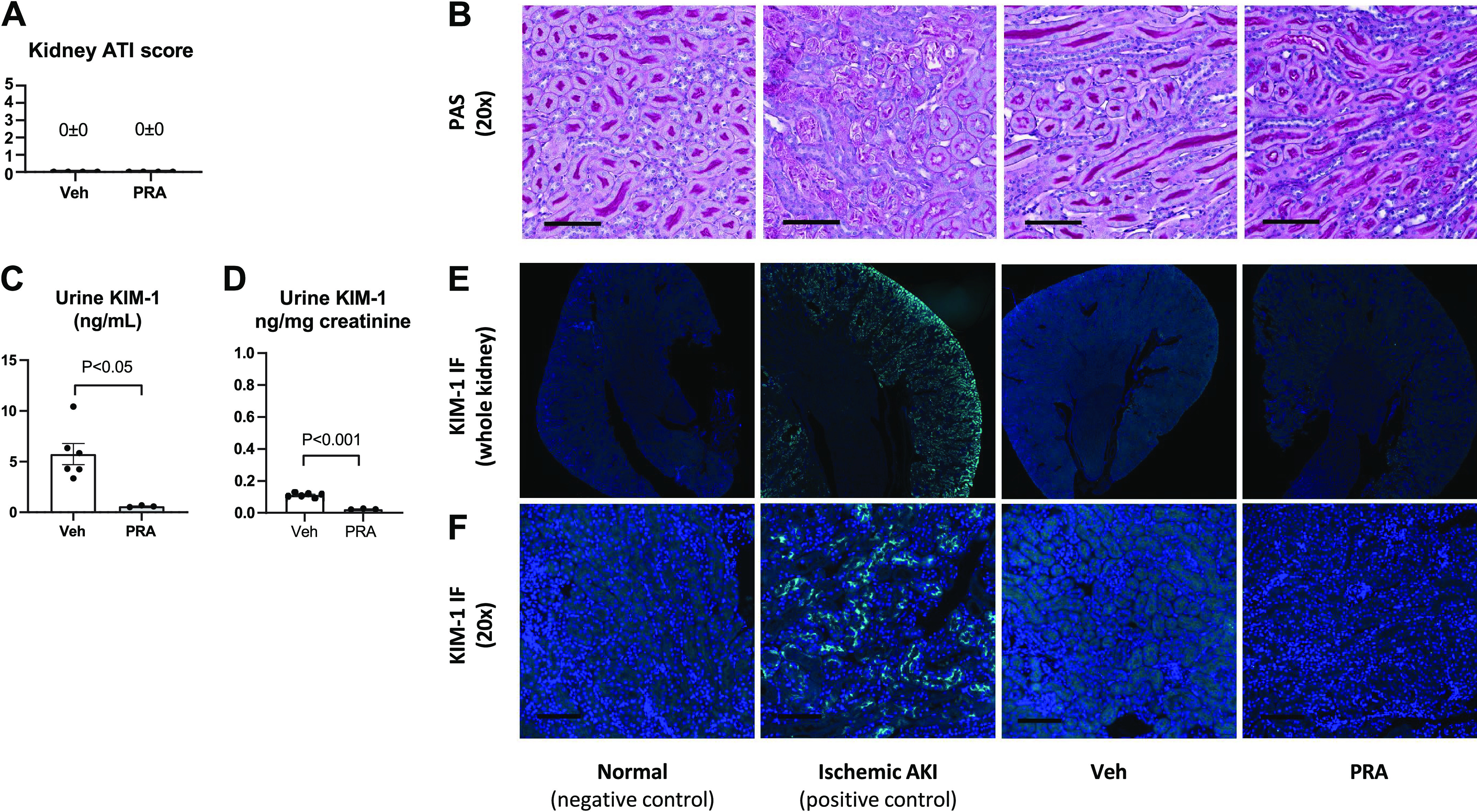
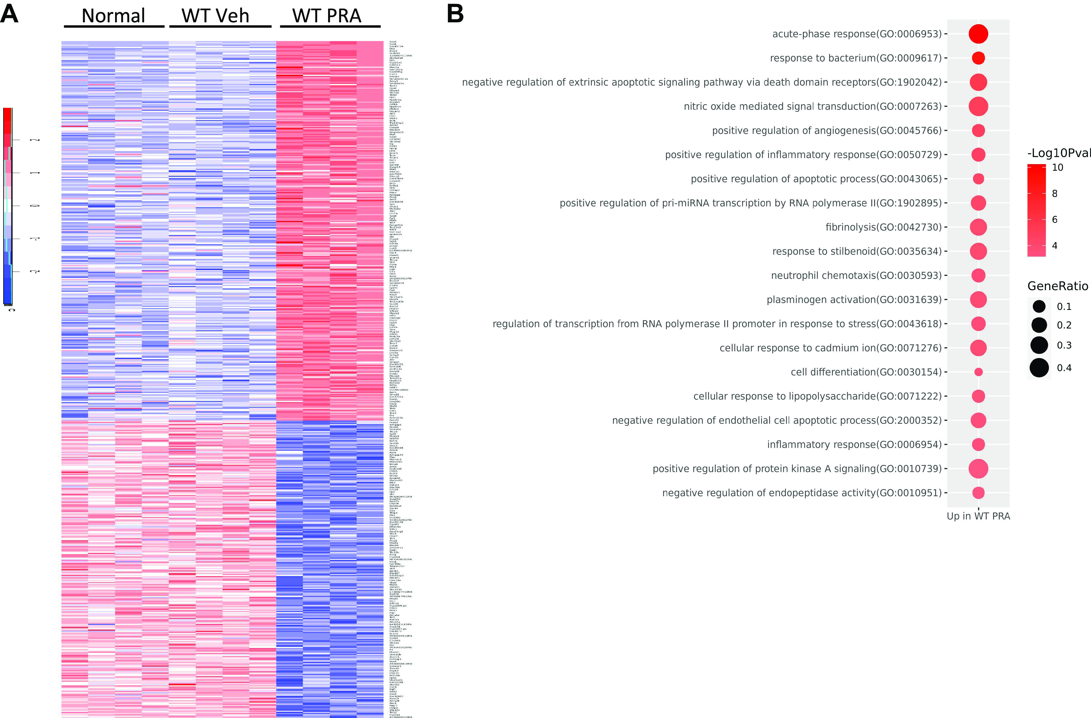
Prerenal azotemia (PRA) is a major cause of acute kidney injury and uncommonly studied in preclinical models. We sought to develop and characterize a novel model of PRA that meets the clinical definition: acute loss of glomerular filtration rate (GFR) that returns to baseline with resuscitation. Adult male C57BL/6J wild-type (WT) and IL-6-/- mice were studied. Intraperitoneal furosemide (4 mg) or vehicle was administered at time = 0 and 3 h to induce PRA from volume loss. Resuscitation began at 6 h with 1 mL intraperitoneal saline for four times for 36 h. Six hours after furosemide administration, measured glomerular filtration rate was 25% of baseline and returned to baseline after saline resuscitation at 48 h. After 6 h of PRA, plasma interleukin (IL)-6 was significantly increased, kidney and liver histology were normal, kidney and liver lactate were normal, and kidney injury molecule-1 immunofluorescence was negative. There were 327 differentially regulated genes upregulated in the liver, and the acute phase response was the most significantly upregulated pathway; 84 of the upregulated genes (25%) were suppressed in IL-6-/- mice, and the acute phase response was the most significantly suppressed pathway. Significantly upregulated genes and their proteins were also investigated and included serum amyloid A2, serum amyloid A1, lipocalin 2, chemokine (C-X-C motif) ligand 1, and haptoglobin; hepatic gene expression and plasma protein levels were all increased in wild-type PRA and were all reduced in IL-6-/- PRA. This work demonstrates previously unknown systemic effects of PRA that includes IL-6-mediated upregulation of the hepatic acute phase response.NEW & NOTEWORTHY Prerenal azotemia (PRA) accounts for a third of acute kidney injury (AKI) cases yet is rarely studied in preclinical models. We developed a clinically defined murine model of prerenal azotemia characterized by a 75% decrease in measured glomerular filtration rate (GFR), return of measured glomerular filtration rate to baseline with resuscitation, and absent tubular injury. Numerous systemic effects were observed, such as increased plasma interleukin-6 (IL-6) and upregulation of the hepatic acute phase response.

Keywords: NGAL; acute kidney injury; biomarkers; interleukin-6; organ cross talk; volume depletion.


Conflict of interest statement

S.F. receives consulting fees from SeaStar Medical. None of the other authors has any conflicts of interest, financial or otherwise, to disclose.

Figures

None
Graphical abstract
Figure 1.
Figure 1.
Experimental protocol and model of prerenal azotemia (PRA). A: experimental protocol. The glomerular filtration rate (GFR), blood urea nitrogen (BUN), and weight (Wt) were measured serially in the same mouse over time beginning 48 h prior to the first intraperitoneal injection of 4 mg furosemide at time 0. The furosemide injection was repeated 3 h later. Food and water were withheld from time 0 to 6 h. Six hours after the first injection of furosemide, GFR measurement, BUN, and weight were reassessed; this time point is the peak of volume depletion, and average urine output during this time ranges from ∼1.5 to 3.0 mL. Volume resuscitation was performed with intraperitoneal administration of 1 mL of saline at 6, 7, 24, and 28 h after the first dose of furosemide. To assess the success of volume resuscitation, GFR, BUN, and weight were measured 48 h after the first dose of furosemide. The gold standard definition of PRA is a decline in kidney function (i.e., GFR) that returns to baseline within 24−72 h after volume resuscitation or other measures to restore renal hemodynamics. GFR (B), BUN (C), and weight (D) were significantly reduced during PRA compared with baseline and recovery and were similar between baseline and recovery. Thus, volume resuscitation successfully restored kidney function (GFR), BUN, and weight to baseline, thus meeting the clinical definition of PRA. Data are expressed as means ± SE and were analyzed by repeated-measures ANOVA with Tukey’s post hoc analysis; n = 7. NS, not significant.
Figure 2.
Figure 2.
Tubular injury is absent in prerenal azotemia (PRA). Tubular injury was assessed 6 h after vehicle (Veh) treatment or furosemide treatment to induce PRA. No evidence of tubular injury was identified after PRA. A: the histological acute tubular injury (ATI) score was 0 ± 0 with Veh treatment and 0 ± 0 in PRA. B: representative histology demonstrating normal tubular architecture (which is disrupted after ischemic acute kidney injury as a positive control). PAS, periodic acid-Schiff. Urine kidney injury molecule (KIM)-1 (ng/mL; C) and urine KIM-1 corrected for urine creatinine (D) was significantly decreased in PRA vs. Veh treatment. E and F: immunofluorescence for KIM-1 in kidney tissue was absent in both Veh treatment and PRA (and was strongly positive after ischemic acute kidney injury as a positive control). Data are expressed as means ± SE and were analyzed by an unpaired Student’s t test; n = 3–6.
Figure 3.
Figure 3.
Effect of prerenal azotemia (PRA) on plasma electrolytes. Plasma levels of Na+ (A), Cl (B), K+ (C), bicarbonate (D), Ca2+ (E), and Mg2+ (F) were determined in normal mice and in mice 6 h after vehicle (Veh) treatment or furosemide injection to induce PRA. There were no significant differences among groups for Na+, Cl, K+, bicarbonate, Ca2+, or Mg2+. P = not significant (NS) among normal, Veh, and PRA group for Na+, Cl, K+, bicarbonate, Ca2+, and Mg2+. Data are expressed as means ± SE and were analyzed by one-way ANOVA with Tukey’s post hoc multiple-comparisons test; n = 3–5.
Figure 4.
Figure 4.
Plasma interleukin (IL)-6 is increased in prerenal azotemia (PRA). Plasma levels of IL-6 as well as kidney, liver, spleen, and lung gene expression and protein levels of IL-6 were determined in normal mice (no treatment) and in wild-type (WT) mice 6 h after vehicle (Veh) treatment or furosemide treatment to induce PRA. A: plasma levels of IL-6 were significantly increased after PRA. Gene expression of IL-6 in the kidney (B), liver (C), spleen (D), and lung (E) were not significantly different between PRA vs. Veh treatment. Protein levels of IL-6 were slightly elevated in PRA vs. Veh treatment in the kidney (F) but were similar in the liver (G), spleen (H), and lung (I). Data are expressed as means ± SE and were analyzed by a Student’s t test comparing WT Veh and WT PRA groups; n = 4–6. NS, not significant.
Figure 5.
Figure 5.
Recombinant human (rh) interleukin (IL)-6 is elevated in the plasma in mice with prerenal azotemia (PRA) vs. vehicle (Veh) treatment after intravenous injection. A total of 200 ng rhIL-6 was administered by tail vein injection 5 h after the first dose of Veh or furosemide injection to induce PRA. A: plasma levels of rhIL-6 were significantly elevated in PRA vs. Veh treatment. B: glomerular filtration rate (GFR) was significantly decreased in PRA. C: blood urea nitrogen (BUN) was significantly elevated in PRA. Data are expressed as means ± SE and were analyzed by a Student’s t test comparing Veh treatment and PRA; n = 4.
Figure 6.
Figure 6.
Effect of prerenal azotemia (PRA) on kidney function, kidney lactate, and kidney succinate in wild-type (WT) and interleukin (IL)-6-deficient (IL-6−/−) mice. Plasma levels of IL-6, creatinine, and blood urea nitrogen (BUN) as well as kidney levels of the tissue ischemia markers lactate and succinate were determined in normal mice (no treatment), and in WT and IL-6−/− mice 6 h after vehicle (Veh) treatment or furosemide treatment to induce PRA. A: plasma creatinine was slightly increased in the WT PRA group vs. the WT Veh group and was similar between the WT PRA group and IL-6−/− PRA group. B: BUN was significantly increased in the WT PRA group vs. the WT Veh group and was similar in the IL-6−/− PRA group vs. the WT PRA group. C: kidney lactate levels were similar between the WT Veh and WT PRA groups and similar between the WT PRA and IL-6−/− PRA groups. D: kidney succinate levels were similar between the WT Veh and WT PRA groups and similar between the WT PRA and IL-6−/− PRA groups. Data are expressed as means ± SE and were analyzed by one-way ANOVA with Sidak’s post hoc multiple-comparisons test comparing prespecified groups (WT Veh vs. WT PRA, WT Veh vs. IL-6−/− Veh, and WT PRA vs. IL-6−/− PRA); n = 3 or 4. NS, not significant.
Figure 7.
Figure 7.
Prerenal azotemia (PRA) does not cause histological liver injury or affect markers of tissue ischemia in wild-type (WT) or interleukin (IL)-6-deficient (IL-6−/−) mice. Liver histology as well as liver lactate and succinate levels were determined in normal mice and in WT and IL-6−/− mice 6 h after vehicle (Veh) treatment or furosemide injection to induce PRA. A: representative hematoxylin and eosin-stained liver histological images. B: liver histologic injury score demonstrating that there was no significant liver injury in the WT PRA or IL-6−/− PRA groups; specifically, liver histological injury was similar to normal for the WT Veh, IL-6−/− Veh, WT PRA, and IL 6−/− PRA groups. C: liver lactate was not increased in the WT PRA group vs. the WT Veh group, and levels were similar between the WT PRA and IL-6−/− PRA groups. D: liver succinate was not increased in the WT PRA group vs. the WT Veh group, and levels were similar between the WT PRA and IL-6−/− PRA groups. Data are expressed as means ± SE and were analyzed by one-way ANOVA with Tukey’s post hoc multiple comparisons test; n = 3–5. #P = not significant (NS) vs. normal.
Figure 8.
Figure 8.
Multidimensional scaling plot demonstrating that prerenal azotemia (PRA) distinctly affects the gene expression profile in the liver. RNA-sequencing analysis was performed with RNA extracted from liver tissue of wild-type (WT) and interleukin (IL)-6 deficient (IL-6−/−) mice subjected to vehicle (Veh) treatment or furosemide treatment to induce PRA. WT mice that received no treatment (normal) were included as an additional control. A multidimensional scaling plot was generated to globally assess the gene expression profiles of each group. Samples from each treatment group are annotated with colored ellipses. As expected, mice in the normal, WT Veh, and IL-6−/− Veh groups clustered together. WT PRA (in green) distinctly affects gene expression, and this expression was further affected during PRA with IL-6 deficiency (IL-6−/− PRA, in pink). Normal, n = 4; WT Veh, n = 4; WT PRA, n = 4; IL-6−/− Veh, n = 4; IL-6−/− PRA, n = 3.
Figure 9.
Figure 9.
Hepatic RNA sequencing reveals that the acute phase response is the most significantly upregulated pathway after prerenal azotemia (PRA). RNA sequencing analysis of liver tissue was performed in wild-type (WT) mice with no treatment (normal) and 6 h after vehicle treatment (WT Veh) or furosemide treatment to induce WT PRA. A total of 587 genes were differentially expressed in the WT PRA group vs. the WT Veh group, with 327 genes upregulated in the WT PRA group and 260 genes suppressed in the WT PRA group. A: heatmap demonstrating differentially expressed genes in the normal (n = 4), WT Veh (n = 4), and WT PRA (n = 4) groups. Red, upregulated; blue, suppressed. B: Gene Ontology analysis revealed that the acute phase response was the most significantly upregulated pathway in the liver after PRA. The top 20 upregulated pathways in the WT PRA group are shown.
Figure 10.
Figure 10.
Hepatic RNA sequencing reveals that the acute phase response is the most significantly downregulated pathway in interleukin (IL)-6-deficient (IL-6−/−) mice with prerenal azotemia (PRA). RNA-sequencing analysis of liver tissue was performed in wild-type (WT) mice with no treatment (normal) and in WT and IL-6−/− mice 6 h after vehicle (Veh) treatment or furosemide treatment to induce PRA. Gene expression in the WT PRA and IL-6−/− PRA groups were compared. A total of 155 genes were differentially expressed in the WT PRA group vs. the IL-6−/− PRA group, with 33 genes upregulated in the IL-6−/− PRA group and 122 genes suppressed in the WT PRA group. A: heatmap demonstrating the relationship of the 155 differentially expressed genes in the normal (n = 4), WT Veh (n = 4), WT PRA (n = 4), IL-6−/− Veh (n = 4), and IL-6−/− PRA (n = 4) groups. Red, upregulated; blue, suppressed. B: Gene Ontology analysis revealed that the acute phase response was the most significantly downregulated pathway in the liver in the IL-6−/− PRA group. The top 20 downregulated pathways in the IL-6−/− PRA group are shown. C: comparison of the 327 genes that were upregulated in WT PRA group with the 122 genes that were suppressed in the IL-6−/− mice with PRA. A total of 84 genes overlapped between the 2 groups, as shown in the Venn diagram.
Figure 11.
Figure 11.
Hepatic gene expression and plasma protein levels of serum amyloid A2, serum amyloid 1, lipocalin-2 (LCN2), chemokine (C-X-C motif) ligand 1 (CXCL1), and haptoglobin in prerenal azotemia (PRA) in wild-type (WT) and interleukin (IL)-deficient (IL-6−/−) mice are consistent with hepatic RNA sequencing. A–I: hepatic gene expression and plasma protein levels of serum amyloid A2, serum amyloid A1, LCN2, CXCL1, and haptoglobin were determined in mice with no treatment (normal) and in WT and IL-6−/− mice 6 h after vehicle (Veh) treatment or furosemide treatment (PRA). Hepatic gene expression of serum amyloid A2, serum amyloid A1, LCN2, CXCL1, and haptoglobin were significantly increased in the WT PRA group vs. the WT Veh group and were significantly decreased in the IL-6−/− PRA group vs. the WT PRA group. No significant differences were noted between the WT Veh and IL-6−/− Veh groups. Plasma protein levels of serum amyloid A2 and 1 together (by ELISA, which detects both forms), neutrophil gelatinase-associated lipocalin (NGAL; the protein product of LCN2), CXCL1, and haptoglobin were significantly increased in the WT PRA group vs. the WT Veh group and were significantly decreased in the IL-6−/− PRA vs. the WT PRA group. No significant differences were noted between the WT Veh and IL-6−/− Veh groups. Data are expressed as means ± SE and were analyzed by one-way ANOVA with Sidak’s post hoc multiple-comparisons test comparing prespecified groups (WT Veh vs. WT PRA, WT Veh vs. IL-6−/− Veh, and WT PRA vs. IL-6−/− PRA). n = 3–5. NS, not significant.
Figure 12.
Figure 12.
Plasma cytokines in prerenal azotemia (PRA) in wild-type (WT) and interleukin (IL)-6-deficient (IL-6−/−) mice. Plasma levels of IL-6, chemokine (C-X-C motif) ligand 1 (CXCL1), IL-5, tumor necrosis factor (TNF)-α, IL-10, IL-1β, interferon (IFN)-γ, and IL-2 were determined in WT mice with no treatment (normal) and in WT and IL-6−/− mice 6 h after vehicle (Veh) treatment or furosemide treatment to induce PRA. Plasma levels of IL-6 (A), CXCL1 (B), and IL-5 (C) were significantly increased in the WT PRA group vs. the WT Veh group and reduced in the IL-6−/− PRA group vs. the WT PRA group. Plasma levels of TNF-α (D) and IL-10 (E) were significantly increased in the WT PRA group vs. the WT Veh group but were not different in the IL-6−/− PRA group vs. the WT PRA group. Plasma IL-1β (F) and IFN-γ (G) were not significantly different in the WT Veh group vs. the WT PRA group and were not significantly different between the WT PRA and IL-6−/− PRA groups. H: plasma IL-2 was not significantly different between the WT Veh and WT PRA groups and was significantly increased in the IL-6−/− PRA group vs. the WT PRA group. Data are expressed as means ± SE and were analyzed by one-way ANOVA with a Sidak’s post hoc multiple-comparisons test comparing prespecified groups (WT Veh vs. WT PRA, WT Veh vs. IL-6−/− Veh, and WT PRA vs. IL-6−/− PRA); n = 3-4. (Note that plasma IL-6 and CXCL1 are also shown in Figs. 3 and 10, respectively; they are included here to allow comparison of all the cytokines together.) NS, not significant.
Figure 13.
Figure 13.
Kidney gene expression levels of kidney injury molecule (KIM)-1, neutrophil gelatinase-associated lipocalin (NGAL), chemokine (C-X-C motif) ligand 1 (CXCL1), and haptoglobin (Hp) are low in prerenal azotemia (PRA) in wild-type (WT) and interleukin (IL)-6-deficient (IL-6−/−) mice. Kidney gene expression of KIM-1, lipocalin-2 (Lcn2; NGAL), CXCL1, and Hp were determined in mice with no treatment (normal) and in WT and IL-6−/− mice 6 h after vehicle (Veh) treatment or furosemide treatment to induce PRA. A: gene expression of KIM-1 was similar in the WT Veh group vs. the WT PRA group and in the WT PRA group vs. the IL-6−/− PRA group. B: gene expression of Lcn2 was increased in the WT PRA group vs. the WT Veh group and was increased to a similar extent in the IL-6−/− PRA group. C: gene expression of CXCL1 was similar in the WT Veh and WT PRA groups and was significantly increased in the IL-6−/− PRA group vs. the WT PRA group. D: gene expression of Hp was similar in the WT Veh and WT PRA group and was to a similar extant in the WT PRA and IL-6−/− PRA groups. Data are expressed as means ± SE and were analyzed by one-way ANOVA with a Sidak’s post hoc multiple-comparisons test comparing prespecified groups (WT Veh vs. WT PRA, WT Veh vs. IL-6−/− Veh, and WT PRA vs. IL-6−/− PRA); n = 3–5.

Similar articles

Cited by

References

    1. Al-Jaghbeer M, Dealmeida D, Bilderback A, Ambrosino R, Kellum JA. Clinical decision support for in-hospital AKI. J Am Soc Nephrol 29: 654–660, 2018. doi:10.1681/ASN.2017070765. - DOI - PMC - PubMed
    1. Lewington AJP, Cerda J, Mehta RL. Raising awareness of acute kidney injury: a global perspective of a silent killer. Kidney Int 84: 457–467, 2013. doi:10.1038/ki.2013.153. - DOI - PMC - PubMed
    1. Uchino S, Bellomo R, Goldsmith D, Bates S, Ronco C. An assessment of the RIFLE criteria for acute renal failure in hospitalized patients. Crit Care Med 34: 1913–1917, 2006. doi:10.1097/01.CCM.0000224227.70642.4F. - DOI - PubMed
    1. Liaño F, Pascual J; The MADRID Acute Renal Failure Study Group. Epidemiology of acute renal failure: a prospective, multicenter, community-based study. Madrid Acute Renal Failure Study Group. Kidney Int 50: 811–818, 1996. doi:10.1038/ki.1996.380. - DOI - PubMed
    1. Nash K, Hafeez A, Hou S. Hospital-acquired renal insufficiency. Am J Kidney Dis 39: 930–936, 2002. doi:10.1053/ajkd.2002.32766. - DOI - PubMed

Publication types