. 2023 Aug 2;145(30):16458-16463.
doi: 10.1021/jacs.3c02752. Epub 2023 Jul 20.

Cell Surface Labeling and Detection of Protein Tyrosine Kinase 7 via Covalent Aptamers

Affiliations

Cell Surface Labeling and Detection of Protein Tyrosine Kinase 7 via Covalent Aptamers

Savannah Albright et al. J Am Chem Soc. .

Abstract

Covalent aptamers are novel biochemical tools for fast and selective transfer of labels to target proteins. Equipped with cleavable electrophiles, these nucleic acid probes enable the installation of functional handles onto native proteins. The high affinity and specificity with which aptamers bind their selected targets allows for quick, covalent labeling that can compete with nuclease-mediated degradation. Here, we introduce the first application of covalent aptamers to modify a specific cell surface protein through proximity-driven label transfer. We targeted protein tyrosine kinase 7 (PTK7), a prominent cancer marker, and demonstrated aptamer-mediated biotin transfer to specific lysine residues on the extracellular domain of the protein. This allowed for tracking of PTK7 expression, localization, and cellular internalization. These studies validate the programmability of covalent aptamers and highlight their applicability in a cellular context, including protein and small molecule delivery.


Conflict of interest statement

The authors declare no competing financial interest.

Figures

Figure 1
Figure 1
(A) Aptamer-mediated transfer of a chemical motif to the cell surface domain of a protein target. (B) Structures of the alkyne-modified phosphoramidite 2 and the N-acyl sulfonamide biotin-transferring electrophilic warhead 1. (C) Predicted structure of sgc8c, with positions that demonstrated the highest labeling efficiency indicated (8, 27, and 30).
Figure 2
Figure 2
(A) Structure–activity study of aptamers modified with 1 and incubated with PTK7. (B) Dose–response biotinylation of PTK7 by sgc8c(27)-1. (C, D) Dose–response analysis of off-target labeling of BSA and serum-supplemented DMEM by sgc8c(27)-1. (E) Time course of PTK7 biotinylation. Data points represent averages, and error bars are standard deviations from two to three independent experiments.
Figure 3
Figure 3
Proposed model of PTK7 (cyan) with biotinylated lysine residues (magenta), as determined by mass spectrometry. The domain structures were predicted by AlphaFold and oriented based on the mass spectrometry results.
Figure 4
Figure 4
(A) Covalent transfer of biotin from sgc8c(27)-1 to PTK7 or PTK7-CFP expressed on HEK293T cells and pulled-down with streptavidin resin. (B, C) Dose- and time-dependent biotinylation of HEK293T cells expressing PTK7-CFP, respectively. Data points represent averages, and error bars indicate standard deviation of at least two independent experiments.
Figure 5
Figure 5
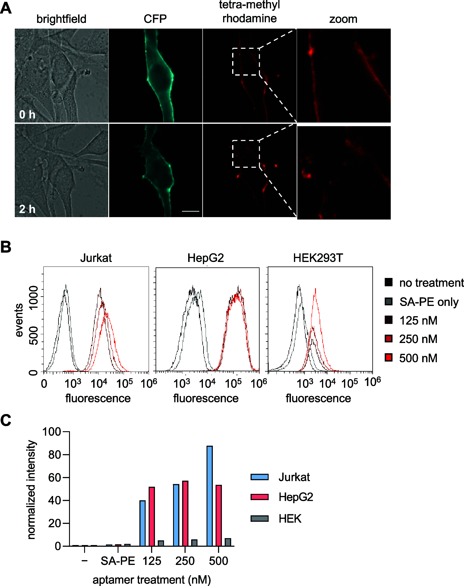
(A) Live cell imaging of NIH3T3 cells expressing PTK7-CFP incubated with sgc8c(27)-1 and stained with NA-TMR. Cells were incubated at 37 °C for 0 h (top) and 2 h (bottom) after NA-TMR staining. The scale bar represents 10 μm. (B) Flow cytometry of PTK7-positive Jurkat cells (left), PTK7-positive HepG2 cells (middle), and PTK7-negative HEK cells (right) incubated with sgc8c(27)-1 and conjugated to SA-PE. (C) Quantification of mean intensity of phycoerythrin fluorescence in flow cytometry. Bars represent an MFI of 50,000 recorded events.

References

    1. Keefe A. D.; Pai S.; Ellington A. Aptamers as therapeutics. Nat. Rev. Drug Discovery 2010, 9, 537–550. 10.1038/nrd3141. - DOI - PMC - PubMed
    1. Shangguan D.; Cao Z.; Meng L.; Mallikaratchy P.; Sefah K.; Wang H.; Li Y.; Tan W. Cell-specific aptamer probes for membrane protein elucidation in cancer cells. J. Proteome Res. 2008, 7, 2133–2139. 10.1021/pr700894d. - DOI - PMC - PubMed
    1. Kroiher M.; Miller M. A.; Steele R. E. Deceiving appearances: signaling by ″dead″ and ″fractured″ receptor protein-tyrosine kinases. BioEssays 2001, 23, 69–76. 10.1002/1521-1878(200101)23:1<69::AID-BIES1009>3.0.CO;2-K. - DOI - PubMed
    1. van der Geer P.; Hunter T.; Lindberg R. A. Receptor protein-tyrosine kinases and their signal transduction pathways. Annu. Rev. Cell Biol. 1994, 10, 251–337. 10.1146/annurev.cb.10.110194.001343. - DOI - PubMed
    1. Lhoumeau A.-C.; Martinez S.; Prébet T.; Borg J.-P.. The PTK7 Receptor Family. In Receptor Tyrosine Kinases: Family and Subfamilies; Wheeler D. L.; Yarden Y., Eds.; Springer International Publishing, 2015; pp. 539–558, 10.1007/978-3-319-11888-8_11. - DOI

Publication types

MeSH terms