. 2023 Sep;299(9):105072.
doi: 10.1016/j.jbc.2023.105072. Epub 2023 Jul 18.

A conserved arginine within the αC-helix of Erk1/2 is a latch of autoactivation and of oncogenic capabilities

Affiliations

A conserved arginine within the αC-helix of Erk1/2 is a latch of autoactivation and of oncogenic capabilities

Nadine Soudah et al. J Biol Chem. 2023 Sep.

Abstract

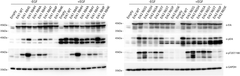
Eukaryotic protein kinases (EPKs) adopt an active conformation following phosphorylation of a particular activation loop residue. Most EPKs spontaneously autophosphorylate this residue. While structure-function relationships of the active conformation are essentially understood, those of the "prone-to-autophosphorylate" conformation are unclear. Here, we propose that a site within the αC-helix of EPKs, occupied by Arg in the mitogen-activated protein kinase (MAPK) Erk1/2 (Arg84/65), impacts spontaneous autophosphorylation. MAPKs lack spontaneous autoactivation, but we found that converting Arg84/65 of Erk1/2 to various residues enables spontaneous autophosphorylation. Furthermore, Erk1 molecules mutated in Arg84 are oncogenic. Arg84/65 thus obstructs the adoption of the "prone-to-autophosphorylate" conformation. All MAPKs harbor an Arg that is equivalent to Arg84/65 of Erks, whereas Arg is rarely found at the equivalent position in other EPKs. We observed that Arg84/65 of Erk1/2 interacts with the DFG motif, suggesting that autophosphorylation may be inhibited by the Arg84/65-DFG interactions. Erk1/2s mutated in Arg84/65 autophosphorylate not only the TEY motif, known as critical for catalysis, but also on Thr207/188. Our MS/MS analysis revealed that a large proportion of the Erk2R65H population is phosphorylated on Thr188 or on Tyr185 + Thr188, and a small fraction is phosphorylated on the TEY motif. No molecules phosphorylated on Thr183 + Thr188 were detected. Thus, phosphorylation of Thr183 and Thr188 is mutually exclusive suggesting that not only TEY-phosphorylated molecules are active but perhaps also those phosphorylated on Tyr185 + Thr188. The effect of mutating Arg84/65 may mimic a physiological scenario in which allosteric effectors cause Erk1/2 activation by autophosphorylation.

Keywords: ERK; MAP kinase; activation loop; active variants; autophosphorylation; eukaryotic protein kinases.


Conflict of interest statement

Conflict of interest The authors declare that they have no conflicts of interest with the contents of this article.

Figures

Figure 1
Figure 1
Mutating Arg84/Arg65 of Erk1/2 to various other residues renders the resulting mutants intrinsically active in vitro. The indicated recombinant purified Erk1/2 mutants were tested in a kinase assay with or without pretreatment with active MEK1. A, fixed volume from each reaction was subjected to SDS-PAGE and stained with Coomassie brilliant blue to verify loading equal amounts of MBP (second row) and was then exposed to a phosphorimager screen to visualize 32P incorporation (first row). Samples from each reaction were also subjected to Western blot analysis and probed with antibodies against total Erk1/2 (third row), phosphorylated(TEY)-Erk1/2 (fourth row), and phosphorylated(Thr207/Thr188)-Erk1/2 (fifth row). B, a portion of each kinase assay reaction was quantified in a β-counter. Quantifications are shown as percentage of the activity of MEK1-activated Erk1/2WT, which was considered as 100% ± standard deviation (100% of Erk1WT = 235,264 cpm; 100% of Erk2WT = 145,430 cpm). Reactions were performed in triplicates. MBP, myelin basic protein.
Figure 2
Figure 2
Erk1/2 molecules mutated in Arg84/Arg65 are spontaneously phosphorylated in HEK293T cells. Expression vectors carrying the indicated Erk1/2 mutants were introduced into HEK293T cells. About 48 h post-transfection, cells were serum starved for 16 h, and EGF was added to the indicated plates (50 ng/ml for 10 min). Protein lysates were then prepared and subjected to Western blot analysis and probed with antibodies against total Erk1/2 (first row), phosphorylated(TEY)-Erk1/2 (second row), phosphorylated(Thr207/Thr188)-Erk1/2 (third row), and GAPDH (fourth row). Note that the externally expressed Erk1 molecules (tagged with polyhistidine that adds just 0.8 kDa to the molecular weight) migrate very close to, almost overlap with, the endogenous Erk1 (left panel). Externally expressed Erk2 molecules, tagged with HA run slower than the endogenous Erk1 and Erk2, as the tag adds 3.2 kDa to the molecular weight (right panel). EGF, epidermal growth factor; HA, hemagglutinin; HEK293T, human embryonic kidney 293T cell line.
Figure 3
Figure 3
Some of the Erk1 molecules mutated at Arg84 oncogenically transform NIH3T3 cells. Expression vectors carrying the indicated Erk1/2 mutants were introduced into NIH3T3 cells. Expression vectors carrying no gene (empty vector) or carrying Erk1WT were used as negative controls, whereas a vector expressing the oncogenic H-RASG12V was used as a positive control for cell transformation. Cells expressing the indicated vectors were selected by the addition of G418 and fixed and stained with crystal violet 4 weeks after transfection. Statistical analysis revealed that the differences in oncogenic efficiency between Erk1R84S and Erk1R84H mutants were not statistically significant (p value of 0.36). Similarly, no significant differences were observed when comparing Erk1R84A with Erk1R84Y, Erk1R84T, Erk1R84P, and Erk1R84K mutants (average p value of 0.16). However, the remaining samples exhibited statistically significant differences, with an average p value of 0.023.
Figure 4
Figure 4
Various mutants of the yeast MAPK Erk/Mpk1, mutated at Arg68 (but not Mpk1R68Kand Mpk1R68P), are intrinsically active. Plasmids carrying the indicated mutants were introduced to mpk1Δ yeast cells (A) or mkk1Δmkk2Δ cells (B). Transformants were grown to logarithmic phase on SD (−URA) medium and then plated in serial dilutions (1:10) on either SD (−URA) plates or on plates containing YPD supplemented with 16 mM caffeine, as indicated. In panels A1 and B1 mutants were expressed from YEP352, and in panels A2 and B2, mutants were expressed from AES426 expression vector. MAPK, mitogen-activated protein kinase; YPD, yeast extract–peptone–dextrose.
Figure 5
Figure 5
Arg65 of Erk2 may block spontaneous autophosphorylation activity via its interaction with the DFG motif.A, superposition of Arg65 from nine crystal structures (Protein Data Bank codes: 1ERK, 3ERK, 4ERK, 4S31, 4GT3,5UMO, 6RFP, 6FLE, and 4XRL) of ERK2 that were crystallized in the P21 space group and have similar cell parameters. The two extreme conformations are colored in dark purple. B, Arg65 conformation comparison between two Erk2WT structures (Protein Data Bank codes: 5UMO and 4S31). In both conformations, Arg65 interacts with residues of the DFG motif, either Asp165 or Gly167. C, in Erk2R65S, Ser65 does not interact with the DFG. D, in Erk2I84A, also an intrinsically active variant (26), Arg65 does not interact with the DFG motif.
Figure 6
Figure 6
Thr207/188 phosphorylation is independent of TEY motif phosphorylation. The indicated recombinant purified Erk1 mutants were tested in a cold kinase assay with or without active MEK1. Fixed volume from each reaction was subjected to Western blot analysis and probed with antibodies against total Erk1/2 (first row), phosphorylated(TEY)-Erk1/2 (second row), and phosphorylated(Thr207/Thr188)-Erk1/2 (third row).
Figure 7
Figure 7
An Arg residue, equivalent to Arg84/65 of Erk1/2, is invariantly conserved in MAPKs, found in all MAPKs in nature, and is restricted to MAPKs.A, a WebLogo depicting the MSA of the α-C helix of all MAPKs. The y-axis represents the probability score. The x-axis displays the position of amino acid in the MSA. The αC-helix Arg is highlighted in light purple. Note that it is present in all MAPKs. B, structure-based MSA found in Kincore was used to align mammalian MAPKs showing 100% conservation of the αC-helix Arg (highlighted in light purple). C, structure-based MSA of MAPKs found in lower eukaryotes also showing conservation of the αC-helix Arg (highlighted in light purple). D, WebLogos depicting the MSA of the different EPK families. The position equivalent to Arg84/65 of Erk1/2 in each family in highlighted in light purple. EPK, eukaryotic protein kinase; MAPK, mitogen-activated protein kinase; MSA, multiple sequence alignment.
Figure 8
Figure 8
Mutating Arg66 in the yeast MAPK p38/Hog1 affects specifically intrinsic autophosphorylation but not catalysis.A, pbs2Δ cells harboring the indicated expression plasmids were plated in five serial dilutions on plates containing YPD supplemented with 0.8 M or 1 M NaCl. All strains were also plated on plates containing SD (−URA) medium with no NaCl. B, as in A but in hog1Δ cells. C, protein lysates prepared from pbs2Δ cells harboring the indicated plasmids, and exposed or not exposed to 0.77 M NaCl, were subjected to a Western blot analysis and probed using the indicated antibodies. MAPK, mitogen-activated protein kinase; YPD, yeast extract–peptone–dextrose.
Figure 9
Figure 9
Arg67 and Arg69 are essential for autophosphorylation of p38β and JNK2α2, respectively.A, the indicated, recombinant, purified p38 molecules were tested in an in vitro kinase assay. A fixed volume from each reaction was subjected to SDS-PAGE and exposed to an X-ray film to visualize 32P incorporation (first row). Samples were also subjected to Western blot analysis and probed with antibodies against the polyhistidine, targeting the His tag on the p38β proteins (second row), and phosphorylated(TGY) p38 (third row). p38βY69H molecules, mutated in the adjacent residue of Arg67, were used as a control for the specificity of the Arg67 function. B, HEK293T cells were transfected with the indicated vectors. About 48 h post-transfection, cells were irradiated, or not, with UV radiation and lyzed 1 h later. Protein lysates were subjected to Western blot analysis using the indicated antibodies. C, JNK2α2WT and JNK2α2R69S molecules were tested in an in vitro kinase assay using GST-c-Jun as a substrate, with or without pretreatment with active MKK7. Reaction mixtures were separated on SDS-PAGE and exposed to an X-ray film. GST, glutathione-S-transferase; HEK293T, human embryonic kidney 293T cell line; JNK, c-Jun N-terminal kinase.
Figure 10
Figure 10
Analysis of crystal structures of p38s and JNKs reveals an association between interaction of MAPK-αc-helix-Arg with DFG and spontaneous autophosphorylation capability.A, amino group of Arg69 (colored in purple) of JNK1 (Protein Data Bank [PDB] code: 4L7F) interacts with the carboxyl group of Asp169 (colored in green) of the DFG motif. The distance of the charged interaction between the two atoms is indicated. BD, as in A, but a zoom in into the interaction between the MAPK-αc-Arg and DFG motif of JNK3 (PDB code: 4KKG), p38γ (PDB code: 7CGA), and p38δ (PDB code: 4YNO). E, zoom in into the MAPK-αc-Arg (colored in purple) of JNK2 (PDB code: 3E7O) showing no association with the DFG motif (colored in green), providing a potential analysis for its autophosphorylation activity. F, as in E but the structural analysis of p38β (PDB code: 3GC9). JNK, c-Jun N-terminal kinase; MAPK, mitogen-activated protein kinase.
Supplemental Fig. S1
Relative quantification of the western blot data shown inFigure 2, as measured by ImageJ. A & B) Quantification of phospho-TEY levels. C & D) Quantification of phospho-Thr207/188. Measurements were normalized to phosphorylation levels of measured in cells harboring an empty plasmid.
Supplemental Fig. S2
Expression of Erk1R84Sand Erk1R84Hinduces strong AP-1-mediated transcription in HEK293T cells. Plasmids expressing Erk1R84S and Erk1R84H were co-introduced into HEK293T cells with either 6xAP-1-luciferase (A) or pY2-luciferase (B) reporter genes. Cells were collected 24 h later and assayed for luciferase activity, which was normalized to activity of co-expressed renilla luciferase.
Supplemental Fig. S3
Thr188 is phosphorylated in cancer-derived cell lines. The indicated ancer-derived cell lines were grown to 75% confluency and collected for protein lysate preparation. 20μg of lysates were subjected to Western blot analysis using antibodies against total Erk1/2 (top row), phosphorylated(TEY)-Erk1/2 (second row), phosphorylated(Thr207/Thr188)- Erk1/2 (third row) and GAPDH (bottom row).
Supplemental Fig. S4
Mutating Arg66 in Hog1 and Arg69 in JNK1 does not evoke intrinsic activity. A) pbs2Δ (left panel) or hog1Δ (right panel) cells harboring the indicated plasmids were plated in five decimal dilutions on plates containing YPD supplemented with 1M NaCl. All strains were also plated on plates containing SD(-URA) medium with no NaCl. B) Recombinant, purified Jnk1WT and Jnk1R69S molecules were tested in an in vitro kinase assay using c- Jun as a substrate, with or without pre-treatment with active MKK7. Reaction mixtures were separated on SDS-PAGE and exposed to a X-ray film.

References

    1. Manning G., Whyte D.B., Martinez R., Hunter T., Sudarsanam S. The protein kinase complement of the human genome. Science. 2002;298:1912–1934. - PubMed
    1. Hanks S.K., Hunter T. The eukaryotic protein kinase superfamily: kinase (catalytic) domain structure and classification 1. FASEB J. 1995;9:576–596. - PubMed
    1. Taylor S.S., Keshwani M.M., Steichen J.M., Kornev A.P. Evolution of the eukaryotic protein kinases as dynamic molecular switches. Philosophical Trans. R. Soc. B: Biol. Sci. 2012;367:2517–2528. - PMC - PubMed
    1. Taylor S.S., Wu J., Bruystens J.G.H., Del Rio J.C., Lu T.W., Kornev A.P., et al. From structure to the dynamic regulation of a molecular switch: a journey over 3 decades. J. Biol. Chem. 2021;296 - PMC - PubMed
    1. Beenstock J., Mooshayef N., Engelberg D. How do protein kinases take a selfie (autophosphorylate)? Trends Biochem. Sci. 2016;41:938–953. - PubMed

Publication types

MeSH terms

Substances

LinkOut - more resources