. 2023 Oct;104(4):174-186.
doi: 10.1124/molpharm.123.000710. Epub 2023 Jul 20.

Atypical Chemokine Receptor 3 "Senses" CXC Chemokine Receptor 4 Activation Through GPCR Kinase Phosphorylation

Affiliations

Atypical Chemokine Receptor 3 "Senses" CXC Chemokine Receptor 4 Activation Through GPCR Kinase Phosphorylation

Christopher T Schafer et al. Mol Pharmacol. 2023 Oct.

Abstract

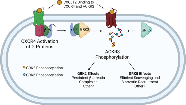
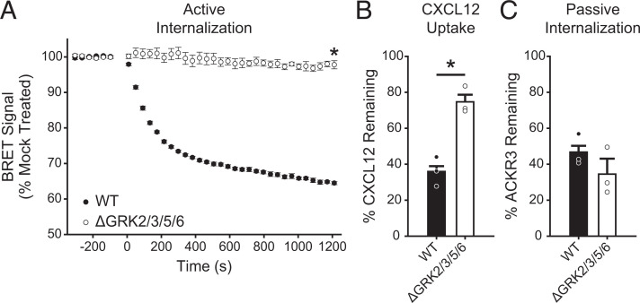
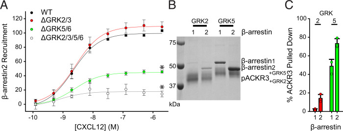
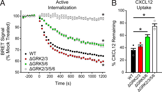
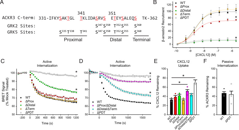
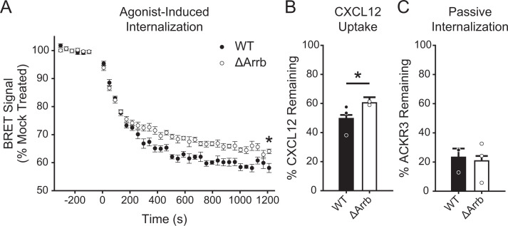
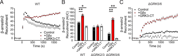
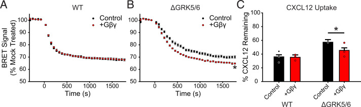
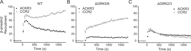
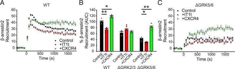
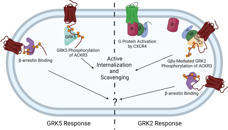
Atypical chemokine receptor 3 (ACKR3) is an arrestin-biased receptor that regulates extracellular chemokine levels through scavenging. The scavenging process restricts the availability of the chemokine agonist CXCL12 for the G protein-coupled receptor (GPCR) CXCR4 and requires phosphorylation of the ACKR3 C-terminus by GPCR kinases (GRKs). ACKR3 is phosphorylated by GRK2 and GRK5, but the mechanisms by which these kinases regulate the receptor are unresolved. Here we determined that GRK5 phosphorylation of ACKR3 results in more efficient chemokine scavenging and β-arrestin recruitment than phosphorylation by GRK2 in HEK293 cells. However, co-activation of CXCR4-enhanced ACKR3 phosphorylation by GRK2 through the liberation of Gβγ, an accessory protein required for efficient GRK2 activity. The results suggest that ACKR3 "senses" CXCR4 activation through a GRK2-dependent crosstalk mechanism, which enables CXCR4 to influence the efficiency of CXCL12 scavenging and β-arrestin recruitment to ACKR3. Surprisingly, we also found that despite the requirement for phosphorylation and the fact that most ligands promote β-arrestin recruitment, β-arrestins are dispensable for ACKR3 internalization and scavenging, suggesting a yet-to-be-determined function for these adapter proteins. Since ACKR3 is also a receptor for CXCL11 and opioid peptides, these data suggest that such crosstalk may also be operative in cells with CXCR3 and opioid receptor co-expression. Additionally, kinase-mediated receptor cross-regulation may be relevant to other atypical and G protein-coupled receptors that share common ligands. SIGNIFICANCE STATEMENT: The atypical receptor ACKR3 indirectly regulates CXCR4-mediated cell migration by scavenging their shared agonist CXCL12. Here, we show that scavenging and β-arrestin recruitment by ACKR3 are primarily dependent on phosphorylation by GRK5. However, we also show that CXCR4 co-activation enhances the contribution of GRK2 by liberating Gβγ. This phosphorylation crosstalk may represent a common feedback mechanism between atypical and G protein-coupled receptors with shared ligands for regulating the efficiency of scavenging or other atypical receptor functions.


Figures

None
Graphical abstract
Fig. 1.
Fig. 1.
GRKs mediate efficient CXCL12-induced internalization and chemokine scavenging by ACKR3. (A) CXCL12-promoted, active internalization following stimulation with 100 nM chemokine at 37°C was monitored by BRET between ACKR3_RlucII and rGFP_CAAX in WT and ΔGRK2/3/5/6 HEK293A cells. (B) Chemokine uptake by WT and ΔGRK2/3/5/6 cells expressing ACKR3 was detected by quantification of the remaining CXCL12 in the media by ELISA. The extent of scavenging was determined by comparison with cells transfected with empty vector. (C) Constitutive cycling, or passive internalization, of ACKR3 in WT and ΔGRK2/3/5/6 cells was quantified by tracking the loss of “prelabeled” receptor by flow cytometry. Errors are reported as standard deviations, and values are the average of three independent experiments performed in triplicate. Individual experiments are presented as points in B and C. Statistical significance was determined by unpaired t test. *P < 0.001.
Fig. 2.
Fig. 2.
GRK5 plays a dominant role in ACKR3 phosphorylation and β-arrestin recruitment in HEK293A cells and with purified components. (A) Recruitment of GFP_ βarr2 to ACKR3_RlucII observed by BRET in ΔGRK HEK293A cell lines across a titration of CXCL12 concentrations. Responses are normalized to WT ACKR3 and are a composite of three independent experiments performed in triplicate. (B) Pulldowns of purified β-arrestin1 and 2 by purified ACKR3 phosphorylated in vitro by either GRK2 or GRK5. (C) Quantification of the amount of β-arrestin pulled down from three independent experiments. The amount of β-arrestin pulled down in C is presented as a percentage of the ACKR3 band density from the same experiment. Errors are reported as standard deviations and significance in A as determined by an extra-sum-of-squares F-test, *P < 0.001. EC50 values were not different among the tested conditions.
Fig. 3.
Fig. 3.
ACKR3 requires GRK5 for efficient active internalization and CXCL12 scavenging in HEK293A cells. (A) Active CXCL12-promoted internalization of ACKR3 monitored by BRET following stimulation with 100 nM CXCL12 in ΔGRK HEK293A cell lines at 37°C. (B) CXCL12 uptake by ACKR3 expressed in ΔGRK cell lines quantified by ELISA and presented as a percentage of identical cells transfected with empty vector. Data are composites of three independent experiments measured in triplicate, errors are reported as standard deviations, and statistical significance was determined by one-way ANOVA followed by a Bonferroni test. *P < 0.001. Data for WT and ΔGRK2/3/5/6 cells are repeated from Fig. 1 for comparison.
Fig. 4.
Fig. 4.
Specific phosphorylation motifs differently contribute to CXCL12 responses by ACKR3 in HEK293A cells. (A) ACKR3 was phosphorylated by either GRK2 or GRK5 in vitro, and the specific sites of modification were determined by mass spectrometry. The detected phosphorylation sites are highlighted in red. Phosphorylated positions in the ACKR3 C-terminus were divided into three clusters and replaced by alanine to produce ΔProx, ΔDistal, ΔTerm, and ΔPDT receptor constructs. (B) Recruitment of GFP_βarr2 to phosphorylation-deficient ACKR3 constructs expressed in WT HEK293A cells and tested across a titration of CXCL12 and detected by BRET. Values represent three independent experiments performed in triplicate and normalized to WT ACKR3 recruitment. (C and D) Active internalization of individual (C) and multiple (D) phosphorylation cluster substitutions tracked by the loss of BRET between ACKR3_RlucII and rGFP_CAAX after CXCL12 addition. (E) CXCL12 uptake by phosphorylation deficient ACKR3 constructs, measured by remaining chemokine and compared with cells transfected with empty vector. (F) Passive agonist-independent internalization of the triple phosphorylation substitution observed by the prelabeled flow cytometry experiment. WT ACKR3 data in B, C, E, and F were repeated from Figs. 1 and 2A for comparison. All error bars represent standard deviations, and statistical significance was determined by (B) the extra sum of squares F-test and (C, D, and E) one-way ANOVA followed by a Bonferroni test. *P < 0.001.
Fig. 5.
Fig. 5.
β-arrestins are dispensable for ACKR3 internalization and CXCL12 scavenging in HEK293 cells. (A) CXCL12-mediated, active internalization in WT and ΔArrb HEK293 cells, measured by decreasing BRET between rGFP_CAAX and ACKR3_RlucII. Note that the cellular background for ΔArrb is HEK293, which leads to slightly different responses compared with the HEK293A cells used in all other figures. (B) Chemokine uptake by WT ACKR3 in ΔArrb and WT HEK293 cells. The remaining chemokine was quantified by ELISA and compared with non-ACKR3 expressing cells. (C) ACKR3 agonist-independent, passive internalization in WT and ΔArrb HEK293 cells. Data presented are the composite of three independent experiments measured in triplicate, and errors represent standard deviations. Statistical significance was determined by an unpaired t test. *P < 0.05.
Fig. 6.
Fig. 6.
GRK2 phosphorylation of ACKR3 is enhanced by expression of Gβγ in HEK293A cells. (A) GFP_βarr2 recruitment to ACKR3_RlucII measured by BRET after 100 nM CXCL12 addition at 0 minute in WT HEK293A cells co-transfected with Gβγ or GRK3-CT. (B) Quantification of Barr2 recruitment in WT, ΔGRK2/3, and ΔGRK5/6 cells with co-transfection with Gβγ or GRK3-CT by integration of the area under the BRET curves after chemokine addition. Areas are normalized to WT ACKR3 recruitment in WT cells without additional treatment. (C) Recruitment of β-arrestin2 to ACKR3 in ΔGRK5/6 cells with Gβγ and GRK3-CT co-expression. Data represent three independent experiments measured in triplicate, and individual experiments are presented as points in (B). Errors are reported as standard deviation, and statistical significance was determined by one-way ANOVA followed by a Bonferroni test. *P < 0.01, **P < 0.001.
Fig. 7.
Fig. 7.
Gβγ co-transfection enhances ACKR3 internalization and CXCL12 scavenging but only in the absence of GRK5/6 in HEK293A cells. (A and B) ACKR3_RlucII active internalization was tracked by rGFP_CAAX BRET with and without co-transfection of Gβγ subunits in WT (A) and ΔGRK5/6 (B) cells following addition of 100 nM CXCL12. Plots are the average of three independent experiments measured in triplicate. (C) CXCL12 uptake in WT and ΔGRK5/6 cells with Gβγ co-transfection. Data from cells without extra Gβγ are repeated here from Fig. 3 for comparison. Errors were reported as standard deviations, and statistical significance was determined by one-way ANOVA followed by a Bonferroni test. *P < 0.001.
Fig. 8.
Fig. 8.
β-arrestin2 recruitment to the canonical GPCR CCR2 is mediated primarily by GRK2/3. A-C) GFP_βarr2 recruitment to ACKR3_RlucII or CCR2_rlucII after stimulation with either 100 nM CXCL12 or CCL2, respectively, was tracked by BRET in WT (A), ΔGRK5/6 (B), and ΔGRK2/3 (C) cells. Data are the average of three independent experiments performed in triplicate, and errors reflect standard deviations.
Fig. 9.
Fig. 9.
Concurrent CXCR4 activation increases ACKR3 phosphorylation by GRK2/3. (A) GFP_βarr2 recruitment to ACKR3_RlucII after 100 nM CXCL12 addition in WT cells treated with 100 µM IT1t for 45 minutes before the experiment or co-transfected with additional CXCR4 DNA. Individual points are the average of three experiments measured in triplicate. (B) Area under the curve analysis of GFP_βarr2 recruitment time courses after CXCL12 addition normalized to WT ACKR3_RlucII in WT cells without treatment. (C) Recruitment time courses of GFP_βarr2 to ACKR3_RlucII by BRET in ΔGRK5/6 cells treated identically to the WT cells described in (A). Errors were represented by standard deviation, and statistical significance was determined by one-way ANOVA followed by a Bonferroni test. *P < 0.005, **P < 0.001.
Fig. 10.
Fig. 10.
ACKR3 GRK specificity is dependent on whether CXCR4 is co-activated. When expressed alone, the atypical receptor is primarily phosphorylated by GRK5, which drives agonist-mediated internalization and scavenging. In the context of CXCR4 co-expression, the co-activation of CXCR4 activates heterotrimeric G proteins and releases Gβγ, which recruits GRK2 and mediates phosphorylation of both receptors. This allows for ACKR3 to sense the activation of CXCR4 and then enhance the scavenging decoy responses of ACKR3. This image does not include the passive constitutive internalization and scavenging pathway. Image created with BioRender.

Update of

References

    1. Aragay AM, Mellado M, Frade JM, Martin AM, Jimenez-Sainz MC, Martinez-A C, Mayor F Jr (1998) Monocyte chemoattractant protein-1-induced CCR2B receptor desensitization mediated by the G protein-coupled receptor kinase 2. Proc Natl Acad Sci USA 95:2985–2990. - PMC - PubMed
    1. Balabanian K, Lagane B, Infantino S, Chow KY, Harriague J, Moepps B, Arenzana-Seisdedos F, Thelen M, Bachelerie F (2005) The chemokine SDF-1/CXCL12 binds to and signals through the orphan receptor RDC1 in T lymphocytes. J Biol Chem 280:35760–35766. - PubMed
    1. Beyett TS, Chen Q, Labudde EJ, Krampen J, Sharma PV, Tesmer JJG (2020) Characterization of a hyperphosphorylated variant of G protein-coupled receptor kinase 5 expressed in E. coli. Protein Expr Purif 168:105547. - PMC - PubMed
    1. Bhandari D, Trejo J, Benovic JL, Marchese A (2007) Arrestin-2 interacts with the ubiquitin-protein isopeptide ligase atrophin-interacting protein 4 and mediates endosomal sorting of the chemokine receptor CXCR4. J Biol Chem 282:36971–36979. - PubMed
    1. Boekhoff I, Inglese J, Schleicher S, Koch WJ, Lefkowitz RJ, Breer H (1994) Olfactory desensitization requires membrane targeting of receptor kinase mediated by beta gamma-subunits of heterotrimeric G proteins. J Biol Chem 269:37–40. - PubMed

Publication types