. 2023 Jul 20;14(1):4384.
doi: 10.1038/s41467-023-40013-6.

A maize epimerase modulates cell wall synthesis and glycosylation during stomatal morphogenesis

Affiliations

A maize epimerase modulates cell wall synthesis and glycosylation during stomatal morphogenesis

Yusen Zhou et al. Nat Commun. .

Abstract

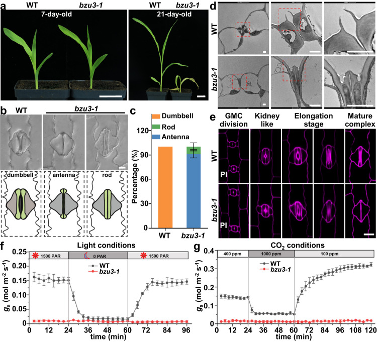
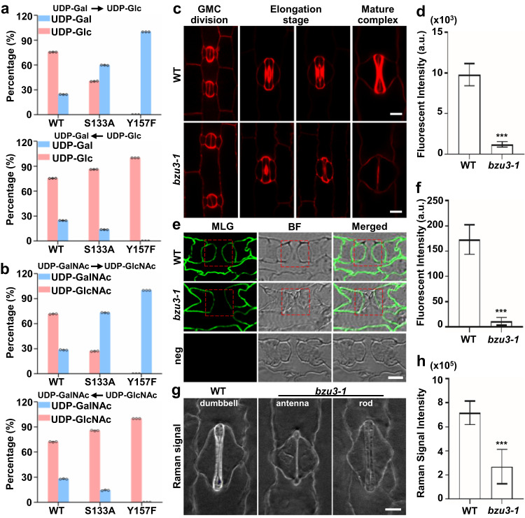
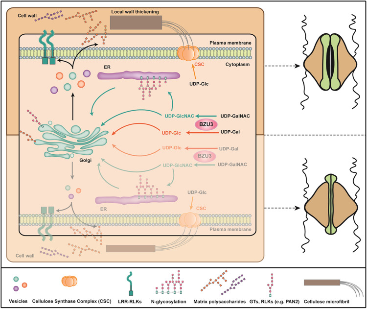
The unique dumbbell-shape of grass guard cells (GCs) is controlled by their cell walls which enable their rapid responses to the environment. The molecular mechanisms regulating the synthesis and assembly of GC walls are as yet unknown. Here we have identified BZU3, a maize gene encoding UDP-glucose 4-epimerase that regulates the supply of UDP-glucose during GC wall synthesis. The BZU3 mutation leads to significant decreases in cellular UDP-glucose levels. Immunofluorescence intensities reporting levels of cellulose and mixed-linkage glucans are reduced in the GCs, resulting in impaired local wall thickening. BZU3 also catalyzes the epimerization of UDP-N-acetylgalactosamine to UDP-N-acetylglucosamine, and the BZU3 mutation affects N-glycosylation of proteins that may be involved in cell wall synthesis and signaling. Our results suggest that the spatiotemporal modulation of BZU3 plays a dual role in controlling cell wall synthesis and glycosylation via controlling UDP-glucose/N-acetylglucosamine homeostasis during stomatal morphogenesis. These findings provide insights into the mechanisms controlling formation of the unique morphology of grass stomata.


Conflict of interest statement

The authors declare no competing interests.

Figures

Fig. 1
Fig. 1. Mutant bzu3 displays abnormal guard cell shapes and is defective in local wall thickening.
a Representative images of 7- and 21-day-old seedlings of WT and bzu3-1. Scale bars, 3 cm. b Representative differential interference contrast (DIC) images (upper panel) and schematic diagram (lower panel) of stomata in WT, bzu3-1 (the second leaf of 7-day-old seedlings). Scale bar, 10 µm. c Proportional phenotypic scoring analysis of stomatal types in WT and bzu3-1. The data was obtained by counting guard cells from ten seedlings of each genotype (WT: n = 1104, bzu3-1: n = 1192). Values represent means ± SD. SD, standard deviation. d Transmission electron microscope observation of stomata longitudinally sectioned of WT and bzu3-1. Red square dashed boxes indicate enlarged regions in the middle and rightmost panels, respectively. Scale bars, 2 µm. e Stomatal staining at different developmental stages in WT and bzu3-1. The cell wall was stained with propidium iodide (PI) and imaged via confocal microscopy. Scale bar, 20 µm. The images were obtained from an approximately 2-3 cm leaf base region taken from the third leaf of 7-day-old seedlings. f Stomatal conductance (gs) in response to changing light conditions. Acclimation began at 1500 units of photosynthetically active radiation (PAR), dropped to 0 PAR, and then increased back to 1500 PAR. The data was obtained from three independent seedlings. Values represent means ± SD. g Stomatal conductance (gs) in response to changing CO2 level. Acclimation began at ambient CO2 conditions (400 ppm), increased to 1000 ppm, and then dropped to 100 ppm. The data was obtained from three independent seedlings. Values represent means ± SD. Source data are provided as a Source Data file.
Fig. 2
Fig. 2. Identification and characterization of BZU3 as the gene responsible for normal guard cell development in maize.
a Fine mapping and cloning of BZU3 based on segregation from population bzu3-1 crossed with B73. BZU3 localizes to the region of bin 1.04 on the long arm of maize Chr 1. The fine-mapping region is 60.67-60.77 Mb and contains only one gene (Zm00001d029151). Chr, Chromosome. b Zm00001d029151 structure with 5’UTR, 9 exons, 8 introns, and 3’UTR. The transposon insertion, deletion mutation and CRISPR-Cas9 mutation positions are indicated. c Stomatal phenotype of B73-329, CRISPR-Cas9-produced mutations (bzu3-3, bzu3-4, and bzu3-5). d Stomatal phenotype of Zheng58, bzu3-2, and the bzu3-2 line complemented by BZU3pro:BZU3-YFP. e BZU3pro:BZU3-YFP expression in bzu3-2 during stomatal development. f, g ZmMUTEpro:BZU3-YFP and ZmFAMApro:YFP-BZU3 remarkably complement homozygous bzu3-2. Representative confocal images of stomatal phenotypes of ZmMUTEpro:BZU3-YFP (f) and ZmFAMApro:YFP-BZU3 (g) complemented bzu3-2. All images were observed from 7-day-old seedlings. YFP fluorescence was observed using confocal microscopy (Zeiss LSM710). Cell walls in the leaf epidermis were stained with PI (propidium iodide) and are indicated in purple. Scale bars, 10 µm.
Fig. 3
Fig. 3. BZU3 catalyzes conversion of UDP-Gal to UDP-Glc in vitro, and bzu3 mutants show defective cell wall synthesis.
a, b Catalytic activity of BZU3 and its variants S133A and Y157F as determined by UDP-Glc, UDP-Gal, UDP-GlcNAc, and UDP-GalNAc. The data was obtained from three biological replicates. Values represent means ± SD. SD, standard deviation. c Representative confocal images of cellulose stained fluorescence of WT and bzu3-1. Cellulose from leaf base were stained using Direct Red 23, with fluorescence being observed using a Zeiss LSM710. Scale bars, 10 µm. d Statistical analysis of Direct Red 23 staining signal intensity in WT and bzu3-1 mature guard cells (c). The signal intensity was obtained from three seedlings of each genotype (n = 32 guard cells). Values represent means ± SD. ***P = 4.998E-42 by Student’s t test (two-sided). SD, standard deviation. e Representative images of MLGs immunolabeling with BG-1 antibody in resin-embedded sections (1.5 μm thick) of leaf base. Labeling was detected with a FITC-labeled secondary antibody and visualized by fluorescence microscopy. neg, negative control. Red square dashed boxes indicate guard cell. Scale bar, 5 µm. f Statistical analysis of MLGs immunolabeling signal shown in (e). The signal intensity was obtained by measuring guard cells from three seedlings of each genotype (n = 30). Values represent means ± SD. ***P = 2.365E-36 by Student’s t-test (two-sided). SD, standard deviation. g Representative images of stimulated Raman scattering microscopy of the cell wall of the epidermis in WT and bzu3-1. The images were produced using Raman spectra with 1100 nm. Scale bar, 10 µm. h Statistical analysis of relative content of cellulose and hemicellulose in stomata of WT and bzu3-1. The signal intensity was obtained by measuring guard cells from five seedlings of each genotype (WT: n = 100, bzu3-1: n = 103) Value represents mean ± SD. ***P = 1.142E-65 by Student’s t-test (two-sided). SD, standard deviation. Source data are provided as a Source Data file.
Fig. 4
Fig. 4. BZU3 catalyzes UDP-Gal epimerization to UDP-Glc in vivo, and UDP-Glc is crucial for maintaining the dumbbell-shape of maize guard cells.
a UPLC-MS detection of UDP-Glc and UDP-Gal in Zheng58 and bzu3-2. 0.1 g leaf base of Zheng58 and bzu3-2. The results were performed three independent biological repeats with similar results. Values represent means ± SD. ***P = 0.0005 of UDP-Glc and ***P = 1.773E-05 of UDP-Gal by Student’s t-test (two-sided). SD, standard deviation. b Phenotypic statistical analysis of bzu3-2 treated with UDP-glucose. The data was obtained by counting guard cells from five seedlings of each genotype (Mock: n = 2901, UDP-Glc: n = 3283). Values represent means ± SD. SD, standard deviation. c Overall structure of BZU3/NAD+/UDP-Glc complex. Cropped view shows the interactions between BZU3, NAD+, and UDP-Glc at the reaction center. d Overall structure of BZU3/NAD+/UDP-GlcNAc complex. Cropped view shows the interactions between BZU3, NAD+, and UDP-GlcNAc at the reaction center. e BZU3pro:BZU3Y157F-YFP failed to rescue the stomatal phenotype in bzu3-2 mutant. The images were obtained from the second leaf of the 7-day-old seedlings. Propidium iodide (PI, purple) was used to stain cell walls. Scale bar, 10 µm. Source data are provided as a Source Data file.
Fig. 5
Fig. 5. Glycoproteomics analysis identifies altered glycosylation in bzu3 mutants.
a The patterns of N-glycans are affected by BZU3 mutation. b Monosaccharides composition of N-glycans is also affected by BZU3 mutation. c Gene ontology enrichment of genes based on the glycoproteomics results. d Site-specific identification of N-glycopeptide PAN2_VLDISTNNFSGPLPAAVSK (01Y(31 F)41Y41M(31 M21Y)(21X)61 M21Y). e Site-specific identification of N-glycopeptide GT14_NFTVNNYLR (01Y41Y41M(31M)(21X)61M). f Site-specific identification of N-glycopeptide FUT_SFPLWNFSFDTRPQ (01Y(31 F)41Y41M(31 M)(21X)61M21Y). Blue square: Y, GlcNAc; Green circle: M, Mannose; Red triangle: F, Fucose; Orange pentagram: X, Xylose. PAN2: Zm00001d007862, GT14: Zm00001d008513, FUT: Zm00001d014505. Source data are provided as a Source Data file.
Fig. 6
Fig. 6. A proposed working model for BZU3.
BZU3 regulates the balance between UDP-Glc and UDP-Gal in maize GCs. The nucleotide sugars are building blocks for the biosynthesis of cellulose and matrix polysaccharides in plant cell wall. BZU3 also catalyzes UDP-GalNAc epimerization to UDP-GlcNAc, the important donor for post-translational modification of protein N-glycosylation. Mutation in BZU3 causes altered glycosylation patterns of many cell wall-related proteins such as glycosyltransferases (GTs) and leucine-rich repeat receptor-like kinases (LRR-RLKs). Thus, BZU3 plays a key role in regulating cell wall synthesis and glycosylation during the morphogenesis of maize GCs.

References

    1. Somerville C, et al. Toward a systems approach to understanding plant cell walls. Science. 2004;306:2206–2211. doi: 10.1126/science.1102765. - DOI - PubMed
    1. Ezquer I, Salameh I, Colombo L, Kalaitzis P. Plant cell walls tackling climate change: insights into plant cell wall remodeling, its regulation, and biotechnological strategies to improve crop adaptations and photosynthesis in response to global warming. Plants (Basel) 2020;9:212. doi: 10.3390/plants9020212. - DOI - PMC - PubMed
    1. Sapala A, et al. Why plants make puzzle cells, and how their shape emerges. Elife. 2018;7:e32794. doi: 10.7554/eLife.32794. - DOI - PMC - PubMed
    1. Lampugnani ER, Khan GA, Somssich M, Persson S. Building a plant cell wall at a glance. J. Cell Sci. 2018;131:jcs207373. doi: 10.1242/jcs.207373. - DOI - PubMed
    1. Anderson CT, Kieber JJ. Dynamic construction, perception, and remodeling of plant cell walls. Annu Rev. Plant Biol. 2020;71:39–69. doi: 10.1146/annurev-arplant-081519-035846. - DOI - PubMed

Publication types