. 2023 Dec;48(13):1931-1940.
doi: 10.1038/s41386-023-01665-6. Epub 2023 Jul 20.

A paraventricular thalamus to insular cortex glutamatergic projection gates "emotional" stress-induced binge eating in females

Affiliations

A paraventricular thalamus to insular cortex glutamatergic projection gates "emotional" stress-induced binge eating in females

Roberta G Anversa et al. Neuropsychopharmacology. 2023 Dec.

Abstract

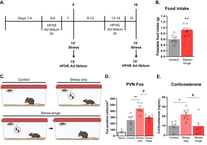
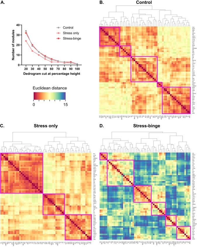
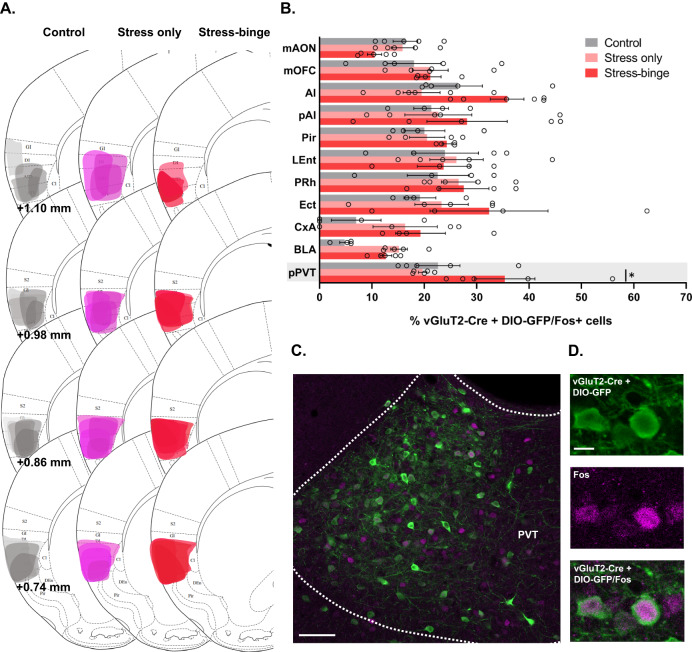
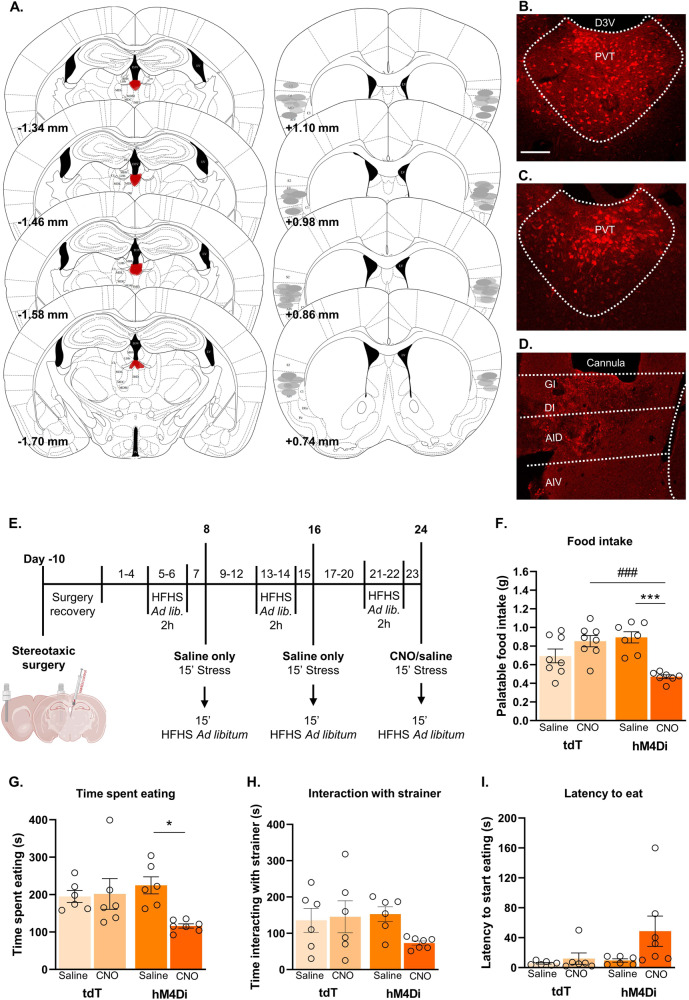
It is well-established that stress and negative affect trigger eating disorder symptoms and that the brains of men and women respond to stress in different ways. Indeed, women suffer disproportionately from emotional or stress-related eating, as well as associated eating disorders such as binge eating disorder. Nevertheless, our understanding of the precise neural circuits driving this maladaptive eating behavior, particularly in women, remains limited. We recently established a clinically relevant model of 'emotional' stress-induced binge eating whereby only female mice display binge eating in response to an acute "emotional" stressor. Here, we combined neuroanatomic, transgenic, immunohistochemical and pathway-specific chemogenetic approaches to investigate whole brain functional architecture associated with stress-induced binge eating in females, focusing on the role of Vglut2 projections from the paraventricular thalamus (PVTVglut2+) to the medial insular cortex in this behavior. Whole brain activation mapping and hierarchical clustering of Euclidean distances revealed distinct patterns of coactivation unique to stress-induced binge eating. At a pathway-specific level, PVTVglut2+ cells projecting to the medial insular cortex were specifically activated in response to stress-induced binge eating. Subsequent chemogenetic inhibition of this pathway suppressed stress-induced binge eating. We have identified a distinct PVTVglut2+ to insular cortex projection as a key driver of "emotional" stress-induced binge eating in female mice, highlighting a novel circuit underpinning this sex-specific behavior.


Conflict of interest statement

The authors declare no competing interests.

Figures

Fig. 1
Fig. 1. Palatable food intake following stress-binge paradigm and validation of stressor.
A Behavioral timeline. B Stressed mice consumed significantly more palatable food in the 15 min following stress compared to control mice who were not subjected to the stressor but had free access to palatable food for the same period. **p = 0.0019, unpaired t-test, two-tailed, df = 13. Data from day 16. C Schematic of distinct behavioral manipulations for control, stress only, and stress-binge groups. D Fos expression following behavioral manipulation across different groups. **p = 0.0061, #p = 0.0420. E Serum corticosterone levels following stress-binge protocol. *p = 0.0178, #p = 0.0148. One-way ANOVA with Tukey post hoc multiple comparisons. Naïve n = 3, control n = 6-7, stress only n = 5, stress-binge n = 7–8. Data are presented as mean ± SEM. 15’, 15 min; 2 h, 2 h; HFHS, high-fat high-sugar.
Fig. 2
Fig. 2. Hierarchical clustering of Euclidean distance matrices for control, stress only and stress-binge groups.
Modules were determined by cutting each dendrogram at 60% of the maximal height and are identified by magenta rectangles. A Number of modules for each group after trimming the hierarchically organized dendrogram at different percentages of the tree height. B Corresponding Euclidean distance of each brain region relative to the others in control mice. Five modules of coactivation were delineated. C Corresponding distance of each brain region relative to the others that were examined in stress only mice. Three modules of co-activation were delineated. D Corresponding distance of each brain region relative to the others that were examined in stress-binge mice. Six modules of co-activation were delineated. ACo Anterior cortical amygdaloid nucleus, AI Anterior insula, AIDV Agranular insular cortex, dorsal-ventral subdivision aPVT, paraventricular nucleus of the thalamus, anterior part, Arc Arcuate hypothalamic nucleus, BLA Basolateral amygdala, BMA Basomedial amygdala, CeA Central nucleus of the amygdala, CM Central medial nucleus of the thalamus, cVMH Ventromedial hypothalamic nucleus, central division; CxA Cortex-amygdala transition zone, DM Dorsomedial hypothalamic nucleus, dmVMH Ventromedial hypothalamic nucleus, dorsomedial division, DP Dorsal peduncular cortex, DR Dorsal raphe, DS Dorsal striatum, DTT Dorsal tenia tecta; dvAI; Ect Ectorhinal cortex, IL Infralimbic cortex, LA Lateral amygdala, lBNST Bed nucleus of stria terminalis, lateral division, lEnt Lateral entorhinal cortex, lHb Lateral habenula, lOFC Orbitofrontal cortex, lateral subdivision, LS Lateral septum, MS Medial septum, mAON Anterior olfactory nucleus, medial part, mBNST Bed nucleus of the stria terminalis, medial division; mCg, cingulate cortex, medial part; MeA Medial nucleus of the amygdala, mHb Medial habenula, mLH Lateral hypothalamus, medial division; mOFC Orbitofrontal cortex, medial subdivision; mPOA, medial preoptic area; NAcC, nucleus accumbens core; NAcSh, nucleus accumbens shell; PAG, periaqueductal gray; pAI, agranular insular cortex, posterior part; pAON, anterior olfactory nucleus, posterior part; PH, posterior hypothalamic area; Pir Piriform cortex, pLH Lateral hypothalamus, posterior division; pPVT, paraventricular thalamus, posterior part; PRh, perirhinal cortex; PrL, prelimbic cortex; PVN, paraventricular nucleus of the hypothalamus, medial parvicellular part; SNc Substantia nigra compacta; SNr, substantia nigra reticulata; VDB, nucleus of the vertical limb of the diagonal band; vlVMH, ventromedial hypothalamic nucleus, ventrolateral division; vOFC, orbitofrontal cortex, ventral subdivision; VP, ventral pallidum; VPM, ventral posteromedial thalamic nucleus; VPMpc, ventral posteromedial thalamic nucleus, parvicellular portion; vSub, ventral subiculum; VTA, ventral tegmental area; ZI, zona incerta.
Fig. 3
Fig. 3. Insular cortexVglut2+ afferents co-localize with Fos protein expression during stress-induced binge eating protocol.
A Representative plots of the spread of the retrograde virus in the injection site for the three groups. B Brain regions with the highest number of Fos-positive Vglut2 projecting neurons to the insular cortex. Increased Fos expression in PVTVglut2 projecting neurons was observed in animals that stress-binged and is highlighted in grey. *p = 0.0308, two-way ANOVA followed by Tukey’s multiple comparisons test. Data are presented as mean ± SEM, n = 4–6 mice per group. C Photomicrograph of the PVT showing Vglut2-Cre projecting neurons (green) and Fos-positive cells nuclei (magenta). Scale bar: 100 μm. D Representative image of PVT immunofluorescence for Vglut2 (green), Fos-protein (magenta), and merged fluorescence. Scale bar: 20 μm. Ai Anterior agranular insular cortex, BLA Basolateral amygdala, CxA Cortex-amygdala transition zone, Ect Ectorhinal cortex, LEnt Lateral entorhinal cortex, mAON Anterior olfactory nucleus, medial portion; mOFC Orbitofrontal cortex, medial portion, PRh Perirhinal cortex, PVT Paraventricular nucleus of the thalamus, pPVT Paraventricular nucleus of the thalamus, posterior portion.
Fig. 4
Fig. 4. Inhibition of PVTVglut2+-insular cortex projections attenuates stress-induced binge eating.
A Inhibitory DREADD injection into the PVT and viral spread (mm from bregma; left) and approximate cannula placements of the injector tips for the insular cortex (right). Representative photomicrographs of Cre-dependent control virus (B) and Cre-dependent hM4Di virus (C) spread in the PVT. D Representative photomicrograph of cannula placement in the insular cortex. E Behavioral testing timeline for chemogenetic manipulations. After stereotaxic surgery for cannulae placement in the insular and viral delivery into the PVT, mice were subjected to the stress protocol, locomotor activity test, food consumption independent of stress test, and light/dark box transition test. F CNO administration significantly reduced the amount of palatable food consumed by hM4Di-expressing mice (main effect of treatment F(1,26) = 5.043, p = 0.0335; interaction F(1,26) = 24.34, p < 0.0001; hM4Di CNO vs hM4Di saline p = 0.0002, hM4Di CNO vs tdT CNO p = 0.0005). A trend was observed in hM4Di CNO vs tdT saline groups, p = 0.056. G hM4Di CNO treated animals spent less time consuming the palatable food post-stress (main effect of treatment, F(1,21) = 0.488, p = 0.0462; interaction, F(1,21) = 5.704, p = 0.0264; posthoc comparison, p = 0.0229 vs hM4Di saline). H No significant differences were seen in time spent interacting with the tea strainer between groups. I No significant differences were seen between groups in the latency to start consuming palatable food once available post-stress. *p < 0.05; ***,###p < 0.005, two-way ANOVA followed by Tukey or Sidak’s multiple comparisons test when applicable. Data = mean ± SEM. tdT saline, n = 6–8; tdT CNO, n = 6–8; hM4Di saline, n = 6–7; hM4Di CNO, n = 7. CNO, clozapine-N-oxide; tdT, tdTomato.
Fig. 5
Fig. 5. Pathway-specific inhibition of PVTVglut2+-insular cortex projections does not impact locomotor activity, anxiety-like behavior or hedonic eating independent of stress.
A Behavioral testing timeline. Locomotor activity was tested on day 26 following infusion of saline or CNO. Palatable food consumption independent of stress was assessed on days 29 and 30 and CNO and saline were delivered across these 2 days in a counterbalanced manner. Light-dark box test was performed on the final day of testing. For locomotor activity, data presented as (B) total distance travelled (cm) (C) ambulatory time (s), (D) ambulatory average speed (cm/s), and (E) number of rearing episodes during locomotor testing period (10 min). F Palatable food consumption independent of stress. No main effect of CNO or activation of hM4Di DREADD was seen under non-stress conditions (two-way RM ANOVA). Data for the light dark/box test as follows: G Latency to enter the light compartment (s). H Total time spent (s) in light compartments. tdT saline n = 6, tdT CNO n = 6, hM4Di saline n = 6, hM4Di CNO n = 7. Data presented as mean ± SEM.

References

    1. Chesney E, Goodwin GM, Fazel S. Risks of all-cause and suicide mortality in mental disorders: a meta-review. World Psychiatry. 2014;13:156–60. - PMC - PubMed
    1. Anversa RG, Muthmainah M, Sketriene D, Gogos A, Sumithran P, Brown RM. A review of sex differences in the mechanisms and drivers of overeating. Front Neuroendocrinol. 2021;63:100941. - PubMed
    1. François M, Fernández-Gayol O, Zeltser LM. A Framework for Developing Translationally Relevant Animal Models of Stress-Induced Changes in Eating Behavior. Biol Psychiatry. 2022;91:888–97. - PMC - PubMed
    1. Substance Abuse and Mental Health Services Administration. National Survey on Drug Use and Health. 2018. https://www.samhsa.gov/data/release/2018-national-survey-drug-use-and-he.... Accessed on 13 Jan 2023.
    1. Kessler RC, Petukhova M, Sampson NA, Zaslavsky AM, Wittchen HU. Twelve-month and lifetime prevalence and lifetime morbid risk of anxiety and mood disorders in the United States. Int J Methods Psychiatr Res. 2012;21:169–84. - PMC - PubMed

Publication types