. 2023 Dec;13(12):3223-3238.
doi: 10.1007/s13346-023-01383-y. Epub 2023 Jul 20.

UV-polymerizable methacrylated gelatin (GelMA)-based hydrogel containing tannic acids for wound healing

Affiliations

UV-polymerizable methacrylated gelatin (GelMA)-based hydrogel containing tannic acids for wound healing

Marismar F do Nascimento et al. Drug Deliv Transl Res. 2023 Dec.

Abstract

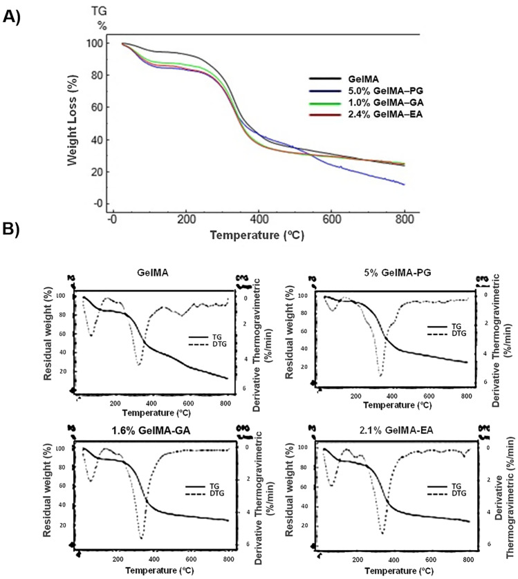
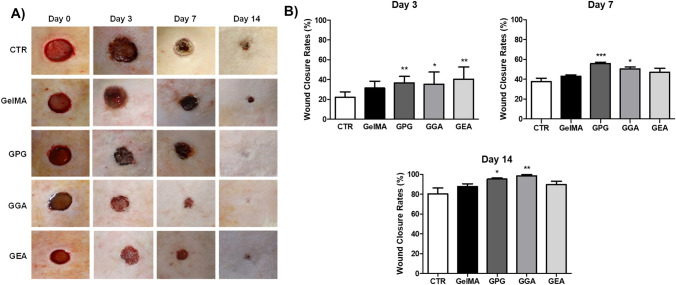
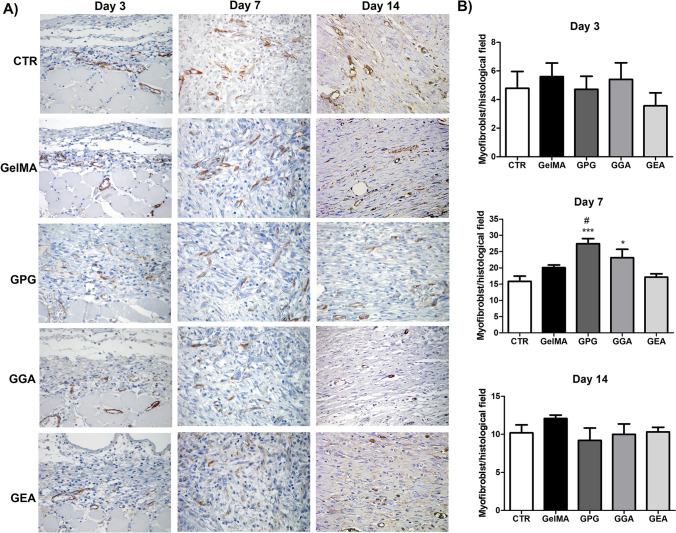
Gelatin-based photopolymerizable methacrylate hydrogel (GelMA) is a promising biomaterial for in situ drug delivery, while aqueous extract of Punica granatum (AEPG) peel fruit rich in gallic acid and ellagic acid is used to improve wound healing. The aim of this study was to develop and analyze the healing properties of GelMA containing AEPG, gallic acid, or ellagic acid in a rodent model. GelMA hydrogels containing 5% AEPG (GelMA-PG), 1.6% gallic acid (GelMA-GA), or 2.1% ellagic acid (GelMA-EA) were produced and their mechanical properties, enzymatic degradation, and thermogravimetric profile determined. Wound closure rates, healing histological grading, and immunohistochemical counts of myofibroblasts were assessed over time. The swelling of hydrogels varied between 50 and 90%, and GelMA exhibited a higher swelling than the other groups. The GPG samples showed higher compression and Young's moduli than GelMA, GGA, and GAE. All samples degraded around 95% in 48 h. GPG and GGA significantly accelerated wound closure, improved collagenization, increased histological grading, and hastened myofibroblast differentiation in comparison to the control, GelMA, and GEA. GelMA containing AEPG (GPG) improved wound healing, and although gallic acid is the major responsible for such biological activity, a potential synergic effect played by other polyphenols present in the extract is evident.

Keywords: GelMA; Gelatin methacryloyl hydrogel; Iridoids; Punica granatum aqueous extract; Wistar rats.


Conflict of interest statement

The authors declare no competing interests.

Figures

Fig. 1
Fig. 1
A Assessment of swelling rate, B compression and Young’s modulus, and C degradability rate and final weight of the hydrogels. Data are expressed as mean ± SD (n = 8). Significant differences in comparison to GelMA are expressed as *p < 0.05, **p < 0.01, and ***p < 0.001; significant differences in comparison to GelMA-PG are expressed as.###p < 0.001 (ANOVA and Tukey’s multiple comparison test)
Fig. 2
Fig. 2
A Thermogravimetric and B derivative thermogravimetric curves of GelMA hydrogel without polyphenolic compounds (GelMA) and containing aqueous extract of Punica granatum (GelMA-PG), gallic acid (GelMA-GA), and ellagic acid (GelMA-EA) and in scan up to 800 °C
Fig. 3
Fig. 3
A Digital photographs of skin wounds in the CTR (control group with unfilled wounds), GelMA (wounds filled with GelMA hydrogel), GPG (wounds filled with GelMA hydrogel containing aqueous extract of Punica granatum), GGA (wounds filled with hydrogel GelMA containing gallic acid), and GEA (wounds filled with GelMA hydrogel containing ellagic acid) and B assessment of wound closure rates over the time-course of wound healing. Data are expressed as mean ± SD. Significant difference compared to CTR are expressed as ***p < 0.001, **p < 0.01, and *p < 0.05 (ANOVA and Tukey’s multiple comparison post-test, n = 6 animals/group)
Fig. 4
Fig. 4
A Hematoxylin–eosin-stained histological slides (400 ×) showing the pathological features observed in the experimental groups over the wound healing time-course characterized by acute inflammation, rich in polymorphonuclear neutrophils (pmn), on day 3, exuberant granulation tissue on day 7, and primary fibrous scar on day 14. Re-epithelization of the wound surfaces (Ep) and the presence of epithelial biddings compatible with rudimentary cutaneous appendages (arrows). B Assessment of histological grading of wound healing over the time-course of the experiment (data expressed as median; interquartile range, n = 6 animals/group and 3 histological slides/animal). C Sirius red-stained histological slides showing the pattern of collagen deposition over the experimental time (polarized light, 100 ×). The birefringence is green for type I collagen and yellow-gold for type I collagen. D Assessment of the mean percentage of collagenization in the wounds over the experimental time (data expressed as mean ± SD, n = 6 animals/group and 3 histological slides/animal). Comparisons between groups of the scores of histological grading of wound healing were performed with Kruskal–Wallis’ test and Dunn’s multiple comparison test), whereas comparisons of the mean percentage of collagenization were performed using ANOVA and Tukey’s multiple comparison test). Significant differences compared to CTR are expressed as *p < 0.05, **p < 0.01, and ***p < 0.001; significant differences compared to GelMA and GEA are expressed as #p < 0.05, ##p < 0.01, and ###p < 0.001. Black bar: 250 µm; white bar: 500 µm
Fig. 5
Fig. 5
A Immunohistochemical detection of α-SMA-positive myofibroblast over the course of wound healing (SABC, 400 ×). Myofibroblasts are identified as spindle-shaped cells exhibiting the brown color of the cytoplasm, typically found along collagen fibrils and fibers. B Assessment of the mean number of myofibroblasts per histological field (0.025 mm2). Data are expressed as mean ± SD. Significant differences compared to CTR are expressed as *p < 0.05 and **p < 0.01; significant differences compared to GelMA and GEA are expressed as.#p < 0.05 (ANOVA and Tukey’s multiple comparison test, n = 6 animals/group and 3 histological slides/animal)

References

    1. Murphree RW. Impairments in skin integrity. Nurs Clin North Am. 2017;52(3):405–417. doi: 10.1016/j.cnur.2017.04.008. - DOI - PubMed
    1. Obagi Z, Damiani G, Grada A, Falanga V. Principles of wound dressings: a review. Surg Technol Int. 2019;35:50–57. - PubMed
    1. Shahnawaz Khan M, Abdelhamid HN, Wu H-F. Near infrared (NIR) laser mediated surface activation of graphene oxide nanoflakes for efficient antibacterial, antifungal and wound healing treatment. Colloids Surf B. 2015;127:281–291. doi: 10.1016/j.colsurfb.2014.12.049. - DOI - PubMed
    1. Hussein KH, Abdelhamid HN, Zou X, Woo H-M. Ultrasonicated graphene oxide enhances bone and skin wound regeneration. Mater Sci Eng, C. 2019;94:484–492. doi: 10.1016/j.msec.2018.09.051. - DOI - PubMed
    1. Soliman M, Sadek AA, Abdelhamid HN, Hussein K. Graphene oxide-cellulose nanocomposite accelerates skin wound healing. Res Vet Sci. 2021;137:262–273. doi: 10.1016/j.rvsc.2021.05.013. - DOI - PubMed

Publication types

LinkOut - more resources