Review
. 2023 Jul 20;25(1):40.
doi: 10.1186/s12968-023-00942-z.

4D Flow cardiovascular magnetic resonance consensus statement: 2023 update

Affiliations
Review

4D Flow cardiovascular magnetic resonance consensus statement: 2023 update

Malenka M Bissell et al. J Cardiovasc Magn Reson. .

Abstract

Hemodynamic assessment is an integral part of the diagnosis and management of cardiovascular disease. Four-dimensional cardiovascular magnetic resonance flow imaging (4D Flow CMR) allows comprehensive and accurate assessment of flow in a single acquisition. This consensus paper is an update from the 2015 '4D Flow CMR Consensus Statement'. We elaborate on 4D Flow CMR sequence options and imaging considerations. The document aims to assist centers starting out with 4D Flow CMR of the heart and great vessels with advice on acquisition parameters, post-processing workflows and integration into clinical practice. Furthermore, we define minimum quality assurance and validation standards for clinical centers. We also address the challenges faced in quality assurance and validation in the research setting. We also include a checklist for recommended publication standards, specifically for 4D Flow CMR. Finally, we discuss the current limitations and the future of 4D Flow CMR. This updated consensus paper will further facilitate widespread adoption of 4D Flow CMR in the clinical workflow across the globe and aid consistently high-quality publication standards.

Keywords: 4D Flow CMR; 4D Flow MRI; Cardiovascular; Clinical; Flow quantification; Flow visualization; Heart disease; Hemodynamics; MR flow imaging; Phase-contrast magnetic resonance imaging; Recommendations.


Conflict of interest statement

No author has and competing interests for this manuscript.

Figures

Fig. 1
Fig. 1
Post-processing of 4D Flow CMR should always include correction for phase offsets and noise masking. Anti-aliasing needs to be performed if aliasing is present in regions of interest. Segmentation can be performed for the whole vessel in 3D or on 2D vessel cross-sections perpendicular to the course of the vessel. Visualization of flow, velocity and advanced parameters is optional but can help identify regions of peak velocities and insufficiencies. Quantification can be performed in 2D cross sections or in regions of the vessel. Parameters can be given averaged over the whole cardiac cycle (e.g. stroke volume) or maximum and minimum parameters (e.g. peak velocity)
Fig. 2
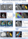
Fig. 2
Valve tracking procedure in 4D flow CMR. In preprocessing phase, velocity data is corrected for aliasing (1), phase offset correction (2) and misregistration (3). Annulus tracking (4) is performed for forward flow and backward flow is obtained by tracking the regurgitant jet (5). Velocity corrections are performed by subtracting through-plane valve motion (6). Then, velocity mapping is performed on the reformatted 2D through-plane velocity images (7). Finally, the net forward volume among the four valves can be used as an internal check for consistency in the analysis (8)
Fig. 3
Fig. 3
Internal consistency of measurements can be checked by comparing flow volumes at different locations in the same vessel or by comparing the sum of branch vessels to the main pulmonary artery

References

    1. Dyverfeldt P, Bissell M, Barker AJ, Bolger AF, Carlhall CJ, Ebbers T, et al. 4D flow cardiovascular magnetic resonance consensus statement. J Cardiovasc Magn Reson. 2015;17:72. doi: 10.1186/s12968-015-0174-5. - DOI - PMC - PubMed
    1. Doyle CM, Orr J, Greenwood JP, Plein S, Tsoumpas C, Bissell MM. Four-dimensional flow magnetic resonance imaging in the assessment of blood flow in the heart and great vessels: a systematic review. J Magn Reson Imaging. 2021 doi: 10.1002/jmri.27874. - DOI - PubMed
    1. Kamphuis VP, van der Palen RLF, de Koning PJH, Elbaz MSM, van der Geest RJ, de Roos A, et al. In-scan and scan-rescan assessment of LV in- and outflow volumes by 4D flow MRI versus 2D planimetry. J Magn Reson Imaging. 2018;47(2):511–522. doi: 10.1002/jmri.25792. - DOI - PMC - PubMed
    1. Kamphuis VP, Westenberg JJM, van der Palen RLF, van den Boogaard PJ, van der Geest RJ, de Roos A, et al. Scan-rescan reproducibility of diastolic left ventricular kinetic energy, viscous energy loss and vorticity assessment using 4D flow MRI: analysis in healthy subjects. Int J Cardiovasc Imaging. 2018 doi: 10.1007/s10554-017-1291-z. - DOI - PubMed
    1. Juffermans JA-O, Westenberg JA-O, van den Boogaard PJ, Roest AAW, van Assen HC, van der Palen RLF, et al. Reproducibility of aorta segmentation on 4D flow MRI in healthy volunteers. J Magn Reson Imaging. 2021 doi: 10.1002/jmri.27431. - DOI - PMC - PubMed

Publication types