Review
. 2023 Jul 20;18(1):49.
doi: 10.1186/s13024-023-00634-3.

Axonal energy metabolism, and the effects in aging and neurodegenerative diseases

Affiliations
Review

Axonal energy metabolism, and the effects in aging and neurodegenerative diseases

Sen Yang et al. Mol Neurodegener. .

Abstract

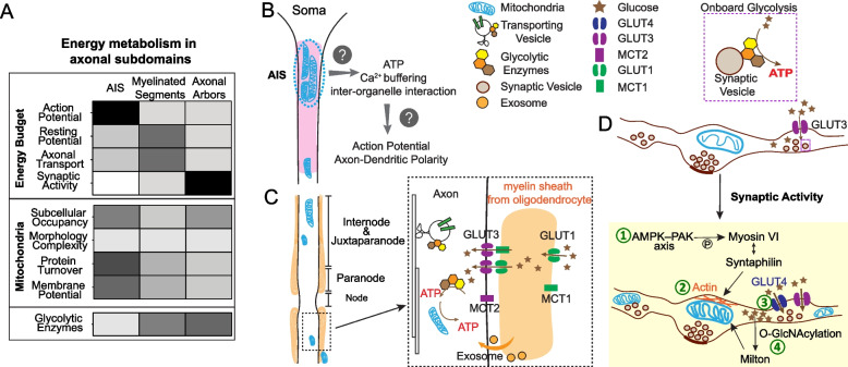
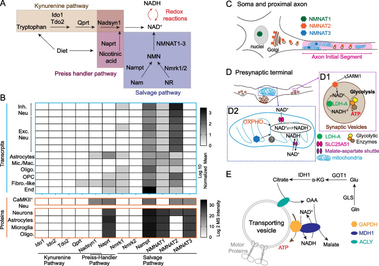
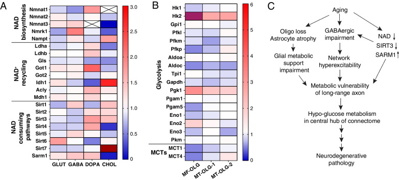
Human studies consistently identify bioenergetic maladaptations in brains upon aging and neurodegenerative disorders of aging (NDAs), such as Alzheimer's disease, Parkinson's disease, Huntington's disease, and Amyotrophic lateral sclerosis. Glucose is the major brain fuel and glucose hypometabolism has been observed in brain regions vulnerable to aging and NDAs. Many neurodegenerative susceptible regions are in the topological central hub of the brain connectome, linked by densely interconnected long-range axons. Axons, key components of the connectome, have high metabolic needs to support neurotransmission and other essential activities. Long-range axons are particularly vulnerable to injury, neurotoxin exposure, protein stress, lysosomal dysfunction, etc. Axonopathy is often an early sign of neurodegeneration. Recent studies ascribe axonal maintenance failures to local bioenergetic dysregulation. With this review, we aim to stimulate research in exploring metabolically oriented neuroprotection strategies to enhance or normalize bioenergetics in NDA models. Here we start by summarizing evidence from human patients and animal models to reveal the correlation between glucose hypometabolism and connectomic disintegration upon aging/NDAs. To encourage mechanistic investigations on how axonal bioenergetic dysregulation occurs during aging/NDAs, we first review the current literature on axonal bioenergetics in distinct axonal subdomains: axon initial segments, myelinated axonal segments, and axonal arbors harboring pre-synaptic boutons. In each subdomain, we focus on the organization, activity-dependent regulation of the bioenergetic system, and external glial support. Second, we review the mechanisms regulating axonal nicotinamide adenine dinucleotide (NAD+) homeostasis, an essential molecule for energy metabolism processes, including NAD+ biosynthetic, recycling, and consuming pathways. Third, we highlight the innate metabolic vulnerability of the brain connectome and discuss its perturbation during aging and NDAs. As axonal bioenergetic deficits are developing into NDAs, especially in asymptomatic phase, they are likely exaggerated further by impaired NAD+ homeostasis, the high energetic cost of neural network hyperactivity, and glial pathology. Future research in interrogating the causal relationship between metabolic vulnerability, axonopathy, amyloid/tau pathology, and cognitive decline will provide fundamental knowledge for developing therapeutic interventions.

Keywords: Aging; Axonal bioenergetics; Axonopathy; Energy metabolism; Glucose; Glycolysis; Mitochondria; NAD; Neurodegeneration; Neuroprotection.


Conflict of interest statement

The authors declare that they have no competing interests.

Figures

Fig. 1
Fig. 1
The spatial organization of glucose metabolism machineries across axon. A. Upper panel: Heat map description of the degree of energy consumption by each axonal activity within three axonal subdomains. Bottom panel: the intensity of each feature/parameter of glucose metabolism machineries within three axonal subdomains. Heat map color from white to dark represents the mild to strong degree/intensity. B.-D. The spatial organization of glucose transporters (GLUTs), monocarboxylic acid transporters (MCTs), glycolytic enzymes, mitochondria, and glial partners in axon initial segment (B), myelinated axonal shaft (C), and axonal arbors (D). D. The activity driven adaption of glucose metabolism machineries in presynaptic compartments. ① Activity dependent recruitment and anchoring of mitochondria to F actin filament at presynaptic sites through the AMPK-PAK-Syntaphilin axis; ② Neuronal stimulation induced mitochondrial ultrastructural changes with wider cristae and more compact or irregular matrices; ③ Surface mobilization of GLUT4, and presumably increased glucose uptake and glucose metabolism; and ④ Increased glucose metabolism through the Hexosamine synthesis pathway to boost O-GlcNAcylation of mitochondrial adaptor protein Milton, which reduce mitochondria mobility
Fig. 2
Fig. 2
The molecular underpinnings of NAD redox potential maintenance in axons. A. A schematic view of NAD biosynthetic pathways. B. Heat map visualization of the abundance of mRNA and proteins of NAD biosynthetic enzymes. Transcript levels are adapted from [259]; protein levels are adapted from [260]. Abbreviations: Inh. Neu, inhibitory neurons; Exc. Neu, excitatory neurons; Mic. Mac., microglia and macrophages; Oligo, oligodendrocytes; OPC, oligodendrocyte precursor cells; Fibro.-like, fibroblast-like cells; End, endothelial cells. C. The subcellular localization of NMNAT1-3 in the soma and proximal axon. D. The subcellular distribution of NMNAT2, NMNAT3, and proteins involved in NAD+/NADH homeostasis in presynaptic boutons. (D1) NMNAT2, LDH-A, and glycolytic enzymes closely attach to synaptic vesicles; NMNAT2 inhibits SARM1 activation and maintains local NAD redox potential together with LDH-A to support “onboard” glycolysis; (D2) NMNAT3 inside mitochondrial matrix maintains local NAD redox potential together with OXPHO units, SLC25A51 (NAD+ transporter), and Malate-Aspartate shuttle. E. A hypothetical model of mitochondrial independent NAD+ recycling through the glutamine carboxylation pathway. MDH1, cytosolic malate dehydrogenase 1; ACLY, ATP citrate lyase; GLS, glutaminase; Gln, glutamine; Glu, glutamate; GOT1, glutamic-oxaloacetic transaminase 1; α-KG, alpha ketoglutarate; IDH1, isocitrate dehydrogenase (NADP+) 1; OAA, oxaloacetate
Fig. 3
Fig. 3
Aging induced bioenergetic maladaptation in mouse brain. A. Transcriptional fold change of enzymes involved in NAD+ redox potential maintenance in glutamatergic (GLUT), GABAergic (GABA), dopaminergic (DOPA) and cholinergic (CHOL) neurons upon aging, adapted from [22]. Sirt7 in aged cholinergic neurons is increased 5.97-fold, labeled in dark red. B. Transcriptional fold change of glycolytic enzymes and monocarboxylic acid transporters (MCTs) in myelin-forming oligodendrocytes (MF-OLG) and mature oligodendrocytes (MT-OLG). (MT-OLG-1 and MT-OLG-2 are two independent repeats in [22]). HK2 in aged myelin-forming oligodendrocytes is increased 9.72-fold, labeled in purple-red. C. A hypothetical model of the sequential events caused by aging that leads to metabolic failure in long-range axonal projections and glucose hypometabolism in central hubs of the brain connectome

References

    1. Erbsloh F, Bernsmeier A, Hillesheim H. The glucose consumption of the brain & its dependence on the liver. Arch Psychiatr Nervenkr Z Gesamte Neurol Psychiatr. 1958;196:611–626. - PubMed
    1. Harris JJ, Attwell D. The energetics of CNS white matter. J Neurosci. 2012;32:356–371. doi: 10.1523/JNEUROSCI.3430-11.2012. - DOI - PMC - PubMed
    1. Yellen G. Fueling thought: management of glycolysis and oxidative phosphorylation in neuronal metabolism. J Cell Biol. 2018;217:2235–2246. doi: 10.1083/jcb.201803152. - DOI - PMC - PubMed
    1. Dienel GA. Brain glucose metabolism: integration of energetics with function. Physiol Rev. 2019;99:949–1045. doi: 10.1152/physrev.00062.2017. - DOI - PubMed
    1. Mergenthaler P, Lindauer U, Dienel GA, Meisel A. Sugar for the brain: the role of glucose in physiological and pathological brain function. Trends Neurosci. 2013;36:587–597. doi: 10.1016/j.tins.2013.07.001. - DOI - PMC - PubMed

Publication types