. 2023 Dec;29(12):3995-4017.
doi: 10.1111/cns.14325. Epub 2023 Jul 20.

Photobiomodulation reduces neuropathic pain after spinal cord injury by downregulating CXCL10 expression

Affiliations

Photobiomodulation reduces neuropathic pain after spinal cord injury by downregulating CXCL10 expression

Zhihao Zhang et al. CNS Neurosci Ther. 2023 Dec.

Abstract

Background: Many studies have recently highlighted the role of photobiomodulation (PBM) in neuropathic pain (NP) relief after spinal cord injury (SCI), suggesting that it may be an effective way to relieve NP after SCI. However, the underlying mechanisms remain unclear. This study aimed to determine the potential mechanisms of PBM in NP relief after SCI.

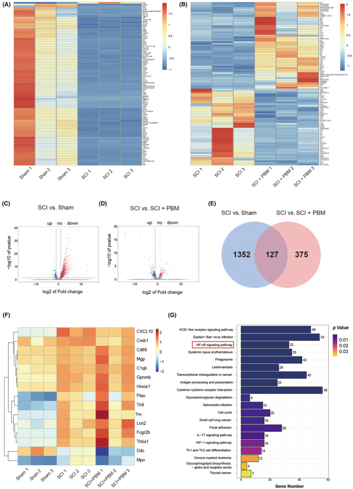
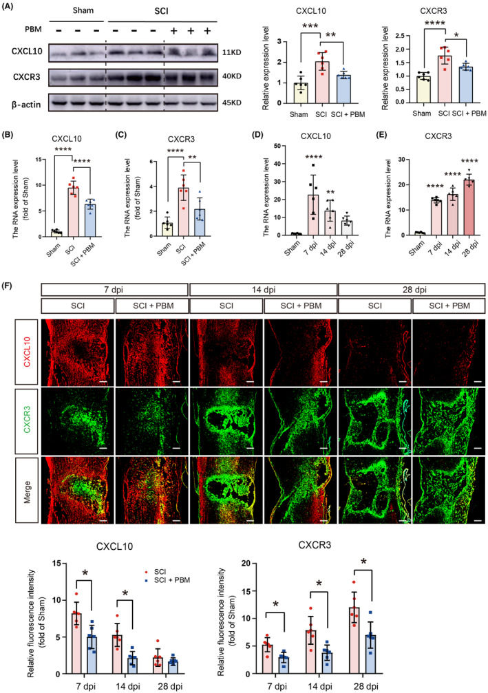
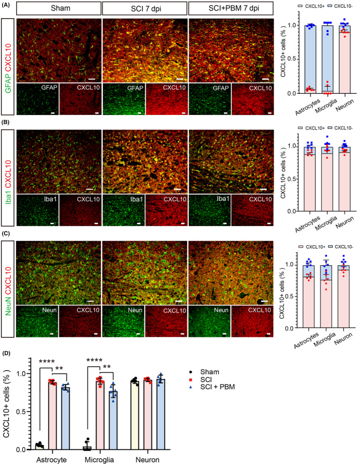
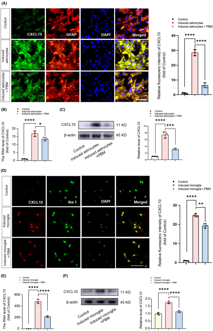
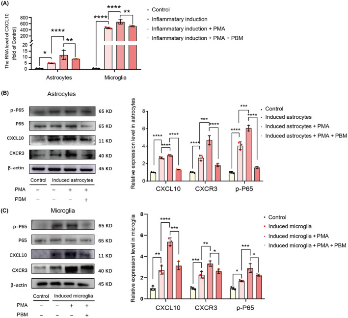
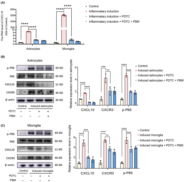
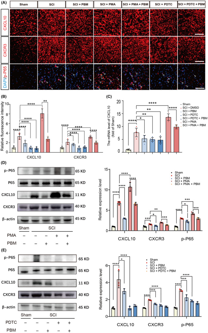
Methods: We performed systematic observations and investigated the mechanism of PBM intervention in NP in rats after SCI. Using transcriptome sequencing, we screened CXCL10 as a possible target molecule for PBM intervention and validated the results in rat tissues using reverse transcription-polymerase chain reaction and western blotting. Using immunofluorescence co-labeling, astrocytes and microglia were identified as the cells responsible for CXCL10 expression. The involvement of the NF-κB pathway in CXCL10 expression was verified using inhibitor pyrrolidine dithiocarbamate (PDTC) and agonist phorbol-12-myristate-13-acetate (PMA), which were further validated by an in vivo injection experiment.

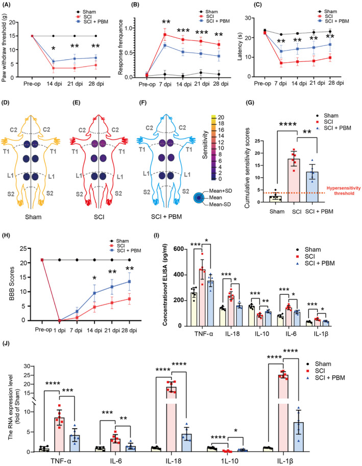
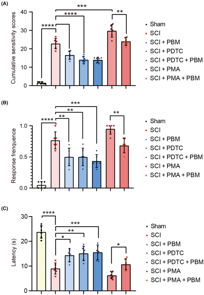
Results: Here, we demonstrated that PBM therapy led to an improvement in NP relative behaviors post-SCI, inhibited the activation of microglia and astrocytes, and decreased the expression level of CXCL10 in glial cells, which was accompanied by mediation of the NF-κB signaling pathway. Photobiomodulation inhibit the activation of the NF-κB pathway and reduce downstream CXCL10 expression. The NF-κB pathway inhibitor PDTC had the same effect as PBM on improving pain in animals with SCI, and the NF-κB pathway promoter PMA could reverse the beneficial effect of PBM.

Conclusions: Our results provide new insights into the mechanisms by which PBM alleviates NP after SCI. We demonstrated that PBM significantly inhibited the activation of microglia and astrocytes and decreased the expression level of CXCL10. These effects appear to be related to the NF-κB signaling pathway. Taken together, our study provides evidence that PBM could be a potentially effective therapy for NP after SCI, CXCL10 and NF-kB signaling pathways might be critical factors in pain relief mediated by PBM after SCI.

Keywords: CXCL10; glia cells; neuropathic pain; photobiomodulation; spinal cord injury.


Conflict of interest statement

The authors declare that they have no conflict of interest.

Figures

FIGURE 1
FIGURE 1
Effect of PBM irradiation on nociceptive hypersensitivity, motor function recovery and expression level of inflammatory factors. The time course of mechanical allodynia (A), cold allodynia (B) and heat hyperalgesia (C) of rats with SCI. The RSSs for the sham group (n = 6) (D), SCI group (n = 6) (E), and SCI + PBM group (n = 6) (F) are shown in the six regions (left and right side above‐level of injury, at the level of injury, and below‐level of injury) on rat dorsal schematic, the gray lines were used to write C2, T1, L1, S2, and midline on that. The number of mean ± SD is corresponding to the colors of the concentric circles on the dorsal schematic, and a color scale depicting the number is shown in the legend. CSSs in the sham animals (n = 6) and SCI animals with (n = 6) or without (n = 6) PBM irradiation are accumulated by RSSs (G); red‐dashed line indicates hypersensitivity threshold (3.76), which is taken from the control group. The BBB score was used to evaluate the recovery of motor function in the sham group (n = 6), SCI group (n = 6) and SCI + PBM group (n = 6) (H). Fold changes in the expression (I) and transcription (J) levels of the representative inflammatory factors associated with the occurrence of pain (TNF‐α, IL‐6, IL‐18, IL‐10, and IL‐1β) in the sham group (n = 6), SCI group (n = 6) and SCI + PBM group (n = 6) at 7 dpi. Data is expressed as mean ± SD, *p < 0.05, **p < 0.01, ***p < 0.001, ****p < 0.0001. BBB, Basso, Beattie, Bresnahan; CSS, cumulative sensitivity score; dpi, days postinjury; PBM, photobiomodulation; RSS, regional sensitivity score; SCI, spinal cord injury; SD, standard deviation.
FIGURE 2
FIGURE 2
RNA‐seq analysis of the effect of PBM on SCI animals at 7 dpi. Gene transcriptional differences in the sham group and SCI group is shown in the heat map (A), with 1242 genes significantly upregulated and 110 genes significantly downregulated (C). Gene transcriptional differences in the SCI and SCI + PBM group is shown in the heat map (B), with 175 genes significantly upregulated and 200 genes significantly downregulated (D). Total number of genes significantly altered after SCI is 1352, and the number of genes could be modified by PBM intervention after SCI is 375. The transcript level of 127 genes significantly altered after SCI and could be modified by PBM intervention (E). Heatmap indicates CXCL10 is the top gene highly upregulated after SCI and marked decreased after PBM irradiation (F). KEGG pathway analysis between the SCI and the sham groups showed that the NF‐κB signaling pathway was dramatically activated after SCI (G). dpi, days postinjury; KEGG, Kyoto Encyclopedia of Genes and Genomes; PBM, photobiomodulation; RNA‐seq, RNA sequencing; SCI, spinal cord injury.
FIGURE 3
FIGURE 3
Spatial and temporal distribution of CXCL10 in rats with SCI and PBM intervention. Representative western blotting results for expression level of CXCL10 and CXCR3 in the sham group (n = 6), SCI group (n = 6), and SCI + PBM group (n = 6) at 7 dpi (A). Transcription levels of CXCL10 (B) and CXCR3 (C) at 7 dpi in the sham group (n = 6), SCI group (n = 6), and SCI + PBM group (n = 6). Quantification of relative expression level of CXCL10 (D) and CXCR3 (E) in the sham group (n = 6) and SCI group at 7, 14 and 28 dpi (n = 6 for each time point). Representative image of immunofluorescence stain for CXCL10, CXCR3 in the sham group (n = 6 for each time point), SCI group (n = 6 for each time point), and SCI + PBM group (n = 6 for each time point) at 7, 14 and 28 dpi. Scale bar for all pictures: 200 μm (F). Data is expressed as mean ± SD, *p < 0.05, **p < 0.01, ***p < 0.001, ****p < 0.0001. dpi, days postinjury; PBM, photobiomodulation; SCI, spinal cord injury.
FIGURE 4
FIGURE 4
Expression levels of CXCL10 in astrocytes, microglia, and neurons in the sham, SCI, and SCI + PBM groups rats at 7 dpi. Co‐stain of CXCL10 and astroglia marker GFAP (A), microglial marker Iba1 (B), and neuronal marker NeuN (C) in representative views in the sham group (n = 6), SCI group (n = 6), and SCI + PBM group (n = 6) at 7 dpi. Scale bar for all pictures: 50 μm. Quantification of the proportion of CXCL10+ cells in astrocytes, microglia, and neurons in the sham group (n = 6), SCI group (n = 6), and SCI + PBM group (n = 6) at 7 dpi (D). Data is expressed as mean ± SD, **p < 0.01, ****p < 0.0001. dpi, days postinjury; PBM, photobiomodulation; SCI, spinal cord injury; SD, standard deviation.
FIGURE 5
FIGURE 5
PBM inhibited the CXCL10 expression in microglia and astrocytes in vitro. Representative views of immunofluorescence staining for CXCL10 and GFAP in the control, induced astrocytes, and induced astrocytes + PBM groups and the quantification of the fluorescence intensity of CXCL10. Scale bar for all pictures: 200 μm (A). Transcript levels of CXCL10 in the control, induced astrocytes, and induced astrocytes + PBM groups (B). Representative western blotting and quantification of the relative expression level of CXCL10 in the control, induced astrocytes, and induced astrocytes + PBM groups (C). Representative views of immunofluorescence staining for CXCL10 and Iba1 in the control, induced microglia, and induced microglia + PBM groups and the quantification of the fluorescence intensity of CXCL10. Scale bar for all pictures: 200 μm (D). Transcription levels of CXCL10 in the control, induced microglia and induced microglia + PBM groups (E). Representative western blotting and quantification of the relative expression level of CXCL10 in the control, induced microglia, and induced microglia + PBM groups (F). Data is expressed as mean ± SD, *p < 0.05, **p < 0.01, ***p < 0.001, ****p < 0.0001. PBM, photobiomodulation; SCI, spinal cord injury; SD, standard deviation.
FIGURE 6
FIGURE 6
PBM suppressed the upregulating effect of PMA in the expression of CXCL10 in vitro. PMA significantly upregulated the transcription level of CXCL10 and PBM significantly suppressed this effect (A). The representative western blotting and quantification of the relative expression level of p‐P65, CXCL10, and CXCR3 in the control, induced astrocytes/microglia, induced astrocytes/microglia + PMA and induced astrocytes/microglia + PMA + PBM groups indicated the expression level of p‐P65, CXCL10 and CXCR3 in induced astrocytes/microglia + PMA group was markedly downregulated by PBM (B, C). Data is expressed as mean ± SD, *p < 0.05, **p < 0.01, ***p < 0.001, ****p < 0.0001. PBM, photobiomodulation; PMA, phorbol 12‐myristate 13‐acetate; SD, standard deviation.
FIGURE 7
FIGURE 7
Both PDTC and PBM had inhibitory effect in the expression of CXCL10 in vitro. PBM had the same effect as PDTC on downregulating the transcription level of CXCL10 (A). The representative western blotting and quantification of the relative expression level of p‐P65, CXCL10 and CXCR3 in the control, induced astrocytes/microglia, induced astrocytes/microglia + PDTC and induced astrocytes/microglia + PDTC + PBM groups showed that the expression level of p‐P65, CXCL10 and CXCR3 in induced astrocytes/microglia group could be markedly downregulated by both PBM and PDTC (B, C). Data is expressed as mean ± SD, **p < 0.01, ***p < 0.001, ****p < 0.0001. PBM, photobiomodulation; PDTC, ammonium pyrrolidine dithiocarbamate; SCI, spinal cord injury; SD, standard deviation.
FIGURE 8
FIGURE 8
PBM reduces the expression of CXCL10 via NF‐ĸB signaling pathway in vivo. Representative views of immunofluorescence staining of CXCL10, CXCR3, p‐P65 in the sham, SCI, SCI + PBM, SCI + PMA, SCI + PMA + PBM, SCI + PDTC, and SCI + PDTC + PBM groups at 7 dpi (n = 6 for each group). Scale bar for all pictures: 200 μm (A, B). PBM significantly suppressed the transcription levels of CXCL10 in SCI and SCI + PMA groups (n = 6 for each group), and had the same effect with PDTC (C). Representative western blotting results and quantification of the relative expression level of p‐P65, CXCL10 and CXCR3 in the sham, SCI, SCI + PBM, SCI + PMA, SCI + PMA + PBM, SCI + PDTC, and SCI + PDTC + PBM groups (n = 6 for each group) at 7 dpi were demonstrated (D, E). Data is expressed as mean ± SD, *p < 0.05, **p < 0.01, ***p < 0.001, ****p < 0.0001. dpi, days postinjury; PBM, photobiomodulation; PDTC, ammonium pyrrolidine dithiocarbamate; PMA, phorbol 12‐myristate 13‐acetate; SCI, spinal cord injury; SD, standard deviation.
FIGURE 9
FIGURE 9
PBM alleviates NP‐related behaviors via NF‐ĸB signaling pathway in rats. Behavior scores of rats in the sham, SCI, SCI + PBM, SCI + PMA, SCI + PMA + PBM, SCI + PDTC, and SCI + PDTC + PBM groups (n = 6 for each group) at 7 dpi for mechanical allodynia (A), heat hyperalgesia (B), and cold allodynia (C) were investigated. Data is expressed as mean ± SD, *p < 0.05, **p < 0.01, ***p < 0.001, ****p < 0.0001. dpi, days postinjury; NP, neuropathic pain; PBM, photobiomodulation; PDTC, ammonium pyrrolidine dithiocarbamate; PMA, phorbol 12‐myristate 13‐acetate; SCI, spinal cord injury; SD, standard deviation.

Similar articles

Cited by

References

    1. Anjum A, Yazid MD, Fauzi Daud M, et al. Spinal cord injury: pathophysiology, multimolecular interactions, and underlying recovery mechanisms. Int J Mol Sci. 2020;21(20):7533. - PMC - PubMed
    1. Jendelova P. Therapeutic strategies for spinal cord injury. Int J Mol Sci. 2018;19(10):3200. - PMC - PubMed
    1. Burke D, Fullen BM, Stokes D, Lennon O. Neuropathic pain prevalence following spinal cord injury: a systematic review and meta‐analysis. Eur J Pain. 2017;21(1):29‐44. - PubMed
    1. Finnerup N, Norrbrink C, Trok K, et al. Phenotypes and predictors of pain following traumatic spinal cord injury: a prospective study. J Pain. 2014;15(1):40‐48. - PubMed
    1. Burke D, Lennon O, Fullen BM. Quality of life after spinal cord injury: the impact of pain. Eur J Pain. 2018;22(9):1662‐1672. - PubMed

Publication types