. 2023 Oct;102(11):1261-1271.
doi: 10.1177/00220345231185758. Epub 2023 Jul 20.

MEMO1 Is Required for Ameloblast Maturation and Functional Enamel Formation

Affiliations

MEMO1 Is Required for Ameloblast Maturation and Functional Enamel Formation

M Kiel et al. J Dent Res. 2023 Oct.

Abstract

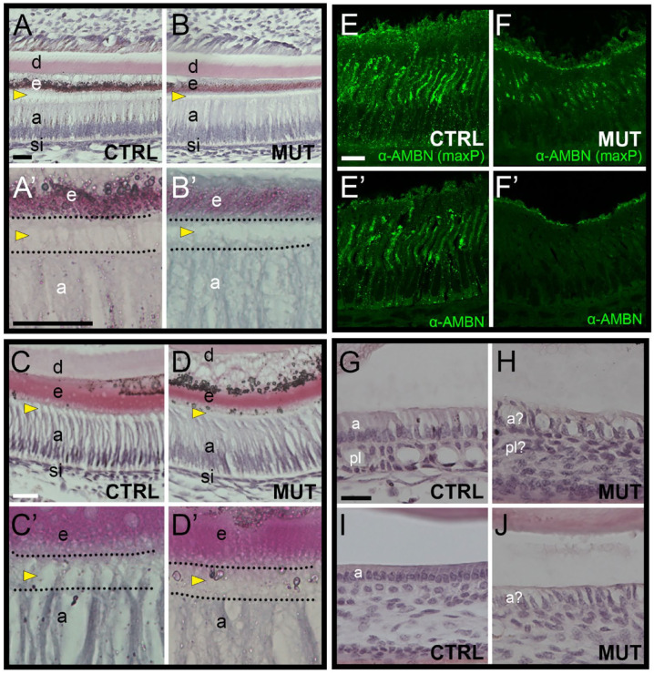
Coordinated mineralization of soft tissue is central to organismal form and function, while dysregulated mineralization underlies several human pathologies. Oral epithelial-derived ameloblasts are polarized, secretory cells responsible for generating enamel, the most mineralized substance in the human body. Defects in ameloblast development result in enamel anomalies, including amelogenesis imperfecta. Identifying proteins critical in ameloblast development can provide insight into specific pathologies associated with enamel-related disorders or, more broadly, mechanisms of mineralization. Previous studies identified a role for MEMO1 in bone mineralization; however, whether MEMO1 functions in the generation of additional mineralized structures remains unknown. Here, we identify a critical role for MEMO1 in enamel mineralization. First, we show that Memo1 is expressed in ameloblasts and, second, that its conditional deletion from ameloblasts results in enamel defects, characterized by a decline in mineral density and tooth integrity. Histology revealed that the mineralization defects in Memo1 mutant ameloblasts correlated with a disruption in ameloblast morphology. Finally, molecular profiling of ameloblasts and their progenitors in Memo1 oral epithelial mutants revealed a disruption to cytoskeletal-associated genes and a reduction in late-stage ameloblast markers, relative to controls. Collectively, our findings integrate MEMO1 into an emerging network of molecules important for ameloblast development and provide a system to further interrogate the relationship of cytoskeletal and amelogenesis-related defects.

Keywords: amelogenesis; amelogenesis imperfecta; cell polarity; cytoskeleton; mineralization; tooth.


Conflict of interest statement

Declaration of Conflicting InterestsThe authors declared no potential conflicts of interest with respect to the research, authorship, and/or publication of this article.

Figures

Figure 1.
Figure 1.
Memo1 is expressed in ameloblasts, and epithelial deletion of MEMO1 results in ectodermal defects, including tooth anomalies. (A) A representation of the lower mandible in a lateral view with lines indicating approximate location of the sections in subsequent panels (B–G). (B–G) β-Galactosidase staining of frontal gross sections (B–E) or cryosections (F, G) through the lower incisor of either a postnatal day 30 mouse harboring a lacZ reporter allele (Memo1tm1b) (B–F) or a negative control (G). White (B–E) or black (F) arrowheads point to the ameloblast layer. Dotted lines in B, F′, F′′, and G demarcate different regions of the incisor. F′ and F′′ are higher-magnification images of the section in F. (H–O) Frontal views of incisors in adolescent (H–K) or adult (L–O) control (H, L, J, N), Memo1;K14CRE (I, M), and Memo1;Pitx2CRE (K, O) mice. Mean age of adult mice is 15.3 mo (L, M) or 12.8 mo (N, O). Red arrowheads in I and K highlight the point of eroded enamel. Yellow arrowheads in M and O highlight fractured teeth. (P) Single weight measurements of age- and sex-matched littermate control (CTRL) and Memo1;K14CRE mutants at various adult time points. a, ameloblast; b, bone; CTRL, control; d, dentin; e, enamel; MUT, mutant; o, odontoblast; p, pulp. Scale bars: B–E = 200 µM; F, G = 100 µM.
Figure 2.
Figure 2.
Compromised enamel in Memo1 oral epithelial mutants. (A, B) Micro–computed tomography (µCT)–based 3-dimensional (A, B) or coronal optical section (A′, B′) centered on the first molar in a control (A) and Memo1;K14CRE (B) adult mouse mandible. Boxed region (A′, B′) shown magnified in inset. The color shaded optical section in A′ and B′ highlights the thresholding applied to define regions of interest for measurements. (C) Boxplot summarizing volumetric quantification of mineral density in incisor enamel, incisor dentin, molar enamel, molar dentin, and alveolar bone from µCT-based scans in control and Memo1;K14CRE adult mandibles. The proximal-distal bounds of the first molar were used as a consistent delimitating factor between samples. (D–G) Lateral scanning electron microscopy images of a control (D, F) or Memo1;K14CRE (E, G) incisor (D, E) and molars (F, G). Note that the third molar was lost during processing of the control sample (F). Red arrow in E indicates abrasion of enamel in Memo1;K14CRE incisor, shown at higher magnification in E′ and E′′ to highlight disordered enamel rods. The asterisk in C indicates a significant P value, using a standard t test, between control and Memo1;K14CRE samples. d, dentin; e, enamel; HA, hydroxyapatite; inc, incisor; m1, first molar; m2, second molar; m3, third molar; mol, molar.
Figure 3.
Figure 3.
Epithelial loss of MEMO1 is associated with altered apical ameloblast projections and an altered basolateral ameloblast–papillary layer interface. (A–D) Hematoxylin and eosin (H&E)–stained paraffin sections of the incisor in P10 control (A, C) or Memo1;Pitx2CRE mutant (B, D) samples in either sagittal (A, B) or frontal (C, D) planes. Sections move from more proximal (A, B) to distal (C, D) locations of secretory ameloblasts (e.g., A and B are at the level of the second molar, while C and D are at the level of the first molar). A′–D′ include higher-magnification images of the ameloblast–enamel interface, with the region between the apical ameloblast and enamel outlined. The yellow arrowheads indicate protrusions found at the apical end of ameloblasts. (E, F) α-Ameloblastin immunofluorescence of control (E) and mutant (F) frontal, lower incisor sections, adjacent to those used for H&E. While E and F are maximum-intensity projections, E′ and F′ highlight a single confocal slice through the section. (G–J) H&E-stained sagittal paraffin sections of the distal incisor in control (G, I) or Memo1;Pitx2CRE mutant (H, J) samples. a, ameloblast; d, dentin; e, enamel; pl, papillary layer; si, stratum intermedium. Scale bars: 25 µM in A, A′, and G; 10 µM in E.
Figure 4.
Figure 4.
Single-cell RNA sequencing of ameloblasts and their progenitors reveals cellular pathology associated with loss of MEMO1 in the oral epithelium. (A) A schematic depicting a lateral view of a hemi-mandible and the region isolated for single-cell RNA sequencing (scRNAseq). (B) UMAP-based projection of the entire scRNAseq data set, with larger groups, based on gene expression profiles, outlined. The epithelial cells (Krt14+), outlined in red, were subset and reclustered in the following panels. (C) UMAP-based projection of the reclustered epithelial cells reveals 9 distinct clusters, splitting into an ameloblast group (i.e., group A) and a “nonameloblast epithelial” group (i.e., group B). Individual clusters were identified based on gene expression profiles. (D) Cell cycle profiles for each of the 9 clusters identified in C. (E) UMAP-based projections, as shown in panel C, but split by genotype. The control UMAP is on the left; the Memo1 mutant UMAP is on the right. Clusters 1 and 8, 3 and 6, and 7 are circled in both UMAPs. (F) Proportion of control (black) and mutant (red) cells identified in each cluster. Clusters 1 and 8 combined, 3 and 6 combined, and 7 are indicated with red, light blue, and blue text and dashed boxes, respectively. The black and red horizontal lines represent expected frequencies for control (56%) and mutant (44%) cells, respectively, if no change is detected and accounting for the number of cells sequenced. (G) Comparison of cell cycle phase between control and mutant samples in each of the 3 clusters with proliferative cells (0 “Krt17+,” 4, 1/8). Clusters 1 and 8 are combined and identified by the red text and dashed box. Red arrows highlight “up” or “down” trends in mutant cells versus control cells for the cell cycle phase listed. (H) Schematic of the mouse incisal cervical loop, from a lateral perspective, and the general cell populations identified by scRNAseq. A rough representation of regions corresponding to clusters 7 and 1/8 are highlighted in light blue or salmon, respectively. a, aboral; d, distal; EA, early ameloblast; exp, expected; IEE, inner enamel epithelium; LA, late ameloblast; Lab, labial; Lin, lingual; o, oral; OEE, outer enamel epithelium; p, proximal; SI, stratum intermedium; SR, stellate reticulum; VEE, ventral enamel epithelium.
Figure 5.
Figure 5.
Molecular defects associated with ameloblast progression and cytoskeletal programs upon epithelial loss of MEMO1. (A) Table summarizing gene set enrichment analysis based on the genes significantly altered between control and mutant cells in the clusters highlighted (1/8, 3/6, and 7). Terms related to the “cytoskeleton” or “biomineralization” have been highlighted. Ann Clu = annotated cluster. (B–G) RNA expression levels of significantly altered genes in mutant versus control samples, derived from either single-cell RNA sequencing analysis of Memo1;Krt14 mutant ameloblasts (B–D, F, G) or bulk real-time reverse transcription polymerase chain reaction (RT-PCR) of Memo1;Pitx2CRE mutant hemi-mandibles (E). The adjacent arrow indicates either down (red arrow) or up (green arrow) regulated genes. Violin plots for single-cell RNA sequencing expression data are grouped and colored by cluster. Boxplots of relative messenger RNA expression in E are based on real-time RT-PCR analysis of complementary DNA synthesized from RNA isolated from the hemimandible of either a sibling control or Memo1;Pitx2CRE mutant. Hemi-mandibles were processed independently from 2 controls and 2 mutants (i.e., 4 samples/group). Note: control samples have been normalized to 1. EA, early ameloblast; LA, late ameloblast; IEE/OEE/SR, inner enamel epithelium/outer enamel epithelium/stellate reticulum.

Similar articles

Cited by

References

    1. Aldred MJ, Savarirayan R, Crawford PJ. 2003. Amelogenesis imperfecta: a classification and catalogue for the 21st century. Oral Dis. 9(1):19–23. - PubMed
    1. Balic A, Thesleff I. 2015. Tissue interactions regulating tooth development and renewal. Curr Top Dev Biol. 115:157–186. - PubMed
    1. Bartlett JD, Dobeck JM, Tye CE, Perez-Moreno M, Stokes N, Reynolds AB, Fuchs E, Skobe Z. 2010. Targeted p120-catenin ablation disrupts dental enamel development. PLoS One. 5(9):e12703. - PMC - PubMed
    1. Benseddik K, Sen Nkwe N, Daou P, Verdier-Pinard P, Badache A. 2013. ErbB2-dependent chemotaxis requires microtubule capture and stabilization coordinated by distinct signaling pathways. PLoS One. 8(1):e55211. - PMC - PubMed
    1. Caton J, Bringas P, Jr, Zeichner-David M. 2005. IGFs increase enamel formation by inducing expression of enamel mineralizing specific genes. Arch Oral Biol. 50(2):123–129. - PubMed