. 2023 Jul 5:13:1149506.
doi: 10.3389/fcimb.2023.1149506. eCollection 2023.

Infiltrated IL-17A-producing gamma delta T cells play a protective role in sepsis-induced liver injury and are regulated by CCR6 and gut commensal microbes

Affiliations

Infiltrated IL-17A-producing gamma delta T cells play a protective role in sepsis-induced liver injury and are regulated by CCR6 and gut commensal microbes

Jian Wan et al. Front Cell Infect Microbiol. .

Abstract

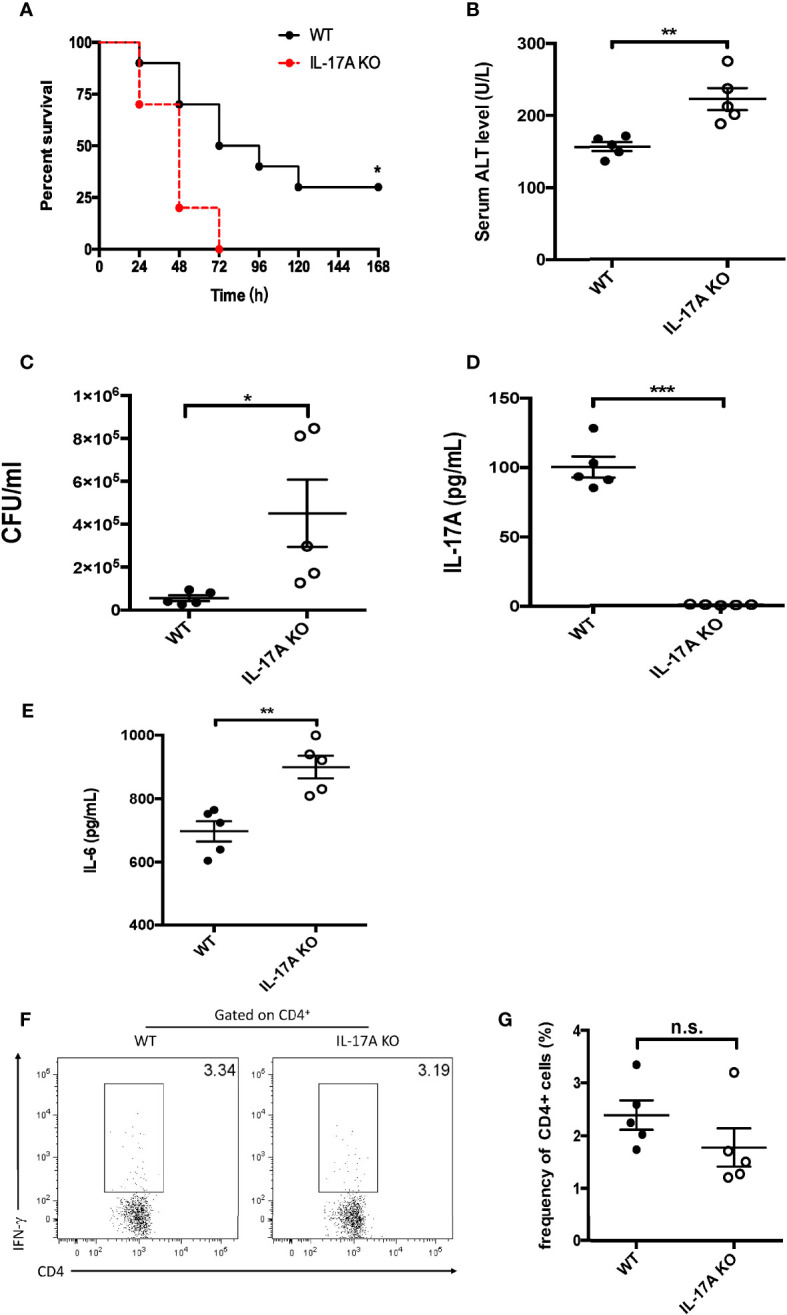
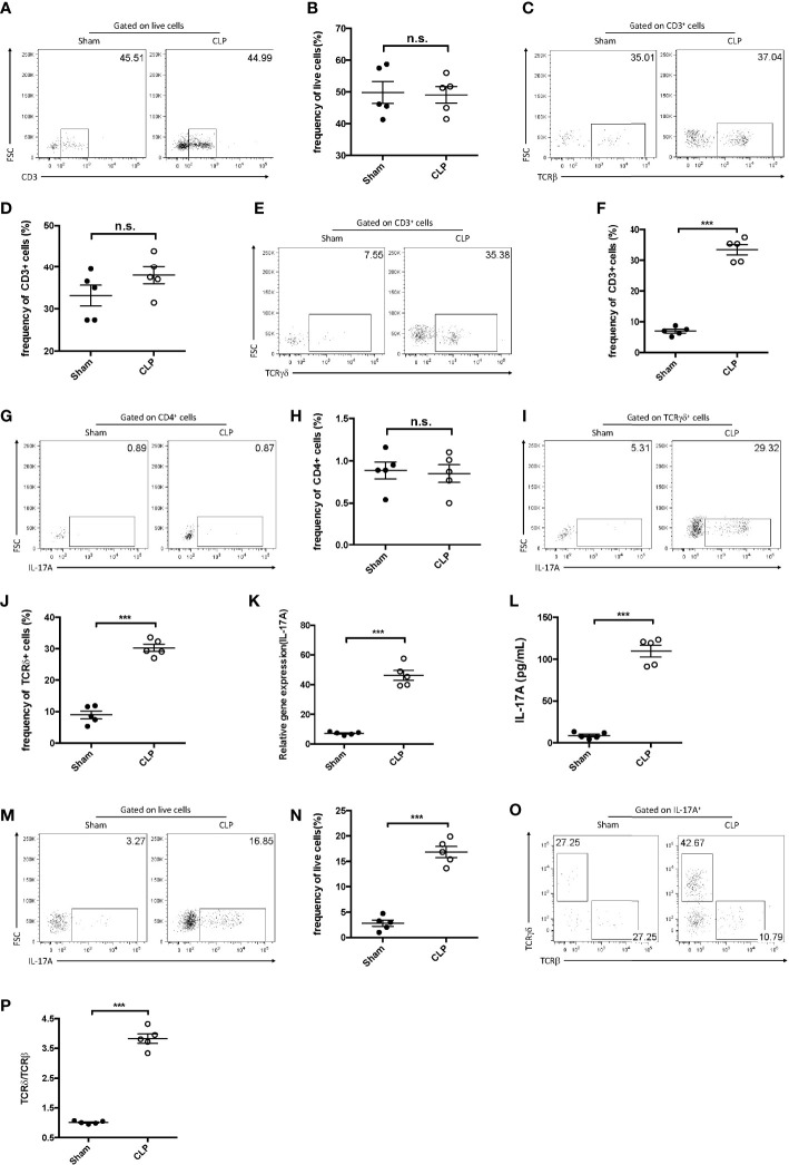
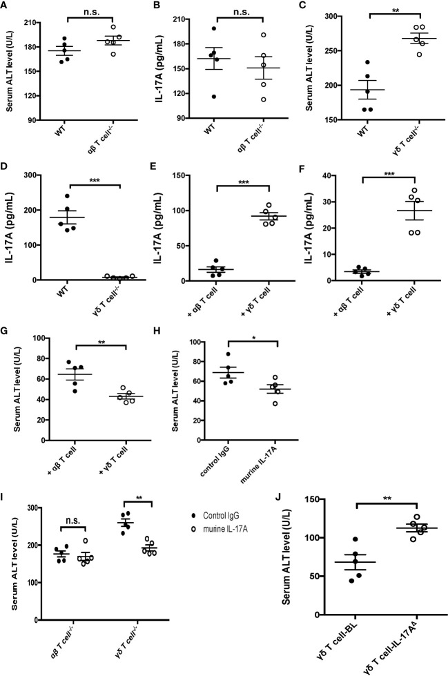
Introduction: Sepsis is a common but serious disease in intensive care units, which may induce multiple organ dysfunctions such as liver injury. Previous studies have demonstrated that gamma delta (γδ) T cells play a protective role in sepsis. However, the function and mechanism of γδ T cells in sepsis-induced liver injury have not been fully elucidated. IL-17A-producing γδ T cells are a newly identified cell subtype.

Methods: We utilized IL-17A-deficient mice to investigate the role of IL-17A-producing γδ T cells in sepsis using the cecum ligation and puncture (CLP) model.

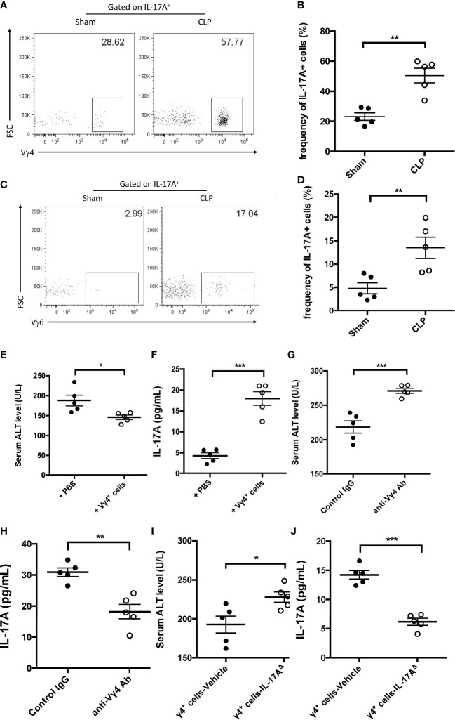
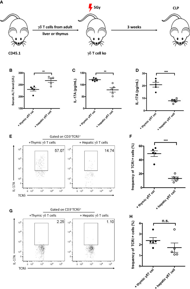
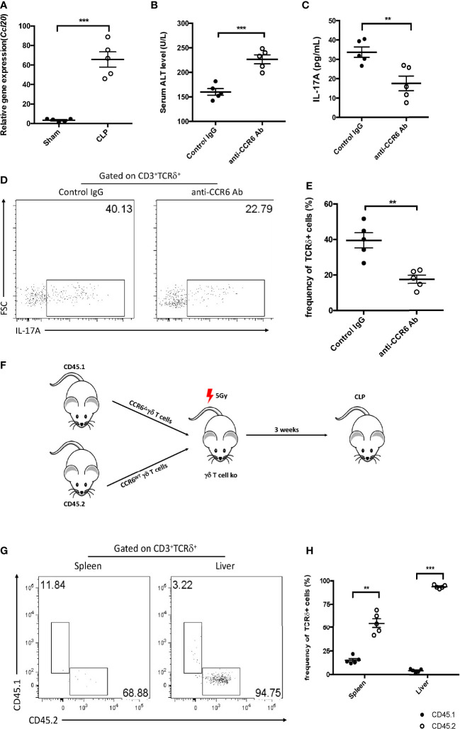
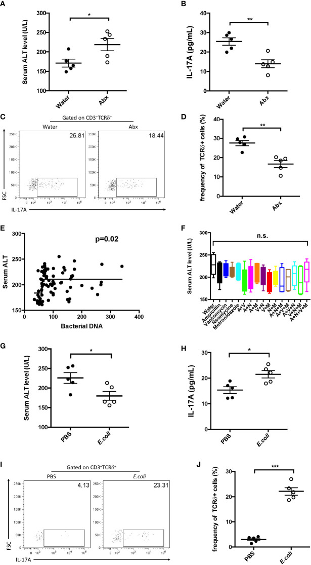
Results: Our findings suggested that these cells were the major source of IL-17A and protected against sepsis-induced liver injury. Flow cytometry analysis revealed that these γδ T cells expressed Vγ4 TCR and migrated into liver from peripheral post CLP, in a CCR6-dependent manner. When CLP mice were treated with anti-CCR6 antibody to block CCR6-CCL20 axis, the recruitment of Vγ4+ γδ T cells was abolished, indicating a CCR6-dependent manner of migration. Interestingly, pseudo germ-free CLP mice treated with antibiotics showed that hepatic IL-17A+ γδ T cells were regulated by gut commensal microbes. E. coli alone were able to restore the protective effect in pseudo germ-free mice by rescuing hepatic IL-17A+ γδ T cell population.

Conclusion: Our research has shown that Vγ4+ IL-17A+ γδ T cells infiltrating into the liver play a crucial role in protecting against sepsis-induced liver injury. This protection was contingent upon the recruitment of CCR6 and regulated by gut commensal microbes.

Keywords: CCR6; IL-17A; gamma delta T cells; liver injury; microbiota; sepsis.


Conflict of interest statement

The authors declare that the research was conducted in the absence of any commercial or financial relationships that could be construed as a potential conflict of interest.

Figures

Figure 1
Figure 1
Characteristics of interleukin 17A (IL-17A) knockout (KO) mice after cecum ligation and puncture (CLP). Wild-type (WT) and IL-17A KO mice were subjected to CLP. (A) Survival curves of WT and IL-17A KO mice subjected to CLP. (B) Serum alanine transaminase (ALT) levels between WT and IL-17A KO mice determined as described post-CLP. (C) Bacterial colony-forming units (CFUs) in the blood between WT and IL-17A KO mice subjected to CLP measured using agarose plates. (D, E) Serum levels of IL-17A and IL-6 between WT and IL-17A KO mice subjected to CLP determined using ELISA. (F) Representative fluorescence-activated cell sorting (FACS) plots of splenic T helper 1 (Th1) cells between WT and IL-17A KO mice subjected to CLP. (G) Statistics of Th1 cells in CD4 T cells. Data are from one intact experiment and representative of three independent experiments (five mice per group). Error bars denote the mean ± SEM. Survival analysis was performed using Mantel–Cox, while the rest was performed using Student’s t-test. n.s., not significant. *p < 0.05, **p < 0.01, ***p < 0.001.
Figure 2
Figure 2
IL-17A+ gamma delta (γδ) T cells were enriched in the liver after cecum ligation and puncture (CLP). C57BL/6 mice were subjected to CLP and analyzed 12 h later. (A, C, E) Representative fluorescence-activated cell sorting (FACS) plots of CD3+ (A), TCRβ+ (C), and TCRγδ+ (E) cells in the liver between sham and CLP mice. (B, D, F) Statistics of CD3+ (B), TCRβ+ (D), and TCRγδ+ (F) cells in (Cheng et al., 2017). (G, I) Representative FACS plots of IL-17A+ cells among the CD4+ cells (G) and the γδ T cells (I) in the liver between sham and CLP mice. (H, J) Statistics of IL-17A+ (H) and IL-17A (J) cells in (Cheng et al., 2017). (K) Transcriptional levels of IL-17A in the liver between sham and CLP mice measured using quantitative PCR (qPCR). (L) Serum levels of IL-17A between sham and CLP mice measured using ELISA. (M) Representative FACS plots of IL-17A+ cells among the total live cells in the liver between sham and CLP mice. (N) Statistics of IL-17A+ cells in (Cheng et al., 2017). (O) Representative FACS plots of TCRβ+ and TCRδ+ cells among the IL-17A+ cells in the liver between sham and CLP mice. (P) Statistics of the TCRδ vs. TCRβ ratio in (Cheng et al., 2017). Data are from one intact experiment and representative of three independent experiments (five mice per group). Error bars denote the mean ± SEM. Statistical analysis was performed using Student’s t-test. n.s., not significant. ***p < 0.001.
Figure 3
Figure 3
Function of IL-17A+ gamma delta (γδ) T cells in cecum ligation and puncture (CLP)-induced liver injury. Wild-type (WT) and alpha beta (αβ) T-cell knockout (KO) mice were subjected to CLP. (A, B) Serum levels of alanine transaminase (ALT) measured as described (A) and of interleukin 17A (IL-17A) determined using ELISA (B). WT and γδ T-cell KO mice were subjected to CLP. (C, D) Serum levels of ALT measured as described (C) and of IL-17A determined using ELISA (D). Rag1 mice were adoptive transferred with αβ or γδ T cells isolated from WT mice and subjected to CLP. (E, F) The levels of IL-17A in the plasma (Cheng et al., 2017) and liver (Cheng et al., 2017) were determined using ELISA. (G) Serum levels of ALT were measured after adoptive transferring. (H) Rag1 mice treated with control immunoglobulin G (IgG) or murine IL-17A through intraperitoneal injection and subjected to CLP. (I) αβ and γδ T-cell KO mice treated with murine IL-17A through intraperitoneal injection of isotype IgG as control and subjected to CLP. (J) Rag1 mice adoptive transferred with BL-vehicle- or BL-IL-17AΔ-transfected γδ T cells and then subjected to CLP. The serum levels of ALT were measured as described. Data are from one intact experiment and representative of three independent experiments (five mice per group). Error bars denote the mean ± SEM. Statistical analysis was performed using Student’s t-test. n.s., not significant. *p < 0.05, **p < 0.01, ***p < 0.001.
Figure 4
Figure 4
Function of Vγ4+ cells in cecum ligation and puncture (CLP)-induced liver injury. (A, C) Representative fluorescence-activated cell sorting (FACS) plots of Vγ4+ (A) and Vγ6+ (C) cells among the IL-17A+ cells between sham and CLP mice. (B, D) Statistics of Vγ4+ (B) and Vγ6+ (D) cells in (Cheng et al., 2017). Rag1 mice were adoptive transferred with Vγ4+ cells and then subjected to CLP. (E) Serum levels of alanine transaminase (ALT) measured as described. (F) Hepatic levels of interleukin 17A (IL-17A) determined using ELISA. C57BL/6 mice were treated with control immunoglobulin G (IgG) or the anti-Vγ4 antibody through intraperitoneal injection and then subjected to CLP. (G) Serum levels of ALT measured as described. (H) Hepatic levels of IL-17A determined using ELISA. Rag1 mice were adoptive transferred with BL-vehicle- or BL-IL-17AΔ-transfected Vg4+ cells and then subjected to CLP. (I) Serum levels of ALT measured as described. (J) Hepatic levels of IL-17A determined using ELISA. Data are from one intact experiment and representative of three independent experiments (five mice per group). Error bars denote the mean ± SEM. Statistical analysis was performed using Student’s t-test. n.s., not significant. *p < 0.05, **p < 0.01, ***p < 0.001.
Figure 5
Figure 5
Gamma delta (γδ) T cells migrated into the liver after cecum ligation and puncture (CLP). (A) Schematic showing the experiment design. Gamma delta (γδ) T cells (1 × 105) sorted from the liver or thymus of CD45.1 mice were intravenously transferred to 5 Gy-irradiated γδ T-cell knockout (KO) mice. Recipients were subjected to CLP 3 weeks later. (B) Serum levels of alanine transaminase (ALT) measured as described. (C, D) The plasma and hepatic levels of interleukin 17A (IL-17A) were determined using ELISA. (E, G) Representative fluorescence-activated cell sorting (FACS) plots of IL-17A+ cells among the TCRδ+ cells in the liver (E) and the spleen (G). (F, H) Statistics of IL-17A+ cells in (Cheng et al., 2017). Data are from one intact experiment and representative of three independent experiments (five mice per group). Error bars denote the mean ± SEM. Statistical analysis was performed using Student’s t-test. **p < 0.01, ***p < 0.001.
Figure 6
Figure 6
Function of CCR6 in gamma delta (γδ) T-cell migration into the liver. (A) Transcriptional levels of Ccl20 in the liver between sham and cecum ligation and puncture (CLP) mice as determined by quantitative PCR (qPCR). C57BL/6 mice were treated with the control immunoglobulin G (IgG) or the anti-CCR6 antibody through intraperitoneal injection and then subjected to CLP. (B) Serum levels of alanine transaminase (ALT) measured as described. (C) Hepatic levels of interleukin 17A (IL-17A) determined using ELISA. (D) Representative fluorescence-activated cell sorting (FACS) plots of IL-17A+ cells among the TCRδ+ cells in the liver. (E) Statistics of IL-17A+ cells in (Cheng et al., 2017). (F) Schematic showing the experiment design. CD45.1+ γδ T cells were transfected with BL-CCR6Δ, while CD45.2+ γδ T cells were transfected with BL-vehicle; subsequently, these two populations were mixed at a ratio of 1:1 and adoptive transferred into 5 Gy-irradiated γδ T-cell knockout (KO) mice as recipients, which were subjected to CLP 3 weeks later. (G) Representative FACS plots of CD45.1+ and CD45.2+ cells from the spleen and liver of recipients. (H) Statistics of CD45.1+ and CD45.2+ cells in (Cheng et al., 2017). Data are from one intact experiment and representative of three independent experiments (five mice per group). Error bars denote the mean ± SEM. Statistical analysis was performed using Student’s t-test. n.s., not significant. **p < 0.01, ***p < 0.001.
Figure 7
Figure 7
Effect of commensal microbes on cecum ligation and puncture (CLP)-induced liver injury. C57BL/6 mice were fed antibiotics for 5 days to deplete the gut microbiota, received fecal suspension for 3 days, and then subjected to CLP. (A) Serum levels of alanine transaminase (ALT) measured as described. (B) Hepatic levels of interleukin 17A (IL-17A) determined using ELISA. (C) Representative fluorescence-activated cell sorting (FACS) plots of IL-17A+ cells among TCRδ+ cells. (D) Statistics of IL-17A+ cells in (Cheng et al., 2017). (E) Pearson’s correlation curves between the serum ALT levels and bacterial DNA load. (F) C57BL/6 mice were fed water containing the following antibiotics: ampicillin (A), vancomycin (V), neomycin (N), and metronidazole (M). Pseudo-germ-free mice received fecal suspension for 3 days and then subjected to CLP. The serum levels of ALT were measured as described. Pseudo-germ-free mice intragastrically received 108 Escherichia coli colony-forming units (CFUs) and then subjected to CLP. (G) Serum levels of ALT measured as described. (H) Hepatic levels of IL-17A determined using ELISA. (I) Representative FACS plots of IL-17A+ cells among TCRδ+ cells. (J) Statistics of IL-17A+ cells in (Cheng et al., 2017). Data are from one intact experiment and representative of three independent experiments (five mice per group). Error bars denote the mean ± SEM. Statistical analysis was performed using Pearson’s correlation or Student’s t-test. *p < 0.05, **p < 0.01, ***p < 0.001.

Similar articles

Cited by

References

    1. Ajuebor M. N., Jin Y., Gremillion G. L., Strieter R. M., Chen Q., Adegboyega P. A. (2008). Gamma delta T cells initiate acute inflammation and injury in adenovirus-infected liver via cytokine-chemokine cross talk. J. Virol. 82 (19), 9564–9576. doi: 10.1128/JVI.00927-08 - DOI - PMC - PubMed
    1. Baggio Savio L. E., Mello P., Figliuolo V. R., de Avelar Almeida T. F., Santana P. T., Oliveira S. D. S., et al. . (2017). CD39 limits P2X7 receptor inflammatory signaling and attenuates sepsis-induced liver injury. J. Hepatol. 67 (4), 716–726. doi: 10.1016/j.jhep.2017.05.021 - DOI - PMC - PubMed
    1. Bonneville M., O'Brien R. L., Born W. K. (2010). Gamma delta T cell effector functions: a blend of innate programming and acquired plasticity. Nat. Rev. Immunol. 10 (7), 467–478. doi: 10.1038/nri2781 - DOI - PubMed
    1. Buford T. W. (2017). (Dis)Trust your gut: the gut microbiome in age-related inflammation, health, and disease. Microbiome 5 (1), 80. doi: 10.1186/s40168-017-0296-0 - DOI - PMC - PubMed
    1. Chassaing B., Etienne-Mesmin L., Gewirtz A. T. (2014). Microbiota-liver axis in hepatic disease. Hepatology 59 (1), 328–339. doi: 10.1002/hep.26494 - DOI - PMC - PubMed

Publication types