Review
. 2022 Jul 1;29(3):140-153.
doi: 10.4078/jrd.2022.29.3.140.

The Mechanism of the NLRP3 Inflammasome Activation and Pathogenic Implication in the Pathogenesis of Gout

Affiliations
Review

The Mechanism of the NLRP3 Inflammasome Activation and Pathogenic Implication in the Pathogenesis of Gout

Seong-Kyu Kim. J Rheum Dis. .

Abstract

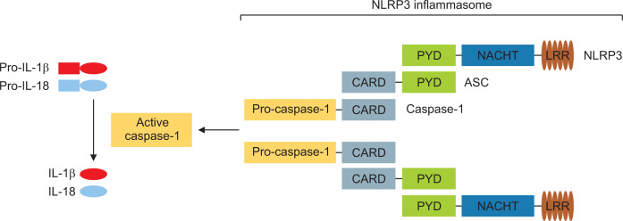
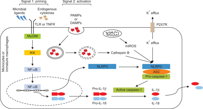
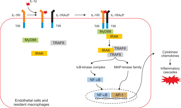
The NACHT, LRR, and PYD-domains-containing protein 3 (NLRP3) inflammasome is an intracellular multi-protein signaling platform that is activated by cytosolic pattern-recognition receptors such as NLRs against endogenous and exogenous pathogens. Once it is activated by a variety of danger signals, recruitment and assembly of NLRP3, ASC, and pro-caspase-1 trigger the processing and release of pro-inflammatory cytokines including interleukin-1β (IL-1β) and IL-18. Multiple intracellular and extracellular structures and molecular mechanisms are involved in NLRP3 inflammasome activation. Gout is an autoinflammatory disease induced by inflammatory response through production of NLRP3 inflammasome-mediated proinflammatory cytokines such as IL-1β by deposition of monosodium urate (MSU) crystals in the articular joints and periarticular structures. NLRP3 inflammasome is considered a main therapeutic target in MSU crystal-induced inflammation in gout. Novel therapeutic strategies have been proposed to control acute flares of gouty arthritis and prophylaxis for gout flares through modulation of the NLRP3/IL-1 axis pathway. This review discusses the basic mechanism of NLRP3 inflammasome activation and the IL-1-induced inflammatory cascade and explains the NLRP3 inflammasome-induced pathogenic role in the pathogenesis of gout.

Keywords: Gout; Inflammasome; Interleukin-1; Monosodium urate; NLRP3.


Conflict of interest statement

CONFLICT OF INTEREST No potential conflict of interest relevant to this article was reported.

Figures

Fig. 1
Fig. 1
Schematic structure of NLRP3 inflammasome complex. The NLRP3 inflammasome complex consists of the NLRP3 scaffold, the ASC adaptor, and pro-caspase-1 proteins. Deletion of the CARD domain and activation of caspase-1 domain of pro-caspase-1 leads to formation of active caspase-1, then resulting in cleavage from pro-IL-1β to IL-1β. ASC: apoptosis-associated speck-like protein, CARD: caspase activation and recruitment domain, IL: interleukin, LRR: leucine-rich repeat, NACHT: neuronal apoptosis inhibitory protein (NAIP), MHC class II transcription activator (CIITA), incompatibility locus protein from Podospora anserina (HET-E), and telomerase-associated protein (TP1), NLRP3: NACHT, LRR, and PYD domains-containing protein 3, PYD: pyrin domain.
Fig. 2
Fig. 2
Signal pathway for NLRP3 inflammasome activation. Activation of the NLRP3 inflammasome is essential for two steps such as priming (signal 1) and activation (signal 2). In priming step, activation of TLR or TNFR after exposure of danger signals facilitates NF-κB signaling pathway for initiating transcription and production of precursor of pro-inflammatory cytokine including pro-IL-1β and pro-IL-18. Next, sensing PAMPs or DAMPs triggers NLRP3 oligomerization and ASC and pro-caspase-1 recruitment (signal 2). Autocleavage and activation of caspase-1 results in maturation and release of pro-inflammatory cytokines IL-1β and IL-18. PAMPs: pathogen-associated molecular patterns, DAMPs: damage-associated molecular patterns, ASC: apoptosis-associated speck-like protein, CARD: caspase activation and recruitment domain, TLR: toll-like receptor, TNFR: tumor necrosis factor receptor, ROS: reactive oxygen species, IKK: IκB kinase, IL: interleukin, NF-κB: nuclear factor-κB, NACHT: neuronal apoptosis inhibitory protein (NAIP), MHC class II transcription activator (CIITA), incompatibility locus protein from Podospora anserina (HET-E), and telomerase-associated protein (TP1), NLRP3: NACHT, LRR, and PYD domains-containing protein 3.
Fig. 3
Fig. 3
Mechanism for IL-1β/IL-1R-related inflammatory signaling pathway. IL-1β/IL-1R complex is generated by the heterodimer signaling receptors and cytokine. The TIR domain, an intercellular signaling protein motif between receptors and adaptors, associates with adaptor MyD88, thus resulting in recruitment of IRAK and dimerized TRAF6. This subsequently activates multiple downstream signaling pathways including IκB-kinase complex and MAPK. Activation of transcription factors including NF-κB and AP-1 results in diverse pro-inflammatory cytokines and chemokines. IL-1β: interleukin-1β, IL-1RI: interleukin-1 receptor type I, TIR: toll/interleukin-1 receptor, MyD88: myeloid differentiation primary response 88, IRAK: interleukin-1 receptor associated kinase, TRAF6: TNF receptor-associated factor 6, TNF: tumor necrosis factor, NF-κB: nuclear factor-κB.

References

    1. Dalbeth N, Gosling AL, Gaffo A, Abhishek A. Gout. Lancet. 2021;397:1843–55. doi: 10.1016/S0140-6736(21)00569-9. Erratum in: Lancet 2021;397:1808. - DOI - PubMed
    1. Busso N, So A. Mechanisms of inflammation in gout. Arthritis Res Ther. 2010;12:206. doi: 10.1186/ar2952. - DOI - PMC - PubMed
    1. Nuki G, Simkin PA. A concise history of gout and hyperuricemia and their treatment. Arthritis Res Ther. 2006;8(Suppl 1):S1. doi: 10.1186/ar1906. - DOI - PMC - PubMed
    1. Freudweiler M. Experimentelle untersuchungen uber das wesen der gichtknoten. Dtsch Arch Klin Med. 1899;63:266–335. German.
    1. McCarty DJ, Hollander JL. Identification of urate crystals in gouty synovial fluid. Ann Intern Med. 1961;54:452–60. doi: 10.7326/0003-4819-54-3-452. - DOI - PubMed

LinkOut - more resources