The role of endobronchial ultrasonography elastography in the diagnosis of hilar and mediastinal lymph nodes
- PMID: 37476908
- PMCID: PMC10388034
- DOI: 10.55730/1300-0144.5634
The role of endobronchial ultrasonography elastography in the diagnosis of hilar and mediastinal lymph nodes
Abstract
Background: Endobronchial ultrasonography (EBUS) is a minimally invasive diagnostic tool in the diagnosis of mediastinal lymph nodes (LNs) and has sonographic features. We aimed to investigate the diagnostic accuracy of EBUS elastography, which evaluates tissue compressibility integrated into EBUS, on malignant vs. benign mediastinal-hilar LNs.
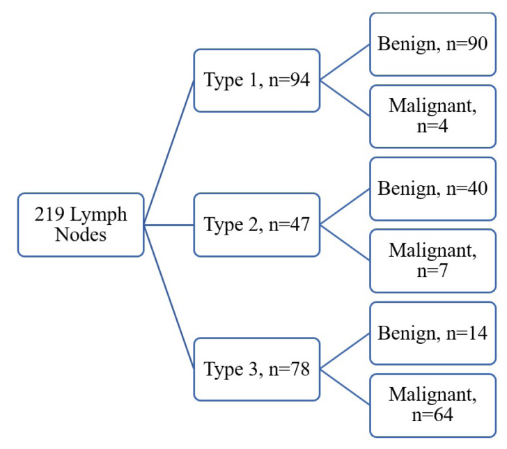
Methods: A single-center, prospective study was conducted at the University of Health Sciences Yedikule Chest Diseases and Thoracic Surgery Training and Research Hospital between 01/10/2019 and 15/11/2019. The features of 219 LNs evaluated by thoracic computed tomography (CT), positron emission tomography (PET)/CT, EBUS sonography and EBUS elastography were recorded. The LNs sampled by EBUS-guided fine needle aspiration were classified according to EBUS elastography color distribution findings as follows: type 1, predominantly nonblue (green, yellow, and red); type 2, part blue, part nonblue; type 3, predominantly blue. The strain ratio (SR) was calculated based on normal tissue with the relevant region.
Results: The average age of 131 patients included in the study was 55.86 ± 13 years, 76 (58%) were male. Two hundred and nineteen lymph nodes were sampled from different stations. Pathological diagnosis of 75 (34.2%) LNs was malignant, the rest was benign. When EBUS B-mode findings and pathological results were compared, sensitivity was 65.33%, specificity 63.19%, positive predictive value (PPV) 48%, negative predictive value (NPV) 77.8%, and diagnostic yield (DY) 64%. When the pathological diagnoses and EBUS elastography findings were compared, while type 1 LNs were considered to be benign and type 3 LNs malignant, sensitivity 94.12%, specificity 86.54%, PPV 82.1%, NPV 95.7%, and DY 89.5%. SR of malignant LNs was significantly higher than benign LNs (p < 0.001). When the classification according to color scale and SR were compared, no difference was found in DY (p = 0.155).
Discussion: The diagnostic accuracy of EBUS elastography is high enough to distinguish malignant LN from benign ones with the SR option. When compared with EBUS-B mode sonographic findings, it was found to have a higher diagnostic yield.
Keywords: Elastography; endobronchial ultrasonography; lymph node; mediastinum; strain ratio.
Figures


Similar articles
-
Diagnostic value of endobronchial ultrasound elastography for differentiating benign and malignant hilar and mediastinal lymph nodes: a systematic review and meta-analysis.Med Ultrason. 2022 Feb 16;24(1):85-94. doi: 10.11152/mu-2971. Epub 2021 Apr 1. Med Ultrason. 2022. PMID: 33793697
-
Added value of combined endobronchial and oesophageal endosonography for mediastinal nodal staging in lung cancer: a systematic review and meta-analysis.Lancet Respir Med. 2016 Dec;4(12):960-968. doi: 10.1016/S2213-2600(16)30317-4. Epub 2016 Oct 20. Lancet Respir Med. 2016. PMID: 27773666
-
Is the diagnostic yield of mediastinal lymph node cryobiopsy (cryoEBUS) better for diagnosing mediastinal node involvement compared to endobronchial ultrasound-guided transbronchial needle aspiration (EBUS-TBNA)? A systematic review.Respir Med. 2023 Nov;218:107389. doi: 10.1016/j.rmed.2023.107389. Epub 2023 Aug 12. Respir Med. 2023. PMID: 37579981
-
Artificial intelligence-assisted endobronchial ultrasound for differentiating between benign and malignant thoracic lymph nodes: a meta-analysis.BMC Pulm Med. 2025 Jul 2;25(1):303. doi: 10.1186/s12890-025-03760-4. BMC Pulm Med. 2025. PMID: 40604844 Free PMC article.
-
PET-CT for assessing mediastinal lymph node involvement in patients with suspected resectable non-small cell lung cancer.Cochrane Database Syst Rev. 2014 Nov 13;2014(11):CD009519. doi: 10.1002/14651858.CD009519.pub2. Cochrane Database Syst Rev. 2014. PMID: 25393718 Free PMC article.
Cited by
-
Clinical utility of artificial intelligence-augmented endobronchial ultrasound elastography in lymph node staging for lung cancer.JTCVS Tech. 2024 Jul 24;27:158-166. doi: 10.1016/j.xjtc.2024.06.024. eCollection 2024 Oct. JTCVS Tech. 2024. PMID: 39478913 Free PMC article.
References
-
- Silvestri GA, Gonzalez AV, Jantz MA, Margolis ML, Gould MK, et al. Methods for staging non-small cell lung cancer: Diagnosis and management of lung cancer, 3rd ed: American College of Chest Physicians evidence-based clinical practice guidelines. Chest. 2013;143(5 Suppl):e211S–e250S. doi: 10.1378/chest.12-2355. - DOI - PubMed
-
- Lilo MT, Allison DB, Younes BK, Cui M, Askin FB, et al. The critical role of EBUS-TBNA cytology in the staging of mediastinal lymph nodes in lung cancer patients: A correlation study with positron emission tomography findings. Cancer Cytopathology. 2017;125(9):717–725. doi: 10.1002/cncy.21886. - DOI - PubMed
-
- Nakajima T, Anayama T, Shingyoji M, Kimura H, Yoshino I, et al. Vascular image patterns of lymph nodes for the prediction of metastatic disease during EBUS-TBNA for mediastinal staging of lung cancer. Journal of Thoracic Oncology. 2012;7(6):1009–1014. doi: 10.1097/JTO.0b013e31824cbafa. - DOI - PubMed
MeSH terms
LinkOut - more resources
Full Text Sources
Medical
Research Materials