. 2023 Nov;43(8):1840-1856.
doi: 10.1007/s10875-023-01547-y. Epub 2023 Jul 21.

Immunologic and Genetic Contributors to CD46-Dependent Immune Dysregulation

Affiliations

Immunologic and Genetic Contributors to CD46-Dependent Immune Dysregulation

Benedikt J Meyer et al. J Clin Immunol. 2023 Nov.

Erratum in

  • Correction to: Immunologic and Genetic Contributors to CD46‑Dependent Immune Dysregulation.
    Meyer BJ, Kunz N, Seki S, Higgins R, Ghosh A, Hupfer R, Baldrich A, Hirsiger JR, Jauch AJ, Burgener AV, Lötscher J, Aschwanden M, Dickenmann M, Stegert M, Berger CT, Daikeler T, Heijnen I, Navarini AA, Rudin C, Yamamoto H, Kemper C, Hess C, Recher M. Meyer BJ, et al. J Clin Immunol. 2023 Nov;43(8):1857. doi: 10.1007/s10875-023-01563-y. J Clin Immunol. 2023. PMID: 37572200 Free PMC article. No abstract available.

Abstract

Mutations in CD46 predispose to atypical hemolytic uremic syndrome (aHUS) with low penetrance. Factors driving immune-dysregulatory disease in individual mutation carriers have remained ill-understood. In addition to its role as a negative regulator of the complement system, CD46 modifies T cell-intrinsic metabolic adaptation and cytokine production. Comparative immunologic analysis of diseased vs. healthy CD46 mutation carriers has not been performed in detail yet. In this study, we comprehensively analyzed clinical, molecular, immune-phenotypic, cytokine secretion, immune-metabolic, and genetic profiles in healthy vs. diseased individuals carrying a rare, heterozygous CD46 mutation identified within a large single family. Five out of six studied individuals carried a CD46 gene splice-site mutation causing an in-frame deletion of 21 base pairs. One child suffered from aHUS and his paternal uncle manifested with adult-onset systemic lupus erythematosus (SLE). Three mutation carriers had no clinical evidence of CD46-related disease to date. CD4+ T cell-intrinsic CD46 expression was uniformly 50%-reduced but was comparable in diseased vs. healthy mutation carriers. Reconstitution experiments defined the 21-base pair-deleted CD46 variant as intracellularly-but not surface-expressed and haploinsufficient. Both healthy and diseased mutation carriers displayed reduced CD46-dependent T cell mitochondrial adaptation. Diseased mutation carriers had lower peripheral regulatory T cell (Treg) frequencies and carried potentially epistatic, private rare variants in other inborn errors of immunity (IEI)-associated proinflammatory genes, not found in healthy mutation carriers. In conclusion, low Treg and rare non-CD46 immune-gene variants may contribute to clinically manifest CD46 haploinsufficiency-associated immune-dysregulation.

Keywords: CD46; Inborn errors of immunity; SLE; aHUS; atypical hemolytic uremic syndrome; haploinsufficiency; next-generation sequencing; penetrance; primary immunodeficiency complement; systemic lupus erythematosus.


Conflict of interest statement

The authors declare no competing interests.

Figures

Fig. 1
Fig. 1
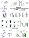
Study design and analyzed CD46 c.475+1G>A mutation. a Visualization of the family tree with several family members carrying a rare CD46 mutation. Circles indicate females, squares indicate male individuals. The numbered individuals [–6] have been analyzed in the current study. The blue colored patient manifested with pediatric-onset aHUS [6], and the green colored individual has the diagnosis of adult-onset SLE [5]. Individuals with grey color carry the mutation but do not manifest CD46 associated immune-dysregulation up to date. b Exons of genomic CD46 schematically depicted (deep blue) with corresponding protein modules of CD46 (bright blue). The c. 475+1G>A point mutation is located at the start of the intron between exon 4 and 5, altering a splice site leading to an in-frame truncated exon 4 (skipped exon 4 part in red) lacking in 21 base pairs (bp). The predicted protein variant has a truncated CCP module 2 (residue deletion represented in orange)
Fig. 2
Fig. 2
Exon 4 partial skipping and transcript coexistence of mutated CD46. a Representative Sanger sequencing of a PCR-amplified CD46-specific sequence using PBMC-derived genomic DNA. Purple arrow shows the heterozygous CD46 intronic mutation detected in five out of six tested family members. b PCR amplification of a CD46-specific sequence using PBMC-derived cDNA revealing two different bands in all tested individuals except family member number 3 which does not carry the CD46 mutation in genomic DNA. Image shown is zoomed for bands and ladder. c Sanger sequencing chromatogram of the bands indicated in b show a skipping of 21 nucleotides in the truncated cDNA band from the del21bp CD46 gene (bottom). d mRNA expression quantitation of WT (top) and del21bp mutant (bottom) CD46 mRNA assessed using PBMC-derived cDNA. Each are normalized as listed on the Y axis. P values represent comparison of all five CD46 mutation carriers and three healthy controls (HCs) by unpaired t tests
Fig. 3
Fig. 3
del21bp CD46 is a loss-of-expression mutation and lacks dominant negativity. a Alignment (top) of exon skipping-mediated CD46 protein 6+1-residue deletion flanking glutamic acid (Glu) 158 and its orientation on a reported WT CD46 structure (middle) appended with a magnified view (bottom). Purple boxes show orientation of a CCP domain-conserved disulfide bond between deleted cysteine residue 157 and cysteine residue 127. Structure adopted/downloaded from Alphafold2 (https://alphafold.ebi.ac.uk/entry/P15529). b+c) Flow-cytometric analysis of CD46 surface expression on freshly purified CD4+ T cells ex vivo. Representative histogram plots are shown in b and MFIs are depicted in c. d Schematic of CD46-specific monoclonal antibody (mAb) binding sites and CD46 surface binding signal intensity in mutation carrier 5 (SLE) in comparison with unrelated healthy donor controls. Dotted line for 8E2 mAb indicates incomplete epitope identification in literature. Fluorescence deviation values compared with intra-patient isotype control staining are normalized against mean of healthy controls for each. CCP1-specific binding is shown in gray and CCP2/3/4 binding are shown in white for mutation carrier 5. e Representative flow-cytometric plot of transient CD46 overexpression via WT versus del21bp CD46 vectors by transfection of a CD46-knocked-out human myeloid HAP1 cell line. f Comparison of WT versus del21bp CD46 protein surface and intracellular expression positivity. Analyzed by unpaired t tests. g ColabFold v1.5.2-based structure prediction of del21bp CD46-variant protein (right) in comparison with queried/predetermined WT CD46 structure (left). Black wedges show a CCP3/CCP4-junction bending contrasting CCP1/CCP2/CCP3 alignment (highlighted in dotted line). Green wedge shows a de novo impairment of CCP2/CCP3. h CD46 expression upon WT and del21bp mutant CD46 co-transfection (1:1 ratio). P values represent Tukey’s post hoc multiple comparison tests of one-way ANOVA. Representative of two experiments performed in quadruplicate (f+h)
Fig. 4
Fig. 4
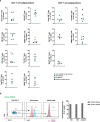
Immune cell subpopulations in heterozygous CD46 c.475+1G>A mutation carriers. a CD4+ and CD8+ T cell subpopulations as indicated of all tested family members are depicted. The blue and green dots represent the aHUS and SLE patient, respectively. Closed symbols represent mutation carriers while the open symbol represents the family member lacking the mutated allele. The lines mark normal reference values. b Left: FoxP3 transcription factor positivity in mutation carrier 5 (SLE) CD4+ T cell subpopulations. Right: subpopulation FoxP3 positivity aligned with two unrelated healthy controls. Values are subtracted for positivity in isotype control staining
Fig. 5
Fig. 5
Impaired CD46-dependent immunometabolic adaptation in heterozygous CD46 c.475+1G>A mutation carriers. a CD46 expression (MFI) on CD4+ T cells following in vitro stimulation. Purified CD4+ T cells were left non-activated (NA) or stimulated with immobilized agonistic antibodies against CD3 or CD3 + CD28 for 36 hours before flow-cytometric analysis. b Purified CD4+ T cells were either analyzed ex vivo or left non-activated (NA) or stimulated with immobilized agonistic antibodies against CD3, CD3 + CD28 or CD3 + CD46 for 36 h. Flow cytometric analysis of the MFI of mitotracker deep red T cell fluroescence as a marker of mitochondrial membrane potential and the MFI of mitotracker green staining as a marker for mitochondrial mass of CD4+ T cells was performed. The ratio of the two MFI’s represents mitochondrial function per mitochondrial mass. c An explanatory graph showing cellular oxygen consumption rate and the respiratory indices after perturbation with oligomycin, FCCP and rotenone in the Seahorse flux analyzer. d + e) Quantification of T cell intrinsic oxygen consumption. Purified CD4+ T cells were left non-activated (NA) or stimulated with immobilized agonistic antibodies against CD3, CD3 + CD28 or CD3 + CD46. 36 h post stimulation, basal c and ATP-coupled d oxidative phosphorylation (OXPHOS, OCR) in CD4+ T cells was measured on the Seahorse metabolic extracellular flux analyzer. All bars indicate means ± SD. Compared by unpaired t-tests as indicated

References

    1. Yamamoto H, Fara AF, Dasgupta P, Kemper C. CD46: the 'multitasker' of complement proteins. Int J Biochem Cell Biol. 2013;45(12):2808–2820. - PubMed
    1. Liszewski MK, Post TW, Atkinson JP. Membrane cofactor protein (MCP or CD46): newest member of the regulators of complement activation gene cluster. Annu Rev Immunol. 1991;9:431–455. - PubMed
    1. Liszewski MK, Atkinson JP. Complement regulator CD46: genetic variants and disease associations. Hum Genomics. 2015;9:7. - PMC - PubMed
    1. Kemper C, Atkinson JP. T-cell regulation: with complements from innate immunity. Nat Rev Immunol. 2007;7(1):9–18. - PubMed
    1. Kemper C, Chan AC, Green JM, Brett KA, Murphy KM, Atkinson JP. Activation of human CD4+ cells with CD3 and CD46 induces a T-regulatory cell 1 phenotype. Nature. 2003;421(6921):388–392. - PubMed

Publication types