. 2023 Dec;16(6):1287-1302.
doi: 10.1007/s12265-023-10411-8. Epub 2023 Jul 21.

Age and Sex Differences in the Genetics of Cardiomyopathy

Collaborators, Affiliations

Age and Sex Differences in the Genetics of Cardiomyopathy

Oyediran Akinrinade et al. J Cardiovasc Transl Res. 2023 Dec.

Abstract

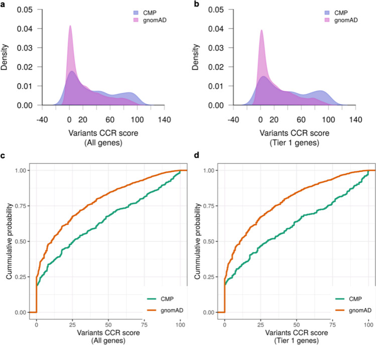
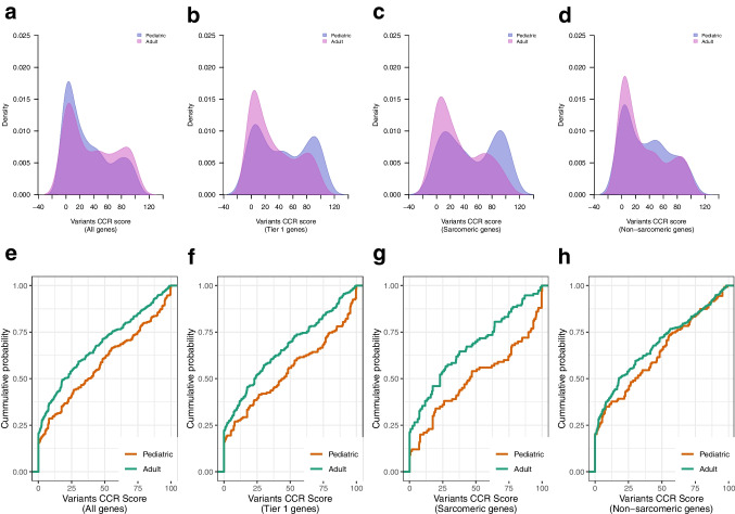
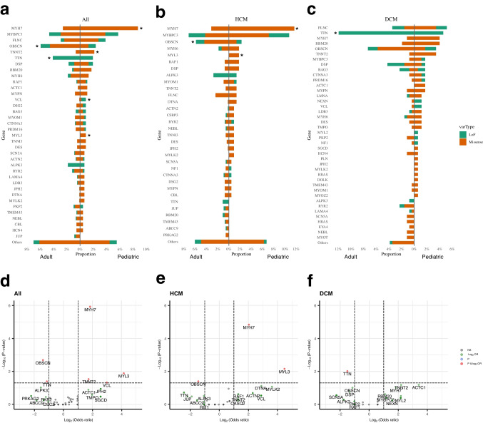
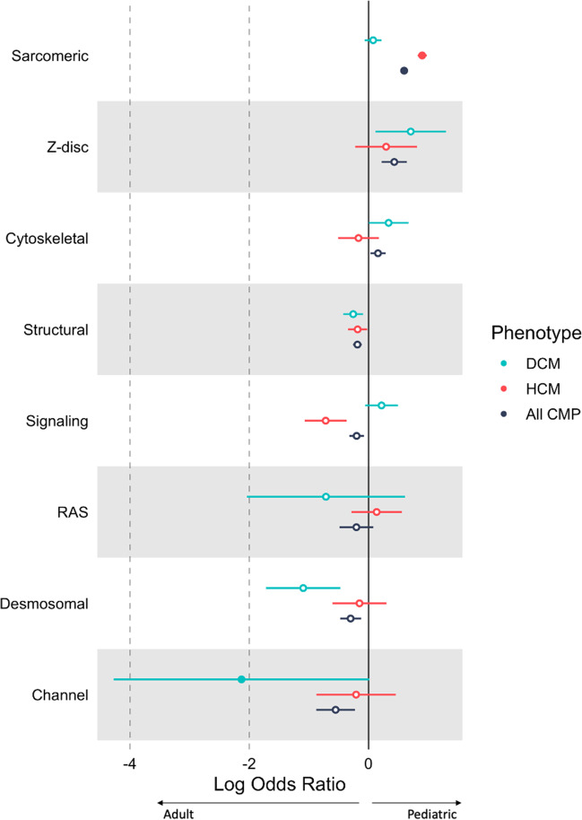
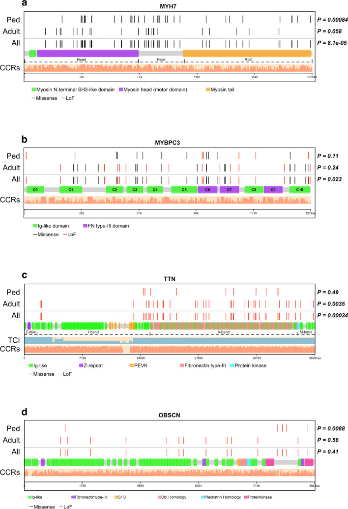
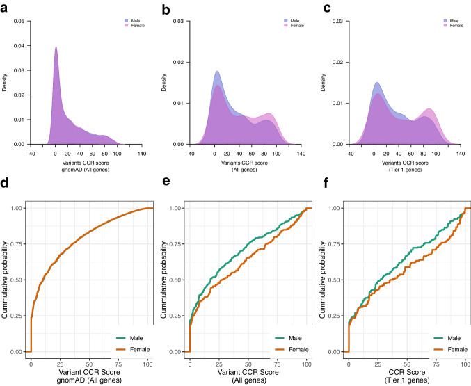
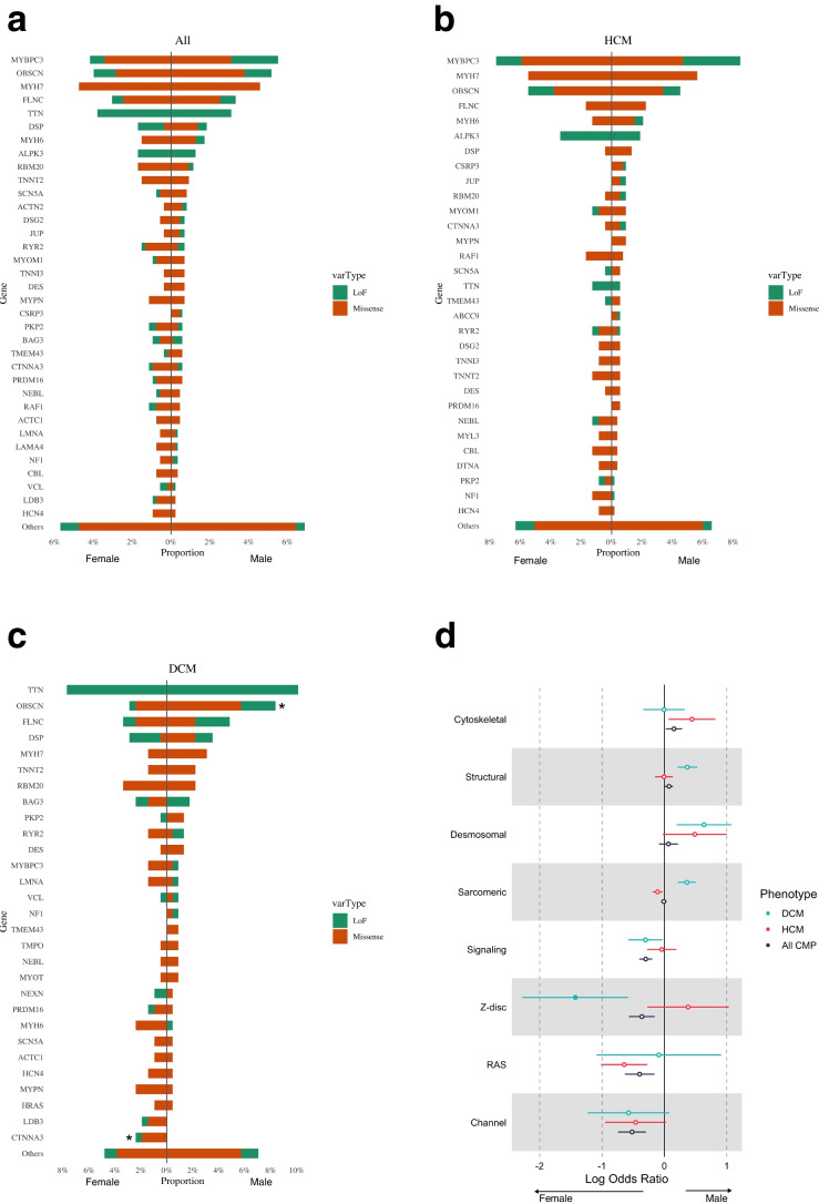
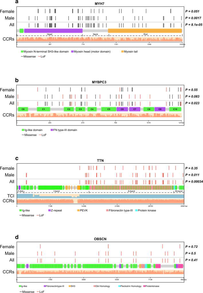
Cardiomyopathy has variable penetrance. We analyzed age and sex-related genetic differences in 1,397 cardiomyopathy patients (Ontario, UK) with whole genome sequencing. Pediatric cases (n = 471) harbored more deleterious protein-coding variants in Tier 1 cardiomyopathy genes compared to adults (n = 926) (34.6% vs 25.9% respectively, p = 0.0015), with variant enrichment in constrained coding regions. Pediatric patients had a higher burden of sarcomere and lower burden of channelopathy gene variants compared to adults. Specifically, pediatric patients had more MYH7 and MYL3 variants in hypertrophic cardiomyopathy, and fewer TTN truncating variants in dilated cardiomyopathy. MYH7 variants clustered in the myosin head and neck domains in children. OBSCN was a top mutated gene in adults, enriched for protein-truncating variants. In dilated cardiomyopathy, female patients had a higher burden of z-disc gene variants compared to males. Genetic differences may explain age and sex-related variability in cardiomyopathy penetrance. Genotype-guided predictions of age of onset can inform pre-test genetic counseling. Pediatric cardiomyopathy patients were more likely to be genotype-positive than adults with a higher burden of variants in MYH7, MYL3, TNNT2, VCL. Adults had a higher burden of OBSCN and TTN variants. Females with dilated cardiomyopathy (DCM) had a higher burden of z-disc gene variants compared to males.

Keywords: Age; Cardiomyopathy; Genetic; Genomic constraints; Sex; Whole genome sequencing.


Conflict of interest statement

SM is on the Advisory Board of Bristol Myers Squibb, and Tenaya Therapeutics. The remaining authors have nothing to disclose.

Figures

Fig. 1
Fig. 1
CCR score of deleterious variants in cases and controls. (a, b) Density plots of CCR scores (blue = cardiomyopathy; pink = gnomAD), and (c, d) Empirical cumulative probability distribution of CCR scores of deleterious variants identified in cardiomyopathy cases (n = 1397) (green) and in gnomAD reference controls (n = 125,748) (orange). Variant CCR scores were higher in cardiomyopathy cases compared to gnomAD reference controls across all cardiomyopathy genes (KS test p < 2.2 × 10–16) and in the subset of Tier 1 genes (KS test p < 2.2 × 10–16). CCR, constrained coding regions; CMP, cardiomyopathy; gnomAD, Genome Aggregation Database; KS, Kolmogorov–Smirnov test
Fig. 2
Fig. 2
CCR score of deleterious variants stratified by age. (a-d) Density plots of CCR scores (blue = pediatric; pink = adult cardiomyopathy), and (e–h) Empirical cumulative probability distribution of CCR scores of deleterious variants identified in pediatric (n = 471) (orange) and adult cardiomyopathy cases (n = 921) (green). Variant CCR scores were higher in pediatric versus adult cardiomyopathy cases across all cardiomyopathy genes (KS test p = 0.0025), in the subset of Tier 1 genes (p = 0.0047) and in sarcomere genes (p = 0.0048), but not different in non-sarcomere genes (p = 0.096). CCR, constrained coding regions; CMP, cardiomyopathy; KS, Kolmogorov–Smirnov test
Fig. 3
Fig. 3
Genes harboring deleterious variants stratified by age. Bar plots showing genes harboring deleterious variants in patients with (a) any cardiomyopathy (n = 1397), (b) only hypertrophic cardiomyopathy (n = 768), and (c) only dilated cardiomyopathy (n = 435). *p < 0.05 between pediatric and adult patients by Fisher test. (d-f) Volcano plots showing log odds ratio of the frequency of variants between pediatric and adult patients with (d) any cardiomyopathy, (e) hypertrophic cardiomyopathy only, and (f) dilated cardiomyopathy only. MYH7, TNNT2, MYL3 and VCL were more frequently mutated in pediatric patients, and TTN and OBSCN were more frequently mutated in adult patients. (orange bar = missense variants, green bar = loss of function variants). NS, no significant difference (grey); log2 OR, genes meeting log2 (odds ratio) > 1 between pediatric and adult (green) (vertical lines); p, genes meeting p < 0.05 between pediatric and adult (blue) (horizontal line); p & log2 OR—genes meeting p < 0.05 with at least 1.5 odds ratio (red)
Fig. 4
Fig. 4
Gene categories harboring rare deleterious variants stratified by age. The forest plot shows the log odds ratio comparing proportion of variant carriers between 471 pediatric and 926 adult patients for different gene categories across cardiomyopathy subtypes. Sarcomere genes were more frequently mutated in pediatric HCM cases while channelopathy genes were more frequently mutated in adult DCM patients. Filled circles indicate significant differences between pediatric and adult patients (p < 0.05). CMP, cardiomyopathy (black); DCM, dilated cardiomyopathy (blue), HCM, hypertrophic cardiomyopathy (red)
Fig. 5
Fig. 5
Location of deleterious variants within protein domains. Each protein is linearly depicted with uniprot domain information. Non-random distribution of variants within each protein was assessed using Kolmogorov–Smirnov goodness-of-fit test. Light salmon bars show the constrained coding region scores across the entire protein. (a) MYH7 showing head, neck and rod/tail regions. Unlike variants in adult patients, deleterious variants in pediatric patients were non-uniformly distributed with clustering within head and neck domains and fewer variants in the tail domain (p = 0.00084 vs gnomAD distribution). (b) MYBPC3 showing the immunoglobulin and fibronectin type 3 domains. Deleterious variants clustered in C5, C7, and C10 domains (p = 0.023), and this distribution did not differ between pediatric (n = 471) and adult (n = 926) patients. (c) TTN showing the four regions and all domains. The light blue bars under titin show the transcript count index. TTN truncating variants clustered in the A-band domain with non-uniform distribution in adult patients (p = 0.0035). (d) OBSCN domains. Deleterious variants clustered around the protein kinase domains of OBSCN in pediatric patients (p = 0.0088). CCR, Constrained coding region; TCI, transcript count index
Fig. 6
Fig. 6
CCR score of deleterious variants in male and female cases and controls. (a-c) Density plots of deleterious variant CCR scores (blue = male, pink = female), and (d-f) Empirical cumulative probability distribution of deleterious variant CCR scores in 1397 cardiomyopathy cases (871 males, 526 females) and in 125,748 gnomAD reference population stratified by sex (green = male; orange = female). There was no difference in variant CCR scores between males and females (a, d) for all genes in gnomAD reference population (p = 0.92), (b, e) for all genes in cardiomyopathy cases (KS test p = 0.068), and (c, f) for Tier 1 genes in cardiomyopathy cases (p = 0.16). CCR, constrained coding regions; CMP, cardiomyopathy; gnomAD, Genome Aggregation Database
Fig. 7
Fig. 7
Genes and gene categories harboring deleterious variants stratified by sex. (a) Bar plots showing no difference in burden of rare deleterious variants in cardiomyopathy genes between male and female patients (871 males, 526 females) across all cardiomyopathy types, (b) in HCM patients, and (c) in DCM patients (orange = missense variants, green = loss of function variants). (d) The forest plot shows the log odds ratio comparing proportion of variant carriers between male and female patients across different gene categories in DCM, HCM and overall cohort. Z-disc genes were enriched for variants in female compared to male DCM patients. Filled circles indicate significant differences between male and female patients (p < 0.05). CMP, cardiomyopathy (black); DCM, dilated cardiomyopathy (blue), HCM, hypertrophic cardiomyopathy (red); LoF, loss of function
Fig. 8
Fig. 8
Location of deleterious variants within protein domains stratified by sex. Each protein is linearly depicted with uniprot domain information. Non-random distribution of variants within each protein was assessed using Kolmogorov–Smirnov goodness-of-fit test. Light salmon bars show the constrained coding region scores across the entire protein. (a) MYH7 showing head, neck and rod/tail regions. Deleterious variants were non-uniformly distributed with clustering within head and neck domains and fewer variants in the tail domain. (b) MYBPC3 showing the immunoglobulin and fibronectin type 3 domains. Deleterious variant distribution did not differ between male and female patients. (c) TTN showing the four regions and all domains. The light blue bars under titin show the transcript count index. TTN truncating variants clustered in the A-band with non-uniform distribution in male patients (p = 0.011). (d) OBSCN LoF variants were uniformly distributed across protein domains in male and female patients. CCR, Constrained coding region; TCI, transcript count index; LoF, loss of function

References

    1. Lee TM, Hsu DT, Kantor P, Towbin JA, Ware SM, Colan SD, Chung WK, Jefferies JL, Rossano JW, Castleberry CD, et al. Pediatric Cardiomyopathies. Circ Res. 2017;121:855–873. doi: 10.1161/circresaha.116.309386. - DOI - PMC - PubMed
    1. Lipshultz SE, Law YM, Asante-Korang A, Austin ED, Dipchand AI, Everitt MD, Hsu DT, Lin KY, Price JF, Wilkinson JD, et al. Cardiomyopathy in Children: Classification and Diagnosis: A Scientific Statement From the American Heart Association. Circulation. 2019;140:e9–e68. doi: 10.1161/cir.0000000000000682. - DOI - PubMed
    1. Musunuru K, Hershberger RE, Day SM, Klinedinst NJ, Landstrom AP, Parikh VN, Prakash S, Semsarian C, Sturm AC. Genetic Testing for Inherited Cardiovascular Diseases: A Scientific Statement From the American Heart Association. Circ Genom Precis Med. 2020;13:e000067. doi: 10.1161/hcg.0000000000000067. - DOI - PubMed
    1. Tayal U, Ware JS, Lakdawala NK, Heymans S, Prasad SK. Understanding the genetics of adult-onset dilated cardiomyopathy: what a clinician needs to know. Eur Heart J. 2021;42:2384–2396. doi: 10.1093/eurheartj/ehab286. - DOI - PMC - PubMed
    1. Ware SM, Wilkinson JD, Tariq M, Schubert JA, Sridhar A, Colan SD, Shi L, Canter CE, Hsu DT, Webber SA, et al. Genetic Causes of Cardiomyopathy in Children: First Results From the Pediatric Cardiomyopathy Genes Study. J Am Heart Assoc. 2021;10:e017731. doi: 10.1161/jaha.120.017731. - DOI - PMC - PubMed

Publication types

LinkOut - more resources