

Feasibility of three-dimensional artificial intelligence algorithm integration with intracardiac echocardiography for left atrial imaging during atrial fibrillation catheter ablation
- PMID: 37477946
- PMCID: PMC10403247
- DOI: 10.1093/europace/euad211
Feasibility of three-dimensional artificial intelligence algorithm integration with intracardiac echocardiography for left atrial imaging during atrial fibrillation catheter ablation
Abstract
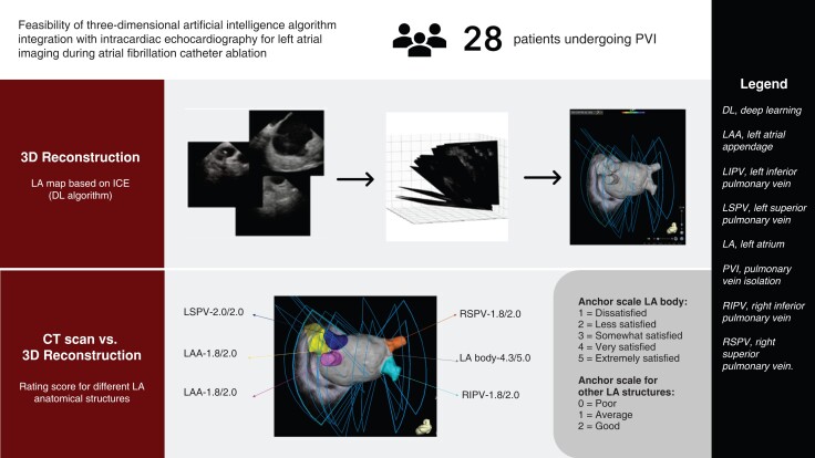
Aims: Intracardiac echocardiography (ICE) is a useful but operator-dependent tool for left atrial (LA) anatomical rendering during atrial fibrillation (AF) ablation. The CARTOSOUND FAM Module, a new deep learning (DL) imaging algorithm, has the potential to overcome this limitation. This study aims to evaluate feasibility of the algorithm compared to cardiac computed tomography (CT) in patients undergoing AF ablation.
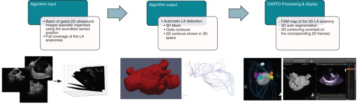
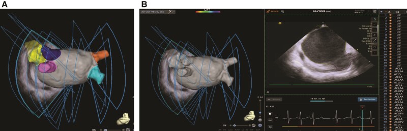
Methods and results: In 28 patients undergoing AF ablation, baseline patient information was recorded, and three-dimensional (3D) shells of LA body and anatomical structures [LA appendage/left superior pulmonary vein/left inferior pulmonary vein/right superior pulmonary vein/right inferior pulmonary vein (RIPV)] were reconstructed using the DL algorithm. The selected ultrasound frames were gated to end-expiration and max LA volume. Ostial diameters of these structures and carina-to-carina distance between left and right pulmonary veins were measured and compared with CT measurements. Anatomical accuracy of the DL algorithm was evaluated by three independent electrophysiologists using a three-anchor scale for LA anatomical structures and a five-anchor scale for LA body. Ablation-related characteristics were summarized. The algorithm generated 3D reconstruction of LA anatomies, and two-dimensional contours overlaid on ultrasound input frames. Average calculation time for LA reconstruction was 65 s. Mean ostial diameters and carina-to-carina distance were all comparable to CT without statistical significance. Ostial diameters and carina-to-carina distance also showed moderate to high correlation (r = 0.52-0.75) except for RIPV (r = 0.20). Qualitative ratings showed good agreement without between-rater differences. Average procedure time was 143.7 ± 43.7 min, with average radiofrequency time 31.6 ± 10.2 min. All patients achieved ablation success, and no immediate complications were observed.
Conclusion: DL algorithm integration with ICE demonstrated considerable accuracy compared to CT and qualitative physician assessment. The feasibility of ICE with this algorithm can potentially further streamline AF ablation workflow.
Keywords: Artificial intelligence; Atrial fibrillation; Catheter ablation; Deep learning; Intracardiac echocardiography.
© The Author(s) 2023. Published by Oxford University Press on behalf of the European Society of Cardiology.
Conflict of interest statement
Conflict of interest: L.D.B. is a consultant for Stereotaxis, Biosense Webster, Boston Scientific, Abbott Medical; has received speaker honoraria/travel from Medtronic, Atricure, Biotronik, Baylis Medical, and Zoll. G.H., D.H., R.A.M., J.A., G.C., and R.U. are employees of Biosense Webster, Inc. The remaining authors report no conflict of interest.
Figures
References
-
- Antolic B, Kajdic N, Vrbajnscak M, Jan M, Zizek D. Integrated 3D intracardiac ultrasound imaging with detailed pulmonary vein delineation guided fluoroless ablation of atrial fibrillation. Pacing Clin Electrophysiol 2021;44:1487–96. - PubMed
-
- Nishiyama T, Katsumata Y, Inagawa K, Kimura T, Nishiyama N, Fukumoto Ket al. . Visualization of the left atrial appendage by phased-array intracardiac echocardiography from the pulmonary artery in patients with atrial fibrillation. Europace 2015;17:546–51. - PubMed
-
- Romero J, Patel K, Briceno D, Alviz I, Tarantino N, Della Rocca DGet al. . Fluoroless atrial fibrillation catheter ablation: technique and clinical outcomes. Card Electrophysiol Clin 2020;12:233–45. - PubMed
-
- Saleh M, Coleman KM, Vaishnav AS, Shein J, Makker P, Skipitaris Net al. . Intracardiac echocardiography guided nonocclusive balloon cryothermal applications to achieve antral isolation during pulmonary vein isolation. J Interv Card Electrophysiol Nov 2021;62:329–36. - PubMed
MeSH terms
LinkOut - more resources
Full Text Sources
Other Literature Sources
Medical
