

Analysis of human invasive cytotrophoblasts demonstrates mosaic aneuploidy
- PMID: 37478076
- PMCID: PMC10361533
- DOI: 10.1371/journal.pone.0284317
Analysis of human invasive cytotrophoblasts demonstrates mosaic aneuploidy
Abstract
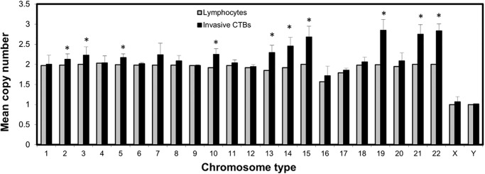
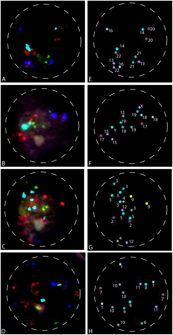
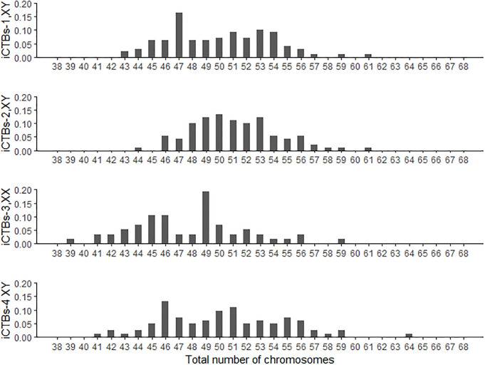
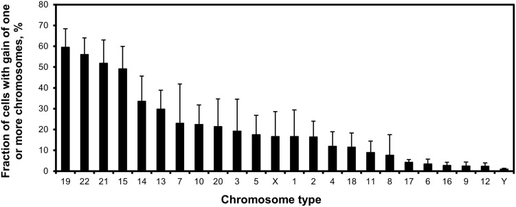
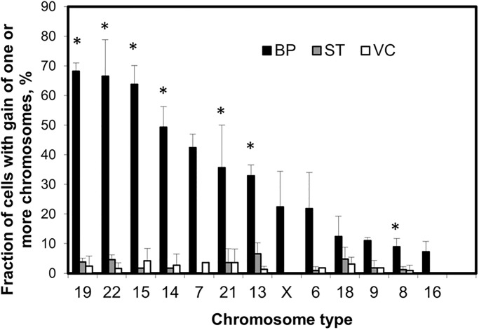
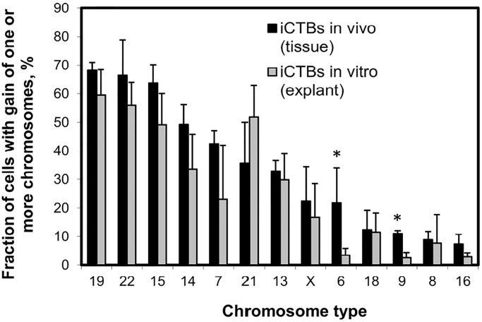
A total of 24 chromosome-specific fluorescence in situ hybridization probes for interphase nucleus analysis were developed to determine the chromosomal content of individual human invasive cytotrophoblasts derived from in vitro cultured assays. At least 75% of invasive cytotrophoblasts were hyperdiploid and the total number of chromosomes ranged from 47 to 61. The results also demonstrated that these hyperdiploid invasive cytotrophoblasts showed significant heterogeneity. The most copy number gains were observed for chromosomes 13, 14, 15, 19, 21, and 22 with average copy number greater than 2.3. A parallel study using primary invasive cytotrophoblasts also showed a similar trend of copy number changes. Conclusively, 24-chromosome analysis of human non-proliferating cytotrophoblasts (interphase nuclei) was achieved. Hyperdiploidy and chromosomal heterogeneity without endoduplication in invasive cytotrophoblasts may suggest a selective advantage for invasion and short lifespan during normal placental development.
Copyright: © 2023 Weier et al. This is an open access article distributed under the terms of the Creative Commons Attribution License, which permits unrestricted use, distribution, and reproduction in any medium, provided the original author and source are credited.
Conflict of interest statement
The authors have declared that no competing interests exist.
Figures
Similar articles
-
Human cytotrophoblasts acquire aneuploidies as they differentiate to an invasive phenotype.Dev Biol. 2005 Mar 15;279(2):420-32. doi: 10.1016/j.ydbio.2004.12.035. Dev Biol. 2005. PMID: 15733669
-
Analysis of human invasive cytotrophoblasts using multicolor fluorescence in situ hybridization.Methods. 2013 Dec 1;64(2):160-8. doi: 10.1016/j.ymeth.2013.05.021. Epub 2013 Jun 6. Methods. 2013. PMID: 23748112
-
Detection of hyperdiploidy and chromosome breakage in interphase human lymphocytes following exposure to the benzene metabolite hydroquinone using multicolor fluorescence in situ hybridization with DNA probes.Mutat Res. 1994 Jul;322(1):9-20. doi: 10.1016/0165-1218(94)90028-0. Mutat Res. 1994. PMID: 7517507
-
Advantages and limitations of using fluorescence in situ hybridization for the detection of aneuploidy in interphase human cells.Mutat Res. 1995 Dec;348(4):153-62. doi: 10.1016/0165-7992(95)90003-9. Mutat Res. 1995. PMID: 8544867 Review.
-
Multicolor detection of every chromosome as a means of detecting mosaicism and nuclear organization in human embryonic nuclei.Panminerva Med. 2016 Jun;58(2):175-90. Epub 2016 Mar 16. Panminerva Med. 2016. PMID: 26982524 Review.
Cited by
-
Chromosomal instability in human trophoblast stem cells and placentas.Nat Commun. 2025 Apr 25;16(1):3918. doi: 10.1038/s41467-025-59245-9. Nat Commun. 2025. PMID: 40280964 Free PMC article.
References
Publication types
MeSH terms
LinkOut - more resources
Full Text Sources
