Review
. 2023 Sep 5;35(9):1500-1518.
doi: 10.1016/j.cmet.2023.06.018. Epub 2023 Jul 20.

The pathogenic "symphony" in type 1 diabetes: A disorder of the immune system, β cells, and exocrine pancreas

Affiliations
Review

The pathogenic "symphony" in type 1 diabetes: A disorder of the immune system, β cells, and exocrine pancreas

Mark A Atkinson et al. Cell Metab. .

Abstract

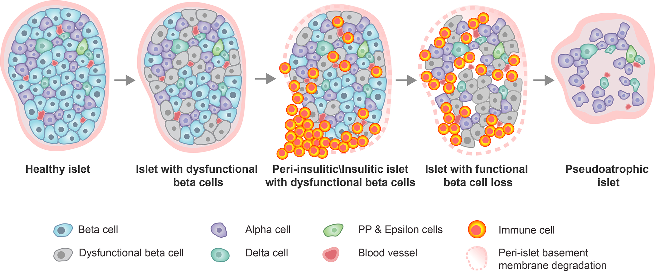
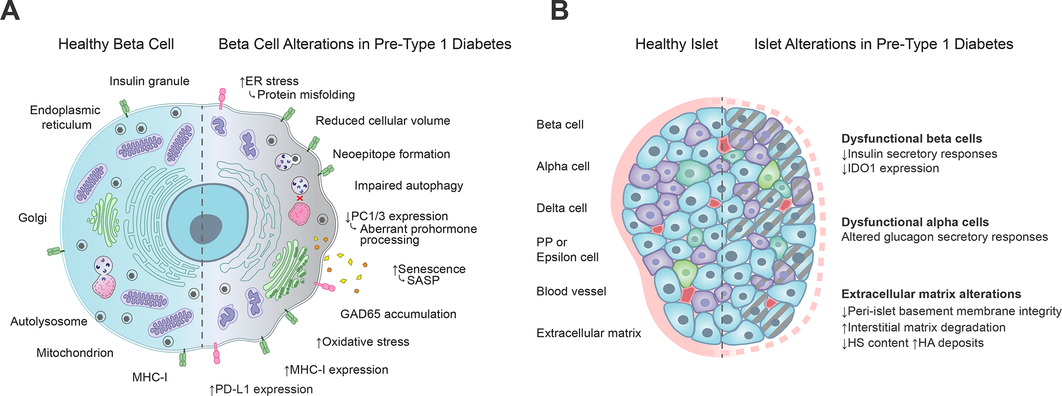
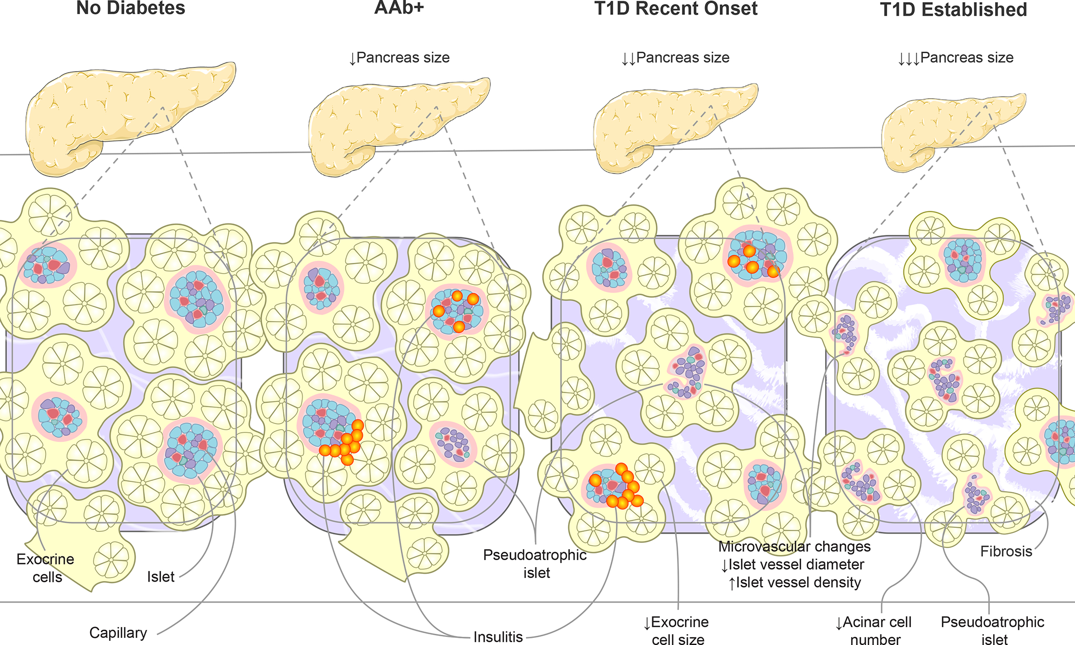
Type 1 diabetes (T1D) is widely considered to result from the autoimmune destruction of insulin-producing β cells. This concept has been a central tenet for decades of attempts seeking to decipher the disorder's pathogenesis and prevent/reverse the disease. Recently, this and many other disease-related notions have come under increasing question, particularly given knowledge gained from analyses of human T1D pancreas. Perhaps most crucial are findings suggesting that a collective of cellular constituents-immune, endocrine, and exocrine in origin-mechanistically coalesce to facilitate T1D. This review considers these emerging concepts, from basic science to clinical research, and identifies several key remaining knowledge voids.

Keywords: autoimmunity; endocrinology; exocrine pancreas; immune cells; inflammation; insulin; insulitis; islet of Langerhans; type 1 diabetes.


Conflict of interest statement

Declaration of interests The authors declare no competing interests.

Figures

Figure 1.
Figure 1.. A re-depiction of the current “staging” model for type 1 diabetes.
Following decades of natural history studies of type 1 diabetes (i.e., monitoring the period prior to and after traditional disease diagnosis), a consensus group within the community of type 1 diabetes developed a model whose intention was to better define risk for disease development. This model was largely developed based on analysis of both immunological (autoantibody type, number) and metabolic markers (response to glucose stimulation). Other considerations not strictly considered in this published model are those of having a relative with the disease, relative relationship, age, and more. Here we depict emerging concepts related to recent studies of pancreatic pathology to improve the staging of type 1 diabetes, as described herein. Adapted from Insel, et al. with insets from Figures 2–3.
Figure 2.
Figure 2.. Histopathologic progression of type 1 diabetes within the human pancreatic islet.
Within an initially healthy islet, a portion of β-cells develop phenotypic and functional impairments. This precipitates immune cell infiltration around (peri-insulitic) and within (insulitic) the islet, coupled with degradation of the peri-islet basement membrane. Following the immune-mediated destruction of functional β-cells, the immune cells exit leaving behind an irregularly shaped, insulin-negative, pseudoatrophic islet characterized by presence of the remaining endocrine cell types (alpha, delta, PP, epsilon), reduced vessel diameter, increased microvascular density, and restored peri-islet basement membrane integrity. PP, pancreatic polypeptide.
Figure 3.
Figure 3.. Alterations associated with type 1 diabetes at the β-cell and whole islet level.
(A) Functional and phenotypic changes within the pancreatic β-cell during type 1 diabetes development include protein misfolding reflective of ER stress, neoepitope formation, impairments in autophagy and prohormone processing, secretion of SASP molecules, accumulation of GAD65 in the Golgi apparatus, mitochondrial oxidative stress, and increased expression of MHC class I and PD-L1 molecules, which respectively serve opposing roles related to T cell activation through antigen presentation and co-inhibitory receptor stimulation (Figure 2). Straight arrows depict increases (up) or decreases (down) in type 1 diabetes as compared to healthy β-cells. Curved arrows depict the functional consequences of those changes. (B) Pancreatic islet alterations during type 1 diabetes development include loss of insulin secretion and IDO1 expression within dysfunctional β-cells, dysfunctional glucagon secretion from alpha cells, and degradation of extracellular matrix comprising the peri-islet basement membrane. Gray dashed lines depict cellular dysfunction. Pink dashed lines depict basement membrane degradation. ER, endoplasmic reticulum; PC1/3, proprotein convertase 1/3; SASP, senescence-associated secretory phenotype; GAD65, glutamic acid decarboxylase 65; MHC, major histocompatibility complex; PD-L1, programmed death ligand 1; IDO1, indoleamine 2, 3-dioxygenase 1; HS, heparan sulphate; HA, hyaluronic acid.
Figure 4.
Figure 4.. Alterations to pancreas size and organization during type 1 diabetes progression.
Disease progression, from AAb seroconversion (stage 1–2) to recent-onset and established type 1 diabetes (stage 3), is associated with increasingly smaller pancreas organ size. Insets show highly vascularized islets surrounded by exocrine cell bundles arranged in acini. Asynchronous islet infiltration and β-cell destruction result in a heterogeneous mixture of normal, insulitic, and pseudoatrophic islets in AAb+ and recent onset type 1 diabetes pancreas while the majority of islets are pseudoatrophic in longstanding type 1 diabetes. Insulin negative pseudoatrophic islets have distinct microvascular alterations including reduced vessel diameter and increase vessel density. Within the exocrine compartment, recent onset type 1 diabetes is associated with smaller exocrine cells, eventually leading to reduced acinar cell number in established disease. The exocrine pancreas is commonly characterized by periductal and interlobular fibrosis after type 1 diabetes onset. Arrows depict direction of change. AAb+, islet autoantibody positive; T1D, type 1 diabetes.

References

    1. Atkinson MA, Eisenbarth GS, and Michels AW (2014). Type 1 diabetes. Lancet 383, 69–82. 10.1016/S0140-6736(13)60591-7. - DOI - PMC - PubMed
    1. Eisenbarth GS (1986). Type I diabetes mellitus. A chronic autoimmune disease. N Engl J Med 314, 1360–1368. 10.1056/NEJM198605223142106. - DOI - PubMed
    1. Insel RA, Dunne JL, Atkinson MA, Chiang JL, Dabelea D, Gottlieb PA, Greenbaum CJ, Herold KC, Krischer JP, Lernmark A, et al. (2015). Staging presymptomatic type 1 diabetes: a scientific statement of JDRF, the Endocrine Society, and the American Diabetes Association. Diabetes Care 38, 1964–1974. 10.2337/dc15-1419. - DOI - PMC - PubMed
    1. Greenbaum C, Lord S, and VanBuecken D (2017). Emerging Concepts on Disease-Modifying Therapies in Type 1 Diabetes. Curr Diab Rep 17, 119. 10.1007/s11892-017-0932-x. - DOI - PubMed
    1. DiMeglio L, Evans-Molina C, and Oram R (2018). Type 1 diabetes. Lancet 391, 2449–2462. 10.1016/S0140-6736(18)31320-5. - DOI - PMC - PubMed

Publication types

LinkOut - more resources