. 2023 Aug 3;83(15):2753-2767.e10.
doi: 10.1016/j.molcel.2023.06.028. Epub 2023 Jul 20.

UBR5 forms ligand-dependent complexes on chromatin to regulate nuclear hormone receptor stability

Affiliations

UBR5 forms ligand-dependent complexes on chromatin to regulate nuclear hormone receptor stability

Jonathan M Tsai et al. Mol Cell. .

Abstract

Nuclear hormone receptors (NRs) are ligand-binding transcription factors that are widely targeted therapeutically. Agonist binding triggers NR activation and subsequent degradation by unknown ligand-dependent ubiquitin ligase machinery. NR degradation is critical for therapeutic efficacy in malignancies that are driven by retinoic acid and estrogen receptors. Here, we demonstrate the ubiquitin ligase UBR5 drives degradation of multiple agonist-bound NRs, including the retinoic acid receptor alpha (RARA), retinoid x receptor alpha (RXRA), glucocorticoid, estrogen, liver-X, progesterone, and vitamin D receptors. We present the high-resolution cryo-EMstructure of full-length human UBR5 and a negative stain model representing its interaction with RARA/RXRA. Agonist ligands induce sequential, mutually exclusive recruitment of nuclear coactivators (NCOAs) and UBR5 to chromatin to regulate transcriptional networks. Other pharmacological ligands such as selective estrogen receptor degraders (SERDs) degrade their receptors through differential recruitment of UBR5 or RNF111. We establish the UBR5 transcriptional regulatory hub as a common mediator and regulator of NR-induced transcription.

Keywords: HECT-E3 ligases; nuclear receptors; protein degradation; structural biology; ubiquitin ligases.


Conflict of interest statement

Declaration of interests B.L.E. has received research funding from Celgene, Deerfield, Novartis, and Calico and consulting fees from GRAIL. He is a member of the Scientific Advisory Board and shareholder for Neomorph Inc., TenSixteen Bio, Skyhawk Therapeutics, and Exo Therapeutics. A.S.S. received consulting fees from Adaptive Technologies. N.H.T. receives funding from the Novartis Research Foundation and is a member of the Scientific Advisory Board of Monte Rosa Therapeutics. K.A.D. is a consultant to Kronos Bio and Neomorph Inc. E.S.F. is a founder, Scientific Advisory Board (SAB) member, and equity holder of Civetta Therapeutics, Lighthorse Therapeutics, Proximity Therapeutics, and Neomorph, Inc. (also board of directors). B.L.E. and M.S. have received research funding from Calico Life Sciences LLC. E.S.F. is an equity holder and SAB member for Avilar Therapeutics and Photys Therapeutics and a consultant to Odyssey, Novartis, Sanofi, EcoR1 Capital, Ajax Therapeutics, and Deerfield. The Fischer lab receives or has received research funding from Deerfield, Novartis, Ajax, Interline, and Astellas.

Figures

Figure 1.
Figure 1.. CRISPR screens identify UBR5 as a principal ligand-dependent regulator of NR degradation
(A) Whole proteomics of NB4 cells treated with 10 mM ATRA for 24 h (n = 3 per condition). (B) Western blotting of PML-RARA, RARA, and HDAC3 in NB4 cells following treatment with ATRA and proteasome inhibitor (MG132). (C) Volcano plots of targeted CRISPR screens highlighting enrichment of UBR5 following ligand treatment of indicated NR fluorescent reporter U937 cell lines. UBR5 is highlighted in red. (D) Western blotting of NB4 cells transduced with shRNAs against luciferase (shCTRL) or UBR5 treated with or without ATRA. (E) Viability by relative cell counts of NB4 cells transduced with control or UBR5 shRNAs (two shRNAs per condition, combined) following treatment with 1 μM ATRA for 24 h. (n = 4, two-sided t test). (F) Bioluminescence resonance energy transfer (BRET) assay of RARA and UBR5, treated ± 9-cis RA. (n = 3 two-sided t test) (G) Western blot showing in vitro FLAG-UBR5 pull-down of purified His-tagged RARA or RARA/RXRA heterodimer in the presence of 50 μM specified retinoid agonist or antagonist. (NS, not significant, *p < 0.05, **p < 0.005, ***p < 0.0005, ****p < 0.00005, error bars represent SEM.)
Figure 2.
Figure 2.. UBR5 competes with nuclear coactivators for the conserved hydrophobic cleft of NRs
(A) Schematic of RARA domain architecture; NTD, N-terminal domain; DBD, DNA-binding domain; LBD, ligand-binding domain. (B) Degradation of RARA reporters containing 4–5 surface-exposed alanine mutations per helix, relative to untreated (DMSO) (n = 3 two-sided t test). See Figure S2 for detailed amino acid substitutions. (C) Degradation of RARA reporters containing glutamate-substituted residues within the H3/H4 hydrophobic cleft. (n = 3 two-sided t test). (D) Western blot showing in vitro FLAG-UBR5 pull-down of purified His-tagged RARA/RXRA heterodimer, with or without single residue substitutions in the hydrophobic cleft and with or without 50 μM ATRA. (E) Competitive nature of RARA binding to UBR5 and NCOA1 demonstrated by UBR5 pull-down with purified proteins. FLAG-UBR5 (15 pmol) was pre-bound to a large excess of His-RARA in the presence of ATRA and excess RARA protein washed away. Increasing amounts of full-length NCOA1 protein were then added to outcompete the UBR5-RARA interaction. NCOA was titrated with a 0.5, 5, and 10× molar excess relative to UBR5. (F) Relative abundances in RARA-GFP IP-MS of NCOR1, NCOA1, and UBR5 normalized to RARA abundance following 0, 4, and 16 h of ATRA treatment. (NS, not significant, *p < 0.05, **p < 0.005, ***p < 0.0005, ****p < 0.00005, error bars are SEM.)
Figure 3.
Figure 3.. Structural and functional characterization of UBR5
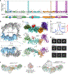
(A) UBR5 tiling screen of RARA-GFP reporter degradation with domain map shown beneath according to residue numbers. Highlighted residues represent guides with over 10-fold enrichment. (B) Cryo-EM density map of UBR5 tetramer, with one monomeric unit colored in light blue, and shown rotated by 90° beneath. (C) Expanded view of the dashed box outlined in (B), showing a surface map at the higher resolution obtained in the dimeric UBR5 map, with domains colored according to the domain architecture above. A cartoon representation of the UBR5 model in the same orientation is shown below. The tandem SBB structure predicted by AlphaFold2 is included for completeness in light gray. (D) Size-exclusion multiple-angle light scattering (SEC-MALS) of full-length and Δtandem UBR5 variant, showing how removal of the tandem SBBs changes the assembly from tetramer to dimer. (E) 2D-class averages for indicated UBR5 variants. Deletion of residues comprising the UBA insertion led to a loss of density in the middle of the ring, whereas deletion of residues comprising the tandem domain led to a dimeric species of UBR5 with altered preferred orientation. Each class represents ~3,000 unique particles. (F) EM density maps of indicated complexes obtained by negative stain EM, limited by resolution to >20 Å. A simulated density map of RARA/RXRA generated by molmap (ChimeraX) and low-pass filtered to 25 Å is shown in center as a reference for the expected additional density size.
Figure 4.
Figure 4.. UBR5 binds RARA on chromatin and regulates transcription
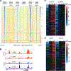
(A) Tornado plots of chromatin immunoprecipitation sequencing (ChIP-seq) targeting RARA or UBR5 with or without ATRA in NB4 cells, clustered by change following ATRA treatment. Plots are centered around peak center and represent log2 fold change over input (n = 2). (B) ChIP-seq track of RARA (red) and UBR5 (blue) peaks with or without 24 h ATRA treatment. (C) RNA sequencing of NB4 cells transduced with shRNAs against luciferase or UBR5, treated with ATRA for 0, 8, 24, and 48 h. Genes were ordered by increasing expression level changes at 24 h following ATRA treatment in shCTRLs with top 200 genes displayed. Heatmap represents centered log2 scale expression (n = 2). (D) RNA sequencing of NB4 cells treated with MG132 or DMSO for 0, 8, and 24 h following ATRA treatment. Genes were ordered by increasing expression level changes at 24 h following ATRA treatment in shCTRLs with top 200 genes displayed. Heatmap represents centered log2 scale expression (n = 2).
Figure 5.
Figure 5.. UBR5 regulates a greater subset of NRs through a common degron
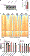
(A) Western blots of A549 cells transduced with shRNAs against luciferase or UBR5 treated ± dexamethasone. (B) Reporter degradation of WT or hydrophobic cleft mutant GFP-GR reporter (n = 3 two-sided t test). (C) Tornado plots of chromatin immunoprecipitation sequencing (ChIP-seq) targeting GR or UBR5 ± dexamethasone in A549 cells, clustered by change following dexamethasone treatment. Plots are centered around peak center and represent log2 fold change over input. (D) Degradation of indicated fluorescent reporter cell lines transduced with shRNAs against luciferase or UBR5 following indicated ligand treatment (ligands: R5020, calcitriol, and levothyroxine [T3]). (n = 3 two-sided t test). (E) Degradation of fluorescent androgen receptor (AR) reporters with substitutions to residues in the hydrophobic cleft swapped to match those residues found in RXRA, GR, RARA, and ER treated with CI-4AS-1. (n = 3 two-sided t test) (NS, not significant, *p < 0.05, **p < 0.005, ***p < 0.0005, ****p < 0.00005), error bars are SEM.
Figure 6.
Figure 6.. Non-endogenous ligands recruit different E3 ligases to induce ER degradation
(A) Western blot showing in vitro FLAG-UBR5 pull-down of purified His-tagged ER in the presence of 50 μM indicated ER ligand. (B) ER reporter degradation following 1 μM of specified SERD treatment. (C) Volcano plots of targeted CRISPR screens highlighting enrichment of UBR5 or RNF111 following SERD treatment of ER fluorescent reporter K562 cell lines. UBR5 is highlighted in blue, RNF111, CRBN, VHL are highlighted in red. (D) Western blots of T47D cells transduced with sgRNAs against RNF111 treated with or without fulvestrant. (E) Co-immunoprecipitations of 293Ts co-transfected with FLAG-RNF111 and ER-GFP treated ± fulvestrant and proteasome inhibitor (MG132) and blotted with antibodies specific for FLAG or GFP as indicated. (NS, not significant, *p < 0.05, **p < 0.005, ***p < 0.0005, ****p < 0.00005, error bars represent SEM.)

Comment in

References

    1. Ismail A, and Nawaz Z (2005). Nuclear hormone receptor degradation and gene transcription: an update. IUBMB Life 57, 483–490. 10.1080/15216540500147163. - DOI - PubMed
    1. Wang L, Oh TG, Magida J, Estepa G, Obayomi SMB, Chong LW, Gatchalian J, Yu RT, Atkins AR, Hargreaves D, et al. (2021). Bromodomain containing 9 (BRD9) regulates macrophage inflammatory responses by potentiating glucocorticoid receptor activity. Proc. Natl. Acad. Sci. USA 118, e2109517118. 10.1073/pnas.2109517118. - DOI - PMC - PubMed
    1. Fu T, Coulter S, Yoshihara E, Oh TG, Fang S, Cayabyab F, Zhu Q, Zhang T, Leblanc M, Liu S, et al. (2019). FXR regulates intestinal cancer stem cell proliferation. Cell 176, 1098–1112.e18. 10.1016/j.cell.2019.01.036. - DOI - PMC - PubMed
    1. Ding N, Yu RT, Subramaniam N, Sherman MH, Wilson C, Rao R, Leblanc M, Coulter S, He M, Scott C, et al. (2013). A vitamin D receptor/SMAD genomic circuit gates hepatic fibrotic response. Cell 153, 601–613. 10.1016/j.cell.2013.03.028. - DOI - PMC - PubMed
    1. Huang P, Chandra V, and Rastinejad F (2010). Structural overview of the nuclear receptor superfamily: insights into physiology and therapeutics. Annu. Rev. Physiol. 72, 247–272. 10.1146/annurev-physiol-021909-135917. - DOI - PMC - PubMed

Publication types