. 2023 Aug 9;31(8):1317-1330.e10.
doi: 10.1016/j.chom.2023.06.013. Epub 2023 Jul 20.

Acetylation of the NS3 helicase by KAT5γ is essential for flavivirus replication

Affiliations

Acetylation of the NS3 helicase by KAT5γ is essential for flavivirus replication

Taryn Serman et al. Cell Host Microbe. .

Abstract

Direct targeting of essential viral enzymes such as proteases, polymerases, and helicases has long been the major focus of antiviral drug design. Although successful for some viral enzymes, targeting viral helicases is notoriously difficult to achieve, demanding alternative strategies. Here, we show that the NS3 helicase of Zika virus (ZIKV) undergoes acetylation in its RNA-binding tunnel. Regulation of the acetylated state of K389 in ZIKV NS3 modulates RNA binding and unwinding and is required for efficient viral replication. NS3 acetylation is mediated by a specific isoform of the host acetyltransferase KAT5 (KAT5γ), which translocates from the nucleus to viral replication complexes upon infection. NS3 acetylation by KAT5γ and its proviral role are also conserved in West Nile virus (WNV), dengue virus (DENV), and yellow fever virus (YFV). Our study provides molecular insight into how a cellular acetyltransferase regulates viral helicase functions, unveiling a previously unknown target for antiviral drug development.

Keywords: NS3 protein; Zika virus; acetylation; flaviviruses; post-translational modifications; viral helicase.


Conflict of interest statement

Declaration of interests The authors declare no competing interests.

Figures
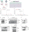
Figure 1.
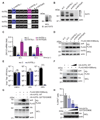
Figure 1.. ZIKV NS3 is robustly acetylated at K389
(A) Schematic representation of the two complementary mass spectrometry (MS) approaches that identified acetylation of the ZIKV NS3 protein. These are MS analyses of (1) FLAG-tagged ZIKV NS3 affinity-purified from HEK293T cells that were transfected for 40 h to express this construct and then treated with IFN-beta for 8 h (left) and (2) anti-pan-acetyl-lysine (anti-acK) immuno-enriched whole-cell lysates (WCLs) of SVGA (astrocytic) cells that were infected for 48 h with ZIKV (strain BRA/2015, MOI 1) (right). (B) ZIKV NS3 domain organization and amino acid sequence. Black lysine residues (bold) indicate the acetylated residues identified by MS analysis of purified FLAG-NS3. Red lysine residues (bold) indicate acetylation sites identified by both MS approaches. (C and D) Tandem mass spectra of the tryptic peptides acK389TFETEFQK (C) and CLacK431PVILDGER (D) of affinity-purified ZIKV NS3 that identified acetylation at K389 and K431, respectively. b- and y-ion designations are shown. (E) Acetylation of FLAG-tagged ZIKV NS3 WT or 6K→R mutant in HEK293T cells that were transfected for 40 h to express these constructs, determined by anti-FLAG pull-down (PD:FLAG) and immunoblot (IB) with anti-acK. WCLs were probed by IB with anti-acK and anti-actin (loading control). (F) Acetylation of FLAG-tagged ZIKV NS3 WT and the indicated single K→R mutants in transiently transfected HEK293T cells, determined as in (E). (G) Acetylation of ZIKV NS3 in SVGA cells that were infected with ZIKV (strain BRA/2015, MOI 0.5) for 60 h, determined by immunoprecipitation (IP) using anti-NS3(ZIKV) and IB with anti-acK. (H) Acetylation of ZIKV NS3 in SVGA cells that were infected with ZIKV (strain BRA/2015, MOI 1) for the indicated times, determined as in (G). (I) Acetylation of GST-fused ZIKV NS3 proteins (strains MR766, H/PF/13, and BRA/2015) in transiently transfected HEK293T cells, determined by GST pull-down (PD:GST) and IB with anti-acK. (J) Acetylation of ZIKV NS3 in SVGA cells that were infected as in (G), assessed by IP with anti-NS3(ZIKV) and IB with an acetyl-K389-NS3(ZIKV)-specific antibody (acK389(NS3)). WCLs were probed by IB with anti-NS3 and anti-actin. Data shown are from two complementary MS screens (B–D) or data are representative of at least two independent experiments (E–J). See also Figure S1.
Figure 2.
Figure 2.. K389 acetylation regulates NS3 RNA binding and unwinding activities and is critical for ZIKV replication
(A) Ribbon representation of the crystal structure of ZIKV NS3 helicase (green) bound to a synthetic ssRNA substrate (orange/blue) (PDB: 5GJB). K389 is indicated in red. (B) Schematic of the experimental approach to examine the in vitro RNA-binding capacity of ZIKV NS3 WT and mutant proteins purified from HEK 293T cells. The in vitro-transcribed synthetic ZIKV RNA substrate “Bio-RNA(ZIKV)” used in this assay comprises the ZIKV 5′UTR, the first 66 nucleotides of the Capsid protein (C66), and the ZIKV 3′UTR. SA, Streptavidin. See also Methods. (C) In vitro RNA-binding ability of purified ZIKV NS3 WT and indicated mutants, determined by Streptavidin pulldown (PD: SA) of Bio-RNA(ZIKV) and IB with anti-NS3(ZIKV). Protein input was determined by IB with anti-NS3(ZIKV). (D) Fluorescence resonance energy transfer (FRET)-based molecular beacon helicase assay measuring real-time unwinding kinetics of Cy3-labeled RNA bound to a Black Hole Quencher (BHQ)-labeled complementary RNA by ZIKV NS3 WT or mutants purified from HEK293T cells. The fluorescent signal was measured every minute and is presented as (Ft-F0)/F0. Ft, fluorescence signal at the indicated time; F0, fluorescence signal at t = 0. (E) qRT-PCR analysis of ZIKV E transcripts in SVGA cells that were transfected with infectious cDNA of ZIKV(WT), ZIKV(K389R), or ZIKV(K389Q) (strain Paraiba_01/2015) for the indicated times. (F) Viral titers in the supernatant of SVGA cells transfected as in (E), determined at the indicated times by plaque assay and presented as PFU/mL. (G) Viral titers across three passages at a fixed MOI (MOI 0.1) in Vero cells that were transfected with infectious cDNA of ZIKV(WT), ZIKV(K389R), or ZIKV(K389Q) (strain Paraiba_01/2015), determined by plaque assay at 72 h.p.i. for each passage. Data are representative of at least two independent experiments (mean ± s.d. of n = 3 biological replicates (E–G)). *p < 0.05, **p < 0.01, ***p < 0.001, ****p < 0.0001 (Student’s t test). ns, not significant. See also Figures S2, S3, and S4.
Figure 3.
Figure 3.. ZIKV NS3 is acetylated by KAT5
(A) qRT-PCR analysis of ZIKV E transcripts in A549 cells that were transfected for 30 h with either non-targeting control siRNA (si.C) or specific siRNAs targeting the indicated human acetyltransferases, and then infected with ZIKV (BRA/2015, MOI 0.001) for 48 h. (B) NS3 acetylation in HEK293T cells that were transfected for 16 h with either si.C or specific siRNAs targeting the indicated genes and then co-transfected with FLAG-NS3 K389only (ZIKV) for 30 h, assessed by PD:FLAG and IB with anti-acK. WCLs were probed by IB with anti-acK and anti-actin (loading control). Knockdown efficiency of the silenced genes was determined in parallel by qRT-PCR (see Figure S5A). (C) Binding of endogenous KAT5 or KAT3B (negative control) and FLAG-tagged ZIKV NS3 in HEK293T cells that were transfected for 40 h with either empty vector or FLAG-NS3 (ZIKV), determined by PD:FLAG and IB with anti-KAT5 or anti-KAT3B: WCLs were probed with the indicated antibodies. (D) Binding of endogenous KAT5 and ZIKV NS3 in A549 cells infected with ZIKV (BRA/2015, MOI 1) for 48 h, assessed by IP with anti-NS3(ZIKV) and IB with anti-KAT5. Asterisk indicates nonspecific band. (E) ZIKV titers in the supernatant of Vero cells that were transfected for 30 h with either si.C or KAT5-specific siRNA (si.KAT5) and then infected with ZIKV (BRA/2015, MOI 0.001) for 48 h, determined by plaque assay. (F) ZIKV titers in the supernatant of HMC3 (microglial) cells that were transfected for 30 h with either si.C or si.KAT5 and then infected with ZIKV (BRA/2015, MOI 0.001) for the indicated times, determined by plaque assay and presented as PFU/mL. (G) ZIKV E transcripts in HMC3 (microglial) cells that were transfected and infected as in (F), determined by qRT-PCR. (H) Representative knockdown efficiency of KAT5 for the experiments in (F and G), determined by qRT-PCR. (I) ZIKV 5′UTR transcripts in A549 cells that were transfected for 30 h with either si.C, si.KAT5, or TMEM41B-specific siRNA (si.TMEM41B) and then infected with ZIKV (BRA/2015, MOI 0.001) for the indicated times, determined by qRT-PCR. (J) Representative knockdown efficiency of the indicated genes for the experiment in (I). Data are representative of at least two independent experiments (mean ± s.d. of n = 3 biological replicates (A, E–J)). *p < 0.05, **p < 0.01, ***p < 0.001, ****p < 0.0001 (Student’s t test). ns, not significant. See also Figure S5.
Figure 4.
Figure 4.. The specific isoform KAT5γ controls ZIKV NS3 acetylation and replication
(A) Left: Schematic representation of the KAT5 gene organization as well as the strategy to silence the four KAT5 splice isoforms in human cells. Right: Representative IB showing the individual KAT5 isoforms (L, α, β and γ) in A549 cells, determined in the WCLs with anti-KAT5. (B) Representative knockdown efficiency of the individual KAT5 isoforms in A549 cells that were transfected for 48 h with either si.C or specific siRNAs targeting the indicated KAT5 isoforms (as depicted in A), determined in the WCL by IB with anti-KAT5. (C) qRT-PCR analysis of ZIKV E transcripts in A549 cells that were transfected for 16 h with the indicated siRNAs and then infected with ZIKV (BRA/2015, MOI 0.001) for the indicated times. (D) Acetylation of FLAG-NS3 K389only (ZIKV) in transiently transfected HEK293T cells that were co-transfected with the indicated siRNAs, determined by PD:FLAG and IB with anti-acK. (E) EMCV, VSV and ZIKV titers in the supernatants of A549 cells that were transfected for 30 h with either si.C or specific siRNA targeting the KAT5 intron (si.KAT5L/γ) and then infected with EMCV (MOI 0.1) for 12 h, VSV (MOI 0.001) for 24 h, or ZIKV (BRA/2015, MOI 0.001) for 48 h, determined by plaque assay and presented as PFU/mL. (F) Acetylation of FLAG-NS3 K389only (ZIKV) in HEK293T cells that were co-transfected for 40 h with either empty vector (–) or increasing amounts of HA-tagged KAT5γ WT, determined by PD:FLAG and IB with anti-acK. (G) Acetylation of FLAG-NS3 K389only (ZIKV) in A549 cells that were co-transfected for 40 h with either empty vector (–) or HA-tagged KAT5γ WT or its dominant-negative mutant (Q377E/G380E), determined by PD:FLAG and IB with anti-acK. (H) Frequency of ZIKV-positive A549 cells that were transfected for 24 h with either empty vector (–) or increasing amounts of HA-tagged KAT5γ Q377E/G380E and then infected with ZIKV (BRA/2015, MOI 0.001) for 48 h, determined by flow cytometry. Representative expression of HA-KAT5γ Q377E/G380E was determined in the WCL by IB with anti-HA. Data are representative of at least two independent experiments (mean ± s.d. of n = 3 biological replicates (C, E and H). *p < 0.05, ***p < 0.001, ****p < 0.0001 (Student’s t test). ns, not significant.
Figure 5.
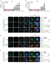
Figure 5.. KAT5γ relocalizes from the nucleus to ZIKV replication complexes
(A and B) KAT5 and ZIKV E transcripts in A549 (A) and SVGA (B) cells that were infected with ZIKV (strain BRA/2015, MOI 0.5) for the indicated times, determined by qRT-PCR. (C) Left: Colocalization of KAT5γ with ZIKV NS3 in A549 cells depleted of all endogenous KAT5 isoforms and stably reconstituted with KAT5γ, assessed by confocal laser scanning microscopy and immunostaining with anti-KAT5 (green) and anti-NS3 (red) at 40 h after ZIKV (strain BRA/2015) infection or mock treatment. Nuclei, DAPI (blue). Scale bar, 10 μm. Numbers (top corner) indicate the percentage of cells showing the respective KAT5γ localization phenotype. Right: Graphs showing relative signal intensity for KAT5γ, NS3, and DAPI along the indicated white lines (Inset). (D) Left: Colocalization of KAT5γ with ZIKV NS4B in A549 cells that were depleted of all endogenous KAT5 isoforms and stably reconstituted with KAT5γ, assessed by confocal laser scanning microscopy and immunostaining with anti-KAT5 (green) and anti-NS4B (red) at 40 h after ZIKV infection or mock treatment. Nuclei, DAPI (blue). Scale bar, 10 μm. Right: Graphs showing relative signal intensity for KAT5γ, NS4B, and DAPI along the indicated white lines (Inset). (E) Left: Colocalization of KAT5γ with dsRNA in A549 cells that were depleted of all endogenous KAT5 isoforms and stably reconstituted with KAT5γ, assessed by confocal laser scanning microscopy and immunostaining with anti-KAT5 (green) and anti-dsRNA (red) at 40 h after ZIKV infection or mock treatment. Nuclei, DAPI (blue). Scale bar, 10 μm. Right: Graphs showing relative signal intensity for KAT5γ, dsRNA and DAPI along the indicated white lines (inset). Data are representative of at least two independent experiments (mean ± s.d. of n = 3 biological replicates (A and B)). See also Figure S6.
Figure 6.
Figure 6.. NS3 acetylation is conserved also for WNV, DENV and YFV
(A) Acetylation of GST-fused NS3 from ZIKV, WNV, DENV, and YFV, or of GST-MDA5-2CARD (GST-MDA5-2C, negative control) in HEK293T cells that were transfected for 40 h to express these constructs, determined by PD:GST and IB with three different anti-acK antibodies (#1, Cell Signaling Technology #9441; #2, Immune Chem #ICP0380; #3, Cell Signaling Technology #9814). (B and C) Acetylation of FLAG-tagged NS3 WT or K389R mutant from WNV (B) or DENV and YFV (C) in transiently transfected HEK293T cells, determined by PD:FLAG and IB with anti-acK. WCLs were probed with the indicated antibodies. (D) Acetylation of WNV NS3 in A549 cells that were infected with WNV (strain NY99, MOI 0.5) for 40 h, determined by IP with anti-acK and IB with anti-NS3. WCLs were probed with the indicated antibodies. (E) Acetylation of DENV NS3 in A549 cells that were infected with DENV (strain 16681, MOI 1) for 30 h, determined as in (D). (F) Acetylation of YFV NS3 in Huh7 cells that were infected with YFV (strain 17D, MOI 0.1) for 72 h, determined as in (D). Data are representative of at least two independent experiments. See also Figure S7.
Figure 7.
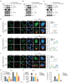
Figure 7.. NS3 acetylation by KAT5γ and its proviral role in WNV, DENV and YFV infection
(A–C) Acetylation of FLAG-tagged WNV NS3 (A), DENV NS3 (B), and YFV NS3 (C) in HEK293T cells that were co-transfected with either empty vector (–) or HA-tagged KAT5γ dominant-negative mutant (Q377E/G380E) for 40 h, determined by PD:FLAG and IB with anti-acK. WCLs were probed with the indicated antibodies. (D–F) Left: Colocalization of KAT5γ with NS3 in A549 cells that were depleted of all endogenous KAT5 isoforms and stably reconstituted with KAT5γ, assessed by confocal microscopy and immunostaining with anti-KAT5 (green) and anti-NS3 (red) at 40 h after WNV (strain NY99) (D), DENV (strain 16681) (E), or YFV (strain 17D) (F) infection, or mock treatment (D–F). Nuclei, DAPI (blue). Scale bars, 10 μm. Numbers (top corner) indicate the percentage of cells showing the respective KAT5γ localization phenotype. Right: Graphs showing relative signal intensity for KAT5γ, NS3, and DAPI along the indicated white lines (Inset) (G) Viral titers in the supernatant of A549 cells that were transfected for 30 h with the indicated siRNAs and then infected with WNV (strain NY99, MOI 0.001) for the indicated times, determined by plaque assay and presented as PFU/mL. (H) DENV NS5 transcripts in A549 cells that were transfected as in (G) and then infected with DENV (strain 16681, MOI 0.001) for 48 h, determined by qRT-PCR. (I) YFV 5UTR transcripts in A549 cells that were transfected as in (G) and then infected with YFV (strain 17D, MOI 0.1) for 48 h, determined by qRT-PCR. Data are representative of at least two independent experiments (mean ± s.d. of n = 3 biological replicates (G–I). *p < 0.05, **<0.01, ****p < 0.0001 (Student’s t test). ns, not significant.

References

    1. Pierson TC, and Diamond MS (2020). The continued threat of emerging flaviviruses. Nat Microbiol 5, 796–812. 10.1038/s41564-020-0714-0. - DOI - PMC - PubMed
    1. Bhatt S, Gething PW, Brady OJ, Messina JP, Farlow AW, Moyes CL, Drake JM, Brownstein JS, Hoen AG, Sankoh O, et al. (2013). The global distribution and burden of dengue. Nature 496, 504–507. 10.1038/nature12060. - DOI - PMC - PubMed
    1. Brasil P, Sequeira PC, Freitas AD, Zogbi HE, Calvet GA, de Souza RV, Siqueira AM, de Mendonca MC, Nogueira RM, de Filippis AM, and Solomon T. (2016). Guillain-Barre syndrome associated with Zika virus infection. Lancet 387, 1482. 10.1016/S0140-6736(16)30058-7. - DOI - PubMed
    1. Maxmen A. (2012). The hidden threat of West Nile virus. Nature 489, 349–350. 10.1038/489349a. - DOI - PubMed
    1. Baharuddin A, Hassan AA, Sheng GC, Nasir SB, Othman S, Yusof R, Othman R, and Rahman NA (2014). Current approaches in antiviral drug discovery against the Flaviviridae family. Curr Pharm Des 20, 3428–3444. 10.2174/13816128113199990635. - DOI - PubMed

LinkOut - more resources