. 2023 Aug 17;30(8):965-975.e6.
doi: 10.1016/j.chembiol.2023.06.018. Epub 2023 Jul 20.

S-Nitrosylation-mediated dysfunction of TCA cycle enzymes in synucleinopathy studied in postmortem human brains and hiPSC-derived neurons

Affiliations

S-Nitrosylation-mediated dysfunction of TCA cycle enzymes in synucleinopathy studied in postmortem human brains and hiPSC-derived neurons

Paschalis-Thomas Doulias et al. Cell Chem Biol. .

Abstract

A causal relationship between mitochondrial metabolic dysfunction and neurodegeneration has been implicated in synucleinopathies, including Parkinson disease (PD) and Lewy body dementia (LBD), but underlying mechanisms are not fully understood. Here, using human induced pluripotent stem cell (hiPSC)-derived neurons with mutation in the gene encoding α-synuclein (αSyn), we report the presence of aberrantly S-nitrosylated proteins, including tricarboxylic acid (TCA) cycle enzymes, resulting in activity inhibition assessed by carbon-labeled metabolic flux experiments. This inhibition principally affects α-ketoglutarate dehydrogenase/succinyl coenzyme-A synthetase, metabolizing α-ketoglutarate to succinate. Notably, human LBD brain manifests a similar pattern of aberrantly S-nitrosylated TCA enzymes, indicating the pathophysiological relevance of these results. Inhibition of mitochondrial energy metabolism in neurons is known to compromise dendritic length and synaptic integrity, eventually leading to neuronal cell death. Our evidence indicates that aberrant S-nitrosylation of TCA cycle enzymes contributes to this bioenergetic failure.


Conflict of interest statement

Declaration of interests The authors declare no competing interests.

Figures
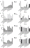
Figure 1.
Figure 1.. Metabolic flux analysis of TCA cycle enzymes
PD A53T-hiN vs. isogenic WT/Control hiN incubated with C13-lactate. Left-hand panels show individual substrates and products; right-hand panels show ratio. (A) Substrates and products for AC/ IDH enzyme reactions. (B-D) Substrates and products for αKGDH enzyme reaction (panel B), for SDH (C), and for MDH (D). Values are mean + SEM, n ≥ 3 experiments. As seen in the A53T PD hiPSC-derived neurons exposed to PQ/MB, but not in isogenic gene-corrected controls, there is a block at the level of αKGDH which is reversed by l-NAME. Abbreviations: cit, citrate; αKG, α-ketoglutarate; ME, molar percent enrichment; MER ratio of substrate/product molar enrichment; succ, succinate, fum, fumarate; mal, malate; OAA, oxaloacetate. Statistical significance, tested by ANOVA followed by a PLSD post hoc test, is presented in the text. See also Figures S1–S3.
Figure 2.
Figure 2.. Schema showing effects of S-nitrosylation of TCA cycle enzymes in isogenic WT and A53T mutant αSYN hiN
A53T-hiN display basal partial inhibition of the TCA cycle at the AC/IDH steps. This is consistent with data that both of these enzymes are S-nitrosylated in A53T-hiN, particularly in the presence of PQ/MB. S-Nitrosylation of αKGDH results in more major enzyme inhibition, as evidenced by reversal by the NOS inhibitor l-NAME, and hence significant kinetic inhibition of flux through the TCA cycle at that point in the cycle. Note that the action of αKGDH also supplies NADH to the ETC for production of ATP. We have previously performed activity assays showing that S-nitrosylation of AC, IDH, αKGDH and other TCA enzymes can inhibit their activity. At the level of MDH and PDH, which were also S-nitrosylated under A53T-PQ/MB conditions, a lesser degree of inhibition was observed. The fold difference in flux rate constants obtained by kinetic modeling (see Methods) in the absence and presence of l-NAME to show enzymes impacted by S-nitrosylation, is shown in parenthesis (with 4x representing a 4-fold increase and 1x representing no significant change).
Figure 3.
Figure 3.. SNO-proteins found under PD conditions (A53T-PQ/MB) in hiN vs. corrected controls
(A) Venn diagram of SNO-proteins under A53T-PQ/MB conditions vs. corrected control. See also Table S1. (B) GO-term analysis of the differences yielded these highest scoring pathways.
Figure 4.
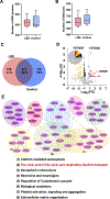
Figure 4.. S-Nitrosoproteome and reactome analysis of affected pathways in LBD vs. control human brains
(A and B) Box and whisker plot of number of SNO-proteins (A) and SNO-sites (B) from each postmortem human brain (n = 6 LBD and 5 controls). Box-and-whiskers plots show the median, interquartile range, and minimum and maximum of SNO-proteins and SNO-peptides found per each group. See also Tables S2–S4. (C) Venn diagram showing the number of SNO-proteins in LBD brains, control brains, and their overlap. (D) Volcano plot showing distribution of upregulated and downregulated SNO-proteins in human LBD brain compared to controls. The predominantly upregulated TCA enzyme, SNO-αKGDH is indicated. (E) Reactome analysis of pathways affected by aberrant protein S-nitrosylation in human LBD brains over controls. Data were analyzed with STRING software.

References

    1. Exner N, Lutz AK, Haass C, and Winklhofer KF (2012). Mitochondrial dysfunction in Parkinson’s disease: molecular mechanisms and pathophysiological consequences. EMBO J. 31, 3038–3062. 10.1038/emboj.2012.170. - DOI - PMC - PubMed
    1. Vazquez-Velez GE, and Zoghbi HY (2021). Parkinson’s disease genetics and pathophysiology. Annu. Rev. Neurosci. 44, 87–108. 10.1146/annurev-neuro-100720-034518. - DOI - PubMed
    1. Cooper O, Seo H, Andrabi S, Guardia-Laguarta C, Graziotto J, Sundberg M, McLean JR, Carrillo-Reid L, Xie Z, Osborn T, et al. (2012). Pharmacological rescue of mitochondrial deficits in iPSC-derived neural cells from patients with familial Parkinson’s disease. Sci. Transl. Med. 4, 141ra190. 10.1126/scitranslmed.3003985. - DOI - PMC - PubMed
    1. Chung CY, Khurana V, Auluck PK, Tardiff DF, Mazzulli JR, Soldner F, Baru V, Lou Y, Freyzon Y, Cho S, et al. (2013). Identification and rescue of α-synuclein toxicity in Parkinson patient-derived neurons. Science 342, 983–987. 10.1126/science.1245296. - DOI - PMC - PubMed
    1. Ryan SD, Dolatabadi N, Chan SF, Zhang X, Akhtar MW, Parker J, Soldner F, Sunico CR, Nagar S, Talantova M, et al. (2013). Isogenic human iPSC Parkinson’s model shows nitrosative stress-induced dysfunction in MEF2-PGC1α transcription. Cell 155, 1351–1364. 10.1016/j.cell.2013.11.009. - DOI - PMC - PubMed

Publication types

MeSH terms