. 2023 Jul 21;28(1):19.
doi: 10.1007/s10911-023-09541-1.

Distinct Requirements for Adaptor Proteins NCK1 and NCK2 in Mammary Gland Development

Affiliations

Distinct Requirements for Adaptor Proteins NCK1 and NCK2 in Mammary Gland Development

Adam P Golding et al. J Mammary Gland Biol Neoplasia. .

Abstract

The adaptor proteins NCK1 and NCK2 are well-established signalling nodes that regulate diverse biological processes including cell proliferation and actin dynamics in many tissue types. Here we have investigated the distribution and function of Nck1 and Nck2 in the developing mouse mammary gland. Using publicly available single-cell RNA sequencing data, we uncovered distinct expression profiles between the two paralogs. Nck1 showed widespread expression in luminal, basal, stromal and endothelial cells, while Nck2 was restricted to luminal and basal cells, with prominent enrichment in hormone-sensing luminal subtypes. Next, using mice with global knockout of Nck1 or Nck2, we assessed mammary gland development during and after puberty (5, 8 and 12 weeks of age). Mice lacking Nck1 or Nck2 displayed significant defects in ductal outgrowth and branching at 5 weeks compared to controls, and the defects persisted in Nck2 knockout mice at 8 weeks before normalizing at 12 weeks. These defects were accompanied by an increase in epithelial cell proliferation at 5 weeks and a decrease at 8 weeks in both Nck1 and Nck2 knockout mice. We also profiled expression of several key genes associated with mammary gland development at these timepoints and detected temporal changes in transcript levels of hormone receptors as well as effectors of cell proliferation and migration in Nck1 and Nck2 knockout mice, in line with the distinct phenotypes observed at 5 and 8 weeks. Together these studies reveal a requirement for NCK proteins in mammary gland morphogenesis, and suggest that deregulation of Nck expression could drive breast cancer progression and metastasis.

Keywords: Actin cytoskeleton; Branching morphogenesis; Knockout mice; Mammary gland development; NCK.


Conflict of interest statement

All authors declare no competing interests.

Figures

Fig. 1
Fig. 1
Nck1 and Nck2 are expressed in various cell types in the developing mammary gland. A, B Single cell RNA sequencing of Nck1 and Nck2 in all cell types of the murine mammary gland (from Schaum et al. 2018) [17]. A Violin plot of Nck1 gene expression in clustered groups defined as luminal epithelial cells, basal cells, stromal cells, and endothelial cells of the mammary gland. B Violin plot of Nck2 gene expression in clustered groups defined in (A). CD (C) Dendrogram showing putative identities of 15 distinct mammary epithelial cell clusters following single cell RNA sequencing of nulliparous “NP”, mid gestation “G”, lactation “L”, and post-involution “PI” mammary glands and (D) t-SNE plot of mammary epithelial cell clusters used to overlay with Nck1 (E) and Nck2 (F) expression (from Bach et al. 2017) [18]. E Nck1 gene expression was detectable in all clusters, with highest levels in the myoepithelial C14 cluster. F High levels of Nck2 gene expression were detectable primarily in luminal hormone sensing progenitors C1 and C2 and hormone sensing differentiated C3 and C4 clusters. G ddPCR analysis of Nck1 and Nck2 abundance, normalized to Hprt/Gapdh, in 5, 8 and 12 week old wildtype mouse mammary glands of the indicated genotype (n = 4). * P < 0.05; by Mann–Whitney test performed independently at each of the respective timepoints (5, 8, and 12 weeks)
Fig. 2
Fig. 2
Loss of Nck1 or Nck2 delays ductal outgrowth and total branch area at 5 and 8 weeks. A Representative wholemount images of L4 mammary gland outgrowth in WT, Nck1KO, and Nck2KO mice at 5 weeks, 8 weeks, and 12 weeks of age. Scale bar 1 mm. B Ductal branch area was quantified using FIJI where a perpendicular line was drawn at the back of the lymph node (LN) and the full branching region extending from here was outlined (n = 5–10 per genotype). C Ductal outgrowth was quantified by averages measuring from the back of the lymph node (LN) to the 3–4 furthest extensions of the ductal network (n = 5–10 mice of each genotype per timepoint). B,C, 1KO: Nck1KO, 2KO: Nck2KO, scale bar 1 mm; * adjusted P < 0.05; ** P < 0.01; **** P < 0.0001; at each time point by one-way ANOVA with post-hoc Dunnett’s test of Nck1KO and Nck2KO compared to WT
Fig. 3
Fig. 3
Nck1 and Nck2 have opposing effects on TEB number at 5 and 8 weeks of age, with no effect on TEB size. A Representative high magnification images of TEBs acquired from mammary gland wholemounts prepared from WT, Nck1KO and Nck2KO mice at 5 weeks (left) and 8 weeks (right). Scale bar 100 μm. B Quantification of total TEB number per mouse at 5-week and 8-week timepoints. TEB minimum size was set at 100 μm to ensure consistency across samples (n = 5–10 mice of each genotype). TEBs undergoing bifurcation were excluded from the analysis. C Quantification of average TEB size per mouse in each genotype at 5-week and 8-week timepoints. D Representative image showing area selected for quantitation of branching rate. Scale bar 1 mm. E Quantification of branching rate (average distance between branches) in 8-week old mice (n = 4–5 mice of each genotype), counted as the total length of the ductal system divided by the number of branch points in the area measured in (D). B,C,E, 1KO: Nck1KO, 2KO: Nck2KO, * adjusted P < 0.05; at each time point by one-way ANOVA with post-hoc Dunnett’s test of Nck1KO and Nck2KO compared to WT
Fig. 4
Fig. 4
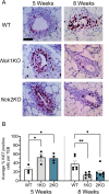
Loss of Nck1 or Nck2 disrupts mammary epithelial cell proliferation. A Representative images of TEB Ki67 immunostaining on mammary gland sections prepared from 5 and 8 week old WT, Nck1KO, and Nck2KO mice. Scale bar 50 μm. B Quantification of average Ki67 positive cell percentage per TEB (n = 5–11 TEBs per mouse) at 5 weeks (n = 3–4 mice per genotype) and 8 weeks (n = 6–7 mice per genotype). 1KO: Nck1KO, 2KO: Nck2KO, * adjusted P < 0.05; ** P < 0.01; at each time point by one-way ANOVA with post-hoc Dunnett’s test of Nck1KO and Nck2KO compared to WT
Fig. 5
Fig. 5
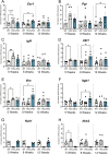
Loss of Nck1 or Nck2 alters gene expression patterns in the developing mammary gland. Quantitative PCR analysis of L3 mammary glands isolated from 5, 8 and 12 week old WT, Nck1KO and Nck2KO mice (n = 5–7 mice of each genotype). mRNA fold changes are shown for genes encoding (A) Estrogen Receptor α (Esr1), B Progesterone receptor (Psr1), C Insulin-like growth factor 1 (Igf1), D Akt1 (Akt1), E Vimentin (Vim), F Integrin β1 (Itgb1), G Nck1 (Nck1) and (H) Nck2 (Nck2). A-H, 1KO: Nck1KO, 2KO: Nck2KO, * adjusted P < 0.05, ** P < 0.01; at each time point by one-way ANOVA with post-hoc Dunnett’s test of Nck1KO and Nck2KO compared to WT
Fig. 6
Fig. 6
Summary of mammary gland phenotypes in Nck1 and Nck2 knockout mice at key stages of development. Mice lacking Nck1 or Nck2 show distinct temporal defects in ductal outgrowth, overall branch area, TEB number and TEB proliferation rates during puberty at 5 weeks and 8 weeks, and the outgrowth and branching phenotypes resolve by 12 weeks

Similar articles

Cited by

References

    1. Inman JL, Robertson C, Mott JD, Bissell MJ. Mammary gland development: cell fate specification, stem cells and the microenvironment. Development. 2015;142:1028–1042. doi: 10.1242/dev.087643. - DOI - PubMed
    1. Paine IS, Lewis MT. The Terminal End Bud: the Little Engine that Could. J Mammary Gland Biol. 2017;22:93–108. doi: 10.1007/s10911-017-9372-0. - DOI - PMC - PubMed
    1. Sternlicht MD, Sunnarborg SW, Kouros-Mehr H, Yu Y, Lee DC, Werb Z. Mammary ductal morphogenesis requires paracrine activation of stromal EGFR via ADAM17-dependent shedding of epithelial amphiregulin. Development. 2005;132:3923–3933. doi: 10.1242/dev.01966. - DOI - PMC - PubMed
    1. Ciarloni L, Mallepell S, Brisken C. Amphiregulin is an essential mediator of estrogen receptor α function in mammary gland development. Proc National Acad Sci. 2007;104:5455–5460. doi: 10.1073/pnas.0611647104. - DOI - PMC - PubMed
    1. Brullo C, Tasso B. New Insights on Fak and Fak Inhibitors. Curr Med Chem. 2021;28:3318–3338. doi: 10.2174/0929867327666201103162239. - DOI - PubMed

Publication types

Grants and funding

LinkOut - more resources