. 2023 Sep;27(18):2730-2743.
doi: 10.1111/jcmm.17860. Epub 2023 Jul 21.

The role of PKN1 in glioma pathogenesis and the antiglioma effect of raloxifene targeting PKN1

Affiliations

The role of PKN1 in glioma pathogenesis and the antiglioma effect of raloxifene targeting PKN1

Yubing Hao et al. J Cell Mol Med. 2023 Sep.

Abstract

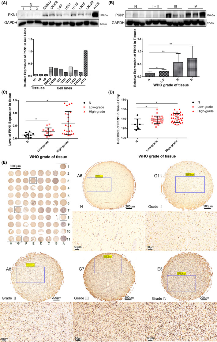
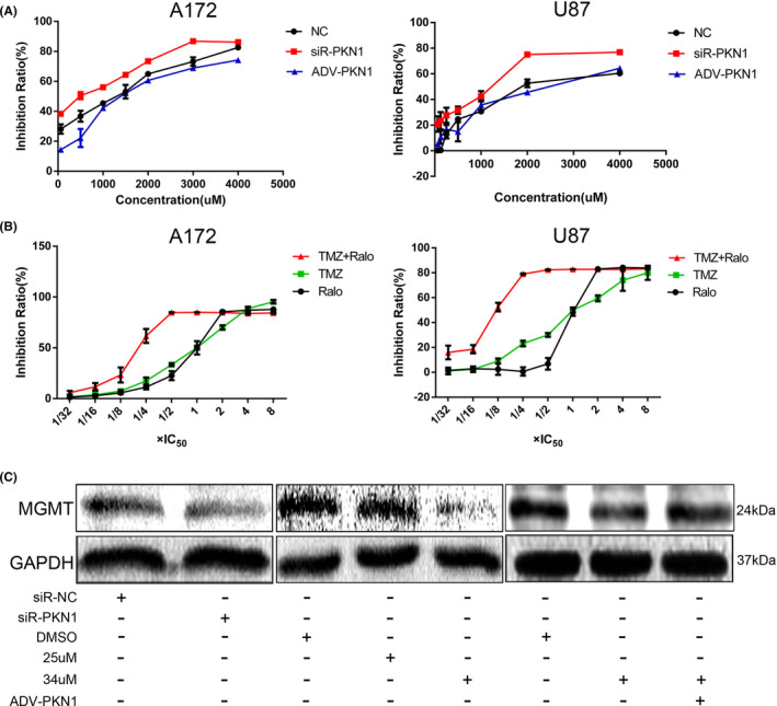
PKN1 (protein kinase N1), a serine/threonine protein kinase family member, is associated with various cancers. However, the role of PKN1 in gliomas has rarely been studied. We suggest that PKN1 expression in glioma specimens is considerably upregulated and positively correlates with the histopathological grading of gliomas. Knocking down PKN1 expression in glioblastoma (GBM) cells inhibits GBM cell proliferation, invasion and migration and promotes apoptosis. In addition, yes-associated protein (YAP) expression, an essential effector of the Hippo pathway contributing to the oncogenic role of gliomagenesis, was also downregulated. In contrast, PKN1 upregulation enhances the malignant characteristics of GBM cells and simultaneously upregulates YAP expression. Therefore, PKN1 is a promising therapeutic target for gliomas. Raloxifene (Ralo), a commonly used selective oestrogen-receptor modulator to treat osteoporosis in postmenopausal women, was predicted to target PKN1 according to the bioinformatics team from the School of Mathematics, Tianjin Nankai University. We showed that Ralo effectively targets PKN1, inhibits GBM cells proliferation and migration and sensitizes GBM cells to the major chemotherapeutic drug, Temozolomide. Ralo also reverses the effect of PKN1 on YAP activation. Thus, we confirm that PKN1 contributes to the pathogenesis of gliomas and may be a potential target for Ralo adjuvant glioma therapy.

Keywords: GBM; PKN1; TMZ; YAP; raloxifene.


Conflict of interest statement

The authors declare that there is no conflict of interest.

Figures

FIGURE 1
FIGURE 1
PKN1 expression in GBM cell lines and glioma tissues (*p < 0.05, **p < 0.01; compared with NB tissues) (A) The protein expression level of PKN1 in GBM cell lines (A172, U87, LN229, LN18, U118, U251, LN308, and SNB19) was detected using western blotting, and the protein rations for all lanes were included in western blots; the NB tissues served as a negative control. (B) The protein expression level of PKN1 in 37 glioma specimens and 10 NB tissues was detected using western blotting, and the protein rations for all lanes were included in western blots. (C) The relative grey value of protein expression of PKN1 in NB and glioma tissues. (D): The H‐SCORE of PKN1 of each tissue dot in the tissue chip. (E): Immunohistochemistry was used to detect PKN1 expression in the brain tissue microarray. 400×, scale bar 50 μm. N: A2, A3, A4, A5, A6; low‐grade (GradeI: G10, G11, H10; Grade II: A8–10, B4–11, C2–6, G9, H2–9, H11); high‐grade (Grade III: G2–4, G5–8; Grade IV: C7–11, D2–11, E2–11, F2–8, F9–11); A1, A7, A11, B2 and B3 are the null‐tissue points.
FIGURE 2
FIGURE 2
Effect of PKN1 knockdown on the biological behaviours of GBM cells (*p < 0.05, **p < 0.01, ***p < 0.001, ****p < 0.0001; compared with the siR‐NC groups). (A) A172 and U87 GBM cells were transfected with siR‐PKN1 followed by western blotting and RT‐PCR (Figure A2) to detect PKN1 expression. The transfection ratios were analysed (Figure A1). (B) Colony formation assay of A172 and U87 cells transfected with siR‐PKN1, scale bar 6 mm. (C) Transwell assay of A172 and U87 cells transfected with siR‐PKN1, 100×, scale bar 200 μm. (D) The cell proliferation of A172 and U87 cells transfected with siR‐PKN1 was detected using the CCK‐8 assay. (E) The wound healing assay of A172 and U87 cells transfected with siR‐PKN1. 100×, scale bar 200 μm. (F) Apoptosis of A172 and U87 cells transfected with siR‐PKN1 was detected by flow cytometry. (G) MMP2, PCNA, and Bcl2 were expressed in A172 and U87 cells transfected with siR‐PKN1, and the loading control was GAPDH.
FIGURE 3
FIGURE 3
Effect of PKN1 overexpression on the biological behaviours of LN229 cells (*p < 0.05, **p < 0.01, ***p < 0.001; compared with the ADV‐NC group). (A) LN229 cells were transfected with different ADV‐PKN1 concentrations (T1, T2, T3, and T4) followed by western blotting and RT‐PCR analysis to detect the PKN1 expression level; GAPDH was the loading control. (B) The CCK‐8 assay examined the LN229 cell proliferation transfected with ADV‐PKN1. (C) The invasive ability of LN229 cells transfected with ADV‐PKN1 detected by the Transwell assay, 100×, scale bar 200 μm. (D) The wound healing assay was used to examine the migration of LN229 cells transfected with ADV‐PKN1. 100×, scale bar 200 μm. (E) LN229 cells transfected with ADV‐PKN1 detected by colony formation assay, scale bar 7 mm. (F) Flow cytometry assayed the apoptosis of LN229 cells transfected with ADV‐PKN1. (G) MMP2, PCNA, and Bcl2 expression was examined using western blotting in LN229 cells transfected with ADV‐PKN1.
FIGURE 4‐1
FIGURE 4‐1
Effect of Ralo on the biological behaviours of GBM cells (*p < 0.05, **p < 0.01, ***p < 0.001, ****p < 0.0001; compared with the DMSO group, # p < 0.05, ## p < 0.01, ### p < 0.001; compared with the ADV‐NC‐DMSO group). (A) The CCK‐8 assay detected the IC50 and IC25 values of Ralo in A172 and U87 cells. (B) Western blotting and RT‐PCR were used to detect PKN1 expression in GBM cells treated with different Ralo concentrations. (C) The migration ability of GBM cells detected using the wound healing assay, 100×, scale bar 200 μm. (D) Proliferation of A172 and U87 cells treated with Ralo was detected using the CCK‐8 assay. (E) Colony formation assay of GBM cells treated with Ralo, scale bar 6 mm. (F) Transwell assay of GBM cells, 100×, scale bar 200 μm.
FIGURE 4‐2
FIGURE 4‐2
(continue Figure 4‐1 ). (A) Apoptosis detected via flow cytometry. (B) MMP2, PCNA, and Bcl2 expression in A172 and U87 cells treated with Ralo were examined using western blotting. (C) Proliferation of A172 and U87 cells treated with Ralo alone and ADV‐PKN1 with Ralo.
FIGURE 5
FIGURE 5
The effect of PKN1 and Ralo treatment on YAP expression in GBM cell lines (*p < 0.05, **p < 0.01; compared with siR‐NC group and DMSO group). (A) After siR‐PKN1 transfection in A172 and U87 cells and ADV‐PKN1 transfection in LN229 cells, the total YAP expression and its distribution in the cytoplasm and nucleus were examined through western blotting. (B) RT‐PCR detected the YAP expression in GBM cells transfected with siR‐PKN1 or ADV‐PKN1. GAPDH was the loading control. (C) RT‐PCR detected the YAP expression in A172 and U87 cells treated with Ralo; GAPDH was the loading control. (D) YAP expression and its distribution in cytoplasm and nucleus after Ralo treatment. (E) ADV‐PKN1 was transfected into Ralo‐treated GBM cells to detect the total YAP expression and its distribution in the cytoplasm and nucleus.
FIGURE 6
FIGURE 6
The effect of PKN1 and Ralo on the IC50 value of TMZ and the effect of Ralo on MGMT expression in A172 cells. (A) The effect of PKN1 knockdown and overexpression on the IC50 value of TMZ in A172 and U87 GBM cells. (B) The effect of Ralo and Ralo combined with TMZ on the IC50 value of TMZ and Ralo in A172 and U87 GBM cells. (C) The effect of PKN1 and Ralo treatment on MGMT expression in A172 GBM cells.
FIGURE 7
FIGURE 7
The conceptual model of the PKN1/YAP axis involves the malignant progression of glioma cells. PKN1 promotes the malignant progression of glioma and the anti‐tumour effect of Ralo by targeting PKN1.

Similar articles

Cited by

References

    1. Young RM, Jamshidi A, Davis G, Sherman JH. Current trends in the surgical management and treatment of adult glioblastoma. Ann Transl Med. 2015;3(9):121. - PMC - PubMed
    1. Louis DN, Perry A, Reifenberger G, et al. The 2016 World Health Organization classification of tumors of the central nervous system: a summary. Acta Neuropathol. 2016;131(6):803‐820. - PubMed
    1. Dolecek TA, Propp JM, Stroup NE, Kruchko C. CBTRUS statistical report: primary brain and central nervous system tumors diagnosed in the United States in 2005‐2009. Neuro Oncol. 2012;14:v1‐v49. - PMC - PubMed
    1. Yasuda H, Yamamoto H, Hanamura K, et al. PKN1 promotes synapse maturation by inhibiting mGluR‐dependent silencing through neuronal glutamate transporter activation. Commun Biol. 2020;3(1):710. - PMC - PubMed
    1. Kim JY, Yu J, Abdulkadir SA, Chakravarti D. KAT8 regulates androgen signaling in prostate cancer cells. Mol Endocrinol. 2016;30(8):925‐936. - PMC - PubMed

Publication types

Substances

LinkOut - more resources