. 2023 Aug:94:104713.
doi: 10.1016/j.ebiom.2023.104713. Epub 2023 Jul 20.

The S1P receptor 1 antagonist Ponesimod reduces TLR4-induced neuroinflammation and increases Aβ clearance in 5XFAD mice

Affiliations

The S1P receptor 1 antagonist Ponesimod reduces TLR4-induced neuroinflammation and increases Aβ clearance in 5XFAD mice

Zhihui Zhu et al. EBioMedicine. 2023 Aug.

Abstract

Background: Previously, we showed that the sphingosine-1-phosphate (S1P) transporter spinster 2 (Spns2) mediates activation of microglia in response to amyloid β peptide (Aβ). Here, we investigated if Ponesimod, a functional S1P receptor 1 (S1PR1) antagonist, prevents Aβ-induced activation of glial cells and Alzheimer's disease (AD) pathology.

Methods: We used primary cultures of glial cells and the 5XFAD mouse model to determine the effect of Aβ and Ponesimod on glial activation, Aβ phagocytosis, cytokine levels and pro-inflammatory signaling pathways, AD pathology, and cognitive performance.
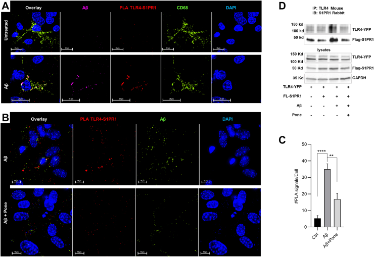
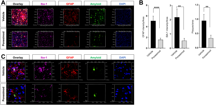
Findings: 42 increased the levels of TLR4 and S1PR1, leading to their complex formation. Ponesimod prevented the increase in TLR4 and S1PR1 levels, as well as the formation of their complex. It also reduced the activation of the pro-inflammatory Stat1 and p38 MAPK signaling pathways, while activating the anti-inflammatory Stat6 pathway. This was consistent with increased phagocytosis of Aβ42 in primary cultured microglia. In 5XFAD mice, Ponesimod decreased the levels of TNF-α and CXCL10, which activate TLR4 and Stat1. It also increased the level of IL-33, an anti-inflammatory cytokine that promotes Aβ42 phagocytosis by microglia. As a result of these changes, Ponesimod decreased the number of Iba-1+ microglia and GFAP+ astrocytes, and the size and number of amyloid plaques, while improving spatial memory as measured in a Y-maze test.

Interpretation: Ponesimod targeting S1PR1 is a promising therapeutic approach to reprogram microglia, reduce neuroinflammation, and increase Aβ clearance in AD.

Funding: NIHR01AG064234, RF1AG078338, R21AG078601, VAI01BX003643.

Keywords: Alzheimer's disease; Neuroinflammation; Phagocytosis; Ponesimod; Sphingosine-1-phosphate; Toll-like receptor 4.


Conflict of interest statement

Declaration of interests The authors declare no competing interests.

Figures

Fig. 1
Fig. 1
Ponesimod prevents Aβ-induced increase of TLR4 and S1PR1 levels. A. Levels of TLR4 and S1PR1 in 5XFAD (N = 3 controls and 3 5XFAD mice, T-test). P < 0.05, ∗∗P < 0.01. B. Levels of TLR4 and S1PR1 in AD patient brain grey matter (N = 5 healthy and 5 AD brains, T-test). Bar graphs show means ± SEM. ∗P < 0.05, ∗∗P < 0.01. C, D. Primary cultures of mixed glia were treated with 1 μM Aβ42 in the presence or absence of 10 nM (in C) or 100 nM Ponesimod (Pone, in C and D) for 24 h. Levels of TLR4 and S1PR1 were determined using immunoblotting. N = 3. One-way ANOVA followed by Turkey's multiple comparisons test.
Fig. 2
Fig. 2
42induces a complex between TLR4 and S1PR1. A, B. Primary cultures of mixed glia cells were treated with 1 μM Aβ42 in the presence or absence of 100 nM Ponesimod (Pone) for 24 h. Proximity ligation assays (PLAs) were performed using anti-TLR4 and S1PR1 antibodies (PLA, red) and co-labeled for Aβ42 (purple in A or green in B) and phagosomes (CD68+, green) visualized with the respective antibodies. The quantitation of PLA signals is shown in C. Data shows means ± SEM. N = 3, One-way ANOVA followed by Turkey's multiple comparisons test, ∗∗P < 0.01, ∗∗∗∗P < 0.0001. D. Hela cells were transfected with TLR4-YFP and Flag-S1PR1 and then treated with 1 μM Aβ42 in the presence or absence of 100 nM Ponesimod (Pone) for 4 h. Cell lysates were incubated with 1 μg of TLR4 mouse antibody to co-immunoprecipitate S1PR1. Captured protein was analyzed by SDS-immunoblotting with anti-S1PR1 rabbit antibody. N = 3.
Fig. 3
Fig. 3
Ponesimod enhances phagocytosis of Aβ42. A. Primary cultures of microglia were treated with 1 μM HiLyte™ Fluor 555-labeled Aβ42 (red) in the presence or absence of 100 nM Ponesimod (Pone) for 24 h. Microglia were fixed and immunolabeled for Iba-1 (purple) and CD-68 (green). Bottom panel shows higher magnification of framed area in top panel. B. BV2 cells were treated with 1 μM HiLyte™ Fluor 555-labeled Aβ42 in the presence or absence of 10 nM or 100 nM Ponesimod (Pone) for 24 h and Aβ42 uptake quantified by flow cytometry. Bar graphs show means ± SEM. N = 3. ∗P < 0.05. C, D. Experiment performed as in A, however, with primary cultures of mixed glia. 200 nM FTY720 (F) was used for comparison with Ponesimod (P). The colocalization of Aβ42 and CD68 in Iba-1(+) cells was quantified using the Pearson's correlation coefficient as shown in (D). N = 3. One-way ANOVA followed by Turkey's multiple comparisons test, ∗∗∗∗P < 0.0001.
Fig. 4
Fig. 4
Ponesimod downregulates neuroinflammatory cell signaling pathways in vitro. A, B. Primary culture of mixed glia were treated with 1 μM Aβ42 in the presence or absence of 10 nM Ponesimod (Pone) for 30 min and 4 h, then protein analyzed by immunoblotting. The phosphorylation of Stat1, Stat3, Stat6 and quantitation are shown in (A). The phosphorylation of ERK, p38 and JNK and quantitation are shown in (B). All data normalized to GAPDH or Actin. Bar graphs show means ± SEM. N = 4. T-test. ∗P < 0.05, ∗∗P < 0.01.
Fig. 5
Fig. 5
Ponesimod decreases neuroinflammatory cytokine levels and increases IL-33 in 5XFAD mice. A, B. Immunoblot analysis of protein from cortical brain tissue of 5XFAD mice with or without Ponesimod treatment. Right panels show quantitation after normalization to GAPDH. Bar graphs show means ± SEM. N = 8 in A (blots showing the other 3 pairs in Supplemental Fig. S1C) and N = 5 in B, T-test. ∗P < 0.05. C. U-plex assay shows the absolute concentration (pg/mg) of CXCL10, TNF-α, IL-1β, IL-6, and IL-33 in mouse brain lysates. Bar graphs show means ± SEM. N = 4. T-test. ∗P < 0.05.
Fig. 6
Fig. 6
Ponesimod ameliorates AD pathology in 5XFAD mice. A. Immunolabeling of Aβ42 (green) and activated astrocytes (GFAP, red) and microglia (Iba-1, purple) using cryosections from the brains of 5XFAD mice with or without Ponesimod (Pone) treatment. B. Quantitation of A. Bars show means ± SEM. T-test. ∗∗P < 0.01, ∗∗∗∗P < 0.0001. C. Images of higher magnification and resolution.
Fig. 7
Fig. 7
Ponesimod improves spatial learning and memory in 5XFAD mice. The 8.5–9 month-old 5XFAD mice were gavaged with vehicle or Ponesimod continuously for four weeks. After completion of treatment, the mice were subjected to a Y-maze test to determine short-term spatial working memory. (A). Schematic of animal treatment, behavioral test and brain tissue collection (top), and the Y-maze (bottom). (B). Spontaneous alternation performance (SAP). Number shows percentage of trials for a mouse visiting 3 different arms in a triad entry. The number of alternations in vehicle-treated- and Ponesimod-treated- 5XFAD mice is shown in (C). (D). Same arm return (SAR) as the percentage of a mouse returning to the same arm in any consecutive entries in a triad entry. (E). Alternative arms return (AAR) as the percentage of a mouse going into alternative arms in a triad entry. (F). Total entries. There are no differences among each group of mice in the total number of entries. All of the statistical analysis was performed by two-way ANOVA tests followed by Turkey's multiple comparisons test, except in (B), which was analyzed with One-sample t-test. (∗∗∗P < 0.001, ∗P < 0.05, WT: N = 7, Vehicle: N = 16. Ponesimod: N = 16).
Fig. 8
Fig. 8
Working model for the effect of Ponesimod on microglial activation and phagocytosis. Aβ42 binds to TLR4 and induces a complex with S1PR1. S1P co-activates the downstream pro-inflammatory cell signaling pathways ERK, p38 MAPK and JNK, which leads to phosphorylation activation of the transcription factors Stat1/3 and induction of neuroinflammation, particularly via gene expression of TNF-α. Ponesimod blocks S1P mediated co-activation of TLR4 and instead, leads to phagocytosis and proteolytic degradation of the receptor complex and Aβ42. In addition to inhibition of the canonical cell signaling for neuroinflammation downstream of TLR4, Ponesimod upregulates microglial phagocytosis via IL-33/Stat6 in a non-canonical cell signaling pathway, thereby contributing to persistent clearance of Aβ and alleviation of AD pathology. Generated using Biorender.
Supplementary Fig. S1
A. Protein levels (immunoblot) of S1PR1 and TLR4 in human brain tissue (white matter). B. IL-33 and TNF-α levels (immunoblot) in vehicle- and Ponesimod-treated 5XFAD brain. C. TLR-4 and S1PR1 levels in the vehicle and Ponesimod-treated 5XFAD brain cortex. D. Phagocytosis of Aβ-555 (1 µM) in microglia prepared from 6-month old 5XFAD brain with or without Ponesimod (100 nM) treatment.
Supplementary Fig. S2
A. Immunolabeling for iNOS (red) and activated microglia (Iba-1, green) using cryosections from the brain of 5XFAD mice with or without Ponesimod treatment.
Supplementary Fig. S3
A. Aβ immunolabeling (red) and activated microglia (Iba-1, purple) in cryosections from the 5XFAD mouse brain with or without Ponesimod. B. Fluoro-jade and Neurotrace co-immunolabeling of neurons, activated microglia (Iba-1, purple) in cryosections from 5XFAD mouse brain with or without Ponesimod. C. Fluoro-jade immunolabeling of neurons (green), Aβ-immunolabeling (red) and Iba-1 labeled activated microglia (Iba-1, purple) in cryosections from 5XFAD mouse brain with or without Ponesimod.
Supplementary Fig. S4A
Immunolabeling for Iba-1 (purple), GFAP (red), and Aβ (green) in cryosections from 5XFAD mouse brain with vehicle (A) or with Ponesimod (B).
Supplementary Fig. S4B
Immunolabeling for Iba-1 (purple), GFAP (red), and Aβ (green) in cryosections from 5XFAD mouse brain with vehicle (A) or with Ponesimod (B).
Blotsfig1
Blotsfig1
Original Western Blots shown in this study (Fig. 1).
Blotsfig2
Blotsfig2
Original Western Blots shown in this study (Fig. 2).
Blotsfig4
Blotsfig4
Original Western Blots shown in this study (Fig. 4).
Blotsfig5
Blotsfig5
Original Western Blots shown in this study (Fig. 5).
BlotsSUPPfig 1
BlotsSUPPfig 1
Original Western Blots shown in this study (Supplementary Figs.).

References

    1. Zhong L., Jiang X., Zhu Z., et al. Lipid transporter Spns2 promotes microglia pro-inflammatory activation in response to amyloid-beta peptide. Glia. 2019;67(3):498–511. - PMC - PubMed
    1. Chen Z., Doyle T.M., Luongo L., et al. Sphingosine-1-phosphate receptor 1 activation in astrocytes contributes to neuropathic pain. Proc Natl Acad Sci U S A. 2019;116(21):10557–10562. - PMC - PubMed
    1. Takabe K., Spiegel S. Export of sphingosine-1-phosphate and cancer progression. J Lipid Res. 2014;55(9):1839–1846. - PMC - PubMed
    1. Maceyka M., Spiegel S. Sphingolipid metabolites in inflammatory disease. Nature. 2014;510(7503):58–67. - PMC - PubMed
    1. Blaho V.A., Hla T. An update on the biology of sphingosine 1-phosphate receptors. J Lipid Res. 2014;55(8):1596–1608. - PMC - PubMed