

Navigation-Assisted Surgery for Locally Advanced Primary and Recurrent Rectal Cancer
- PMID: 37481493
- PMCID: PMC10562504
- DOI: 10.1245/s10434-023-13964-9
Navigation-Assisted Surgery for Locally Advanced Primary and Recurrent Rectal Cancer
Abstract
Background: In some surgical disciplines, navigation-assisted surgery has become standard of care, but in rectal cancer, indications for navigation and the utility of different technologies remain undetermined.
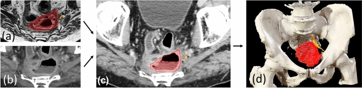
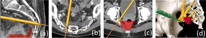
Methods: The NAVI-LARRC prospective study (NCT04512937; IDEAL Stage 2a) evaluated feasibility of navigation in patients with locally advanced primary (LARC) and recurrent rectal cancer (LRRC). Included patients had advanced tumours with high risk of incomplete (R1/R2) resection, and navigation was considered likely to improve the probability of complete resection (R0). Tumours were classified according to pelvic compartmental involvement, as suggested by the Royal Marsden group. The BrainlabTM navigation platform was used for preoperative segmentation of tumour and pelvic anatomy, and for intraoperative navigation with optical tracking. R0 resection rates, surgeons' experiences, and adherence to the preoperative resection plan were assessed.
Results: Seventeen patients with tumours involving the posterior/lateral compartments underwent navigation-assisted procedures. Fifteen patients required abdominosacral resection, and 3 had resection of the sciatic nerve. R0 resection was obtained in 6/8 (75%) LARC and 6/9 (69%) LRRC cases. Preoperative segmentation was time-consuming (median 3.5 h), but intraoperative navigation was accurate. Surgeons reported navigation to be feasible, and adherence to the resection plan was satisfactory.
Conclusions: Navigation-assisted surgery using optical tracking was feasible. The preoperative planning was time-consuming, but intraoperative navigation was accurate and resulted in acceptable R0 resection rates. Selected patients are likely to benefit from navigation-assisted surgery.
Keywords: Feasibility study; Image-guided surgery; Locally advanced rectal cancer; Locally recurrent rectal cancer; Navigation-assisted surgery; Optical tracking.
© 2023. The Author(s).
Conflict of interest statement
The authors declare no conflicts of interest. Arne M. Solbakken was supported by a PhD grant from South-Eastern Norway Regional Health Authority (Grant no. 2019028).
Figures


Comment in
-
The Application Prospect of Navigation-Assisted Surgery for Locally Advanced Primary and Recurrent Rectal Cancer is Broad.Ann Surg Oncol. 2024 Mar;31(3):1702-1703. doi: 10.1245/s10434-023-14699-3. Epub 2023 Dec 6. Ann Surg Oncol. 2024. PMID: 38055091 No abstract available.
References
MeSH terms
Grants and funding
LinkOut - more resources
Full Text Sources
