. 2023 Sep;299(9):105077.
doi: 10.1016/j.jbc.2023.105077. Epub 2023 Jul 21.

A riboside hydrolase that salvages both nucleobases and nicotinamide in the auxotrophic parasite Trichomonas vaginalis

Affiliations

A riboside hydrolase that salvages both nucleobases and nicotinamide in the auxotrophic parasite Trichomonas vaginalis

Marco Patrone et al. J Biol Chem. 2023 Sep.

Abstract

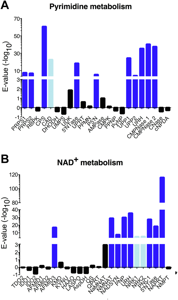
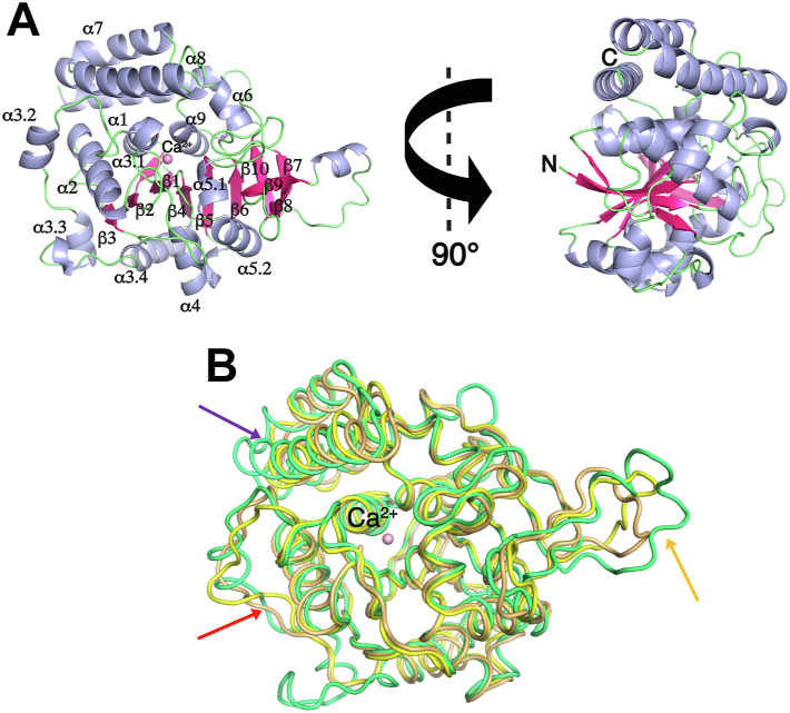
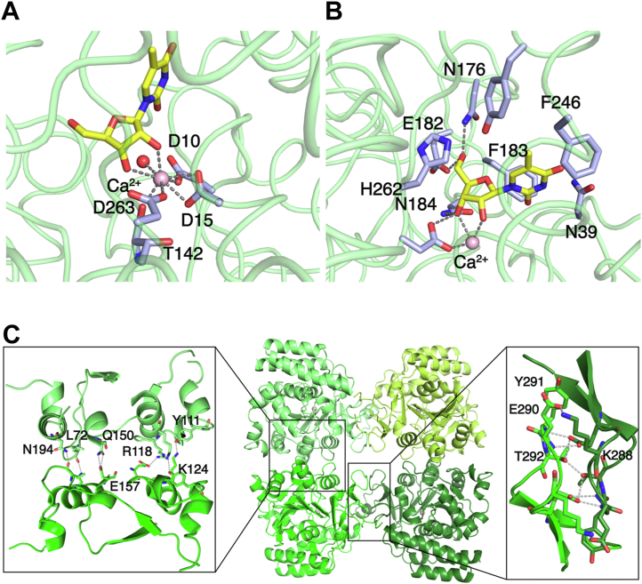
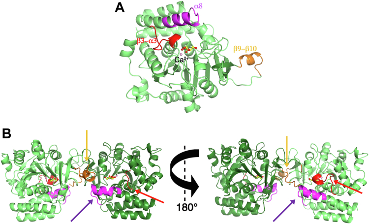
Pathogenic parasites of the Trichomonas genus are causative agents of sexually transmitted diseases affecting millions of individuals worldwide and whose outcome may include stillbirths and enhanced cancer risks and susceptibility to HIV infection. Trichomonas vaginalis relies on imported purine and pyrimidine nucleosides and nucleobases for survival, since it lacks the enzymatic activities necessary for de novo biosynthesis. Here we show that T. vaginalis additionally lacks homologues of the bacterial or mammalian enzymes required for the synthesis of the nicotinamide ring, a crucial component in the redox cofactors NAD+ and NADP. Moreover, we show that a yet fully uncharacterized T. vaginalis protein homologous to bacterial and protozoan nucleoside hydrolases is active as a pyrimidine nucleosidase but shows the highest specificity toward the NAD+ metabolite nicotinamide riboside. Crystal structures of the trichomonal riboside hydrolase in different states reveals novel intermediates along the nucleoside hydrolase-catalyzed hydrolytic reaction, including an unexpected asymmetry in the homotetrameric assembly. The active site structure explains the broad specificity toward different ribosides and offers precise insights for the engineering of specific inhibitors that may simultaneously target different essential pathways in the parasite.

Keywords: NAD; Trichomonas vaginalis; X-ray crystallography; drug design; enzyme structure; pyrimidine.


Conflict of interest statement

Conflict of interest The authors declare that they have no conflicts of interest with the contents of this article.

Figures

Figure 1
Figure 1
Trichomonas vaginalis lacks homologues of enzymes necessary for nucleobase and pyridine synthesis. The indicated enzymes involved in pyrimidine and pyridine metabolism were individually aligned against the T. vaginalis genome using tBLASTn and the returned −log(E-values) are plotted, shown as blue bars when >3. The Expect value E is a measure of the significance of the match. A, identification of pyrimidine-metabolizing enzymes. The human multifunctional CAD protein sequence identified significant homology with a Trichomonas gene in the aspartate carbamoyltransferase domain only, and the corresponding bar is colored light blue. B, identification of NAD+-metabolizing enzymes. The Trichomonas homologue of the human NRK1 and NRK2 kinases, also shown in light blue, is a AAA domain protein with several insertions compared with the human homologue and divergent at the characteristic β-hairpin lining the catalytic site. 5'NTase, 5′-nucleotidase; AFMID, kynurenine formamidase; AMPase, AMP phosphorylase; AspDH, aspartate dehydrogenase; CAD, Gln-dependent carbamoyl-phosphate synthase/aspartate carbamoyltransferase/dihydroorotase; CMPK, cytidine monophosphate kinase; CMPNase, cytidine deaminase 1,2; CNase, cytosine/isoguanine deaminase; cNPDA, 2′,3′-cyclic-nucleotide 2′-phosphodiesterase; CPS, carbamoyl-phosphate synthase-1; DHODH, dihydroorotate dehydrogenase; HAAO, 3-hydroxyanthranilate 3,4-dioxygenase; IDO1/2, indoleamine 2,3-dioxygenase 1,2; KMO, kynurenine 3-monooxygenase; KYNU, kynureninase; LASPO, L-aspartate oxidase; NADSYN, ammonia-dependent NAD+ synthetase; NaMNAT, nicotinate mononucleotide adenylyltransferase; NAPRT, nicotinate phosphoribosyltransferase; NMNAT, nicotinamide/nicotinic acid mononucleotide adenylyltransferase; NMPT, nicotinamide phosphoribosyltransferase; NRK 1,2, nicotinamide riboside kinase 1,2; P5′N, pyrimidine 5′-nucleotidase; PNC1, nicotinamidase 1; PNP, purine nucleoside phosphorylase; PPMN, pyrimidine/purine nucleotide 5′-monophosphate nucleosidase; PPNP, purine/pyrimidine nucleoside phosphorylase; PRPS, ribose-phosphate pyrophosphokinase 1,2; PyNP, pyrimidine nucleoside phosphorylase; QPRT, nicotinate-nucleotide pyrophosphorylase; QS, quinolinate synthase; RBPK, ribose 1,5-bisphosphate phosphokinase; TDO2, tryptophan 2,3-dioxygenase 2; UDK, uridine-cytidine kinase; UMPS, uridine 5′-monophosphate synthase; UPP, uridine phosphorylase 1,2; UPRT, uracil phosphoribosyltransferase; URH, pyrimidine-specific ribonucleoside hydrolase. The gene and corresponding proteins used in the homology search as well as the matches with highest homology in the T. vaginalis genome are listed in Table S1.
Figure 2
Figure 2
Crystal structure of TvRH.A, two orthogonal views showing the overall structure of the TvRH monomer with α-helices shown as light blue ribbons and β-strands as hotpink arrows. Secondary structure elements are labeled following the scheme proposed for NHs (13). Loop regions are shown as light green tubes, and the active site calcium ion as a pink sphere. B, the structures of the individual monomers of TvRH bound to ribose (light green), the C. fasciculata IUNH bound to a transition state–like inhibitor (2MAS, yellow), and the E. coli RihB in complex with the substrate inosine (3B9X, orange) are superimposed. The arrows indicate the region where TvRH is dissimilar, namely, the β3–α3 crossover loop (red), the α7–α8 helices (purple), and the β9–β10 loop (orange).
Figure 3
Figure 3
Structural difference between TvRH and NH proteins.A, octacoordination of the active site Ca2+ ion in TvRH in complex with 5mUR. The coordination geometry is conserved in all structures determined. B, direct interactions between the ribosyl moiety of 5mUR substrate and the TvRH active site. Residues within 5 Å of the substrate are shown as sticks with carbon atoms colored light blue. C, the TvRH homotetrameric structure, with the molecular 2-fold axes aligned horizontally, vertically, and perpendicular to the plane. Details of the interactions at the minor (left) and major (right) interfaces are shown, with interacting residues depicted as sticks and potential hydrogen bonding interactions as dashed lines.
Figure 4
Figure 4
Conformation of TvRH-bound substrates and ligands.A, omit electron density (mFo–DFc, ɸc) contoured at 3.0 σ showing 5mUR bound at the TvRH active site. Polar contacts are shown as black dashed lines. Residues involved in Ca2+ ion (depicted as a light pink sphere in all panels) coordination are detailed in Figure 3B. B, contacts between the 5mUR substrate and TvRH. The β3–α3 is in an open conformation. In (A and B), the amino acid residues that are within 5 Å of the ligand are shown as light blue sticks. C, the structures of the complexes of TvRH with 5mUR (carbon atoms colored green), Crithidia IUNH with pAPIR (PDB code 2MAS, yellow), and E. coli RihB with inosine (PDB code 3B9X, magenta) were superimposed showing the aglycones in both the pAPIR inhibitor and inosine molecules are axially oriented and the β3–α3 loop is closed while the thymine base is oriented axially when bound to the TvRH active site with an open β3–α3 loop. D, the positioning of the ribosyl moiety of 5mUR at the active site differs compared with other homologous ligands bound to the parasitic and enterobacterial NHs, as does the five-membered ring conformation.
Figure 5
Figure 5
Structural rearrangements along the TvRH reaction coordinate.A, contacts between the reaction product ribose and TvRH, underscoring the closed conformation of the β3–α3 loop and the movement of residue Asn39. The amino acid residues that are within 5 Å of the ligand are shown as light blue sticks. B, superposition of TvRH monomers from the 5mUR-bound and ribose-bound crystal structures. The backbone of both monomers is colored green. The regions differing in conformation are colored orange (5mUR complex) and blue (ribose). Arrows indicate the β3–α3 loop (red), α8 helix (purple), and β9–β10 loop (orange), respectively. C, hydrophobic interactions between Ile82 from the β3–α3 loop and a hydrophobic patch on helix α8 stabilize the closed conformation. The ribose molecule and Ca2+ ion are shown for reference. D, binding of glycerol at the TvRH active site mimics part of the ribosyl–enzyme interactions.
Figure 6
Figure 6
Asymmetry in the TvRH dimers.A, the structure of the TvRH monomer shown as a cartoon representation highlighting the β3–α3 loop (red), α8 helix (purple), and β9–β10 loop (orange) that differ in conformation within a TvRH dimer. B, two views of a TvRH dimer obtained through the major interaction surface. When the β3–α3 loop is flexible (left view), the β9–β10 loop is ordered in a helical conformation and the α8 helix is bent away from the active site. When the β3–α3 loop is in a fixed, closed conformation (right) the α8 helix leans toward the active site and the β9–β10 loop is flexible.
Figure 7
Figure 7
Models of Michaelis complexes of TvRH.A, section of the molecular surface of TvRH in the closed conformation from the glycerol-bound structure with NR modeled at the active site based on the C. fasciculata IUNH–inhibitor complex. In the model, residue Asn176 is positioned close to the carboxyamide group of the nicotinamide ring, which is also approached by the side chain of Tyr250. B, same as (A), with a molecule of the substrate UR (right) modeled in the active site. The side chains of His83 and Tyr250 are within hydrogen bonding distance of the O2 and O4 carbonyl oxygens of the uracil base, respectively. In both panels, residues within 5 Å of the modeled substrate molecule are shown as light blue sticks. Parts of the structure have been omitted for clarity. The distances reported in the panels are measured in the model obtained as described and without energetic optimization and are thus not to be interpreted as experimentally determined values.

References

    1. Rowley J., Vander Hoorn S., Korenromp E., Low N., Unemo M., Abu-Raddad L.J., et al. Chlamydia, gonorrhoea, trichomoniasis and syphilis: global prevalence and incidence estimates, 2016. Bull. World Health Organ. 2019;97:548–562P. - PMC - PubMed
    1. Ryan C.M., de Miguel N., Johnson P.J. Trichomonas vaginalis: current understanding of host-parasite interactions. Essays Biochem. 2011;51:161–175. - PMC - PubMed
    1. Dunne R.L., Dunn L.A., Upcroft P., O'Donoghue P.J., Upcroft J.A. Drug resistance in the sexually transmitted protozoan Trichomonas vaginalis. Cell Res. 2003;13:239–249. - PubMed
    1. Heyworth P.G., Gutteridge W.E., Ginger C.D. Purine metabolism in Trichomonas vaginalis. FEBS Lett. 1982;141:106–110. - PubMed
    1. Heyworth P.G., Gutteridge W.E., Ginger C.D. Pyrimidine metabolism in Trichomonas vaginalis. FEBS Lett. 1984;176:55–60. - PubMed

Publication types

LinkOut - more resources