. 2023 Dec;43(12):1289-1311.
doi: 10.1002/cac2.12470. Epub 2023 Jul 22.

An HGF-dependent positive feedback loop between bladder cancer cells and fibroblasts mediates lymphangiogenesis and lymphatic metastasis

Affiliations

An HGF-dependent positive feedback loop between bladder cancer cells and fibroblasts mediates lymphangiogenesis and lymphatic metastasis

Yuting Li et al. Cancer Commun (Lond). 2023 Dec.

Abstract

Background: Cancer-associated fibroblasts (CAFs) play a vital role in facilitating tumor progression through extensive reciprocal interplay with cancer cells. Tumor-derived extracellular vesicles (EVs) are the critical mediators involved in the crosstalk between cancer cells and stromal cells, contributing to the metastasis of cancers. Yet, the biological mechanisms of tumor-derived EVs in triggering CAFs phenotype to stimulate the lymph node (LN) metastasis of bladder cancer (BCa) are largely unknown. Here, we aimed to explore the effects and molecular mechanisms of tumor-derived EV-mediated CAFs phenotype in regulating BCa LN metastasis.

Methods: The high-throughput sequencing was utilized to identify the crucial long non-coding RNA (lncRNA) associated with CAF enrichment in BCa. The functional role of the transition of fibroblasts to CAFs induced by LINC00665-mediated EVs was investigated through the in vitro and in vivo assays. Chromatin isolation by RNA purification assays, fluorescence resonance energy transfer assays, cytokine profiling and patient-derived xenograft (PDX) model were performed to explore the underlying mechanism of LINC00665 in the LN metastasis of BCa.

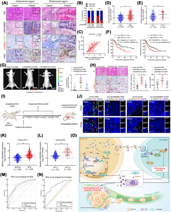
Results: We found that CAFs are widely enriched in the tumor microenvironment of BCa, which correlated with BCa lymphangiogenesis and LN metastasis. We then identified a CAF-associated long non-coding RNA, LINC00665, which acted as a crucial mediator of CAF infiltration in BCa. Clinically, LINC00665 was associated with LN metastasis and poor prognosis in patients with BCa. Mechanistically, LINC00665 transcriptionally upregulated RAB27B expression and induced H3K4me3 modification on the promoter of RAB27B through the recruitment of hnRNPL. Moreover, RAB27B-induced EVs secretion endowed fibroblasts with the CAF phenotype, which reciprocally induced LINC00665 overexpression to form a RAB27B-HGF-c-Myc positive feedback loop, enhancing the lymphangiogenesis and LN metastasis of BCa. Importantly, we demonstrated that blocking EV-transmitted LINC00665 or HGF broke this loop and impaired BCa lymphangiogenesis in a PDX model.

Conclusion: Our study uncovers a precise mechanism that LINC00665 sustains BCa LN metastasis by inducing a RAB27B-HGF-c-Myc positive feedback loop between BCa cells and fibroblasts, suggesting that LINC00665 could be a promising therapeutic target for patients with LN metastatic BCa.

Keywords: Cancer-associated fibroblasts; HGF; bladder cancer; extracellular vesicles; long non-coding RNA; lymph node metastasis; lymphangiogenesis; positive feedback loop.


Conflict of interest statement

The authors declare no potential conflicts of interest.

Figures

FIGURE 1
FIGURE 1
LINC00665 is correlated with CAF‐induced LN metastasis of BCa. (A) tSNE plot of single cells identified by scRNA‐seq and colored by major cell types. (B) tSNE plot of single cells identified by scRNA‐seq and colored by tissue types. (C) tSNE plot of single cells identified by scRNA‐seq and colored by major cell types in non‐invasive BCa tissues. (D) tSNE plot of single cells identified by scRNA‐seq and colored by major cell types in invasive BCa tissues. (E) The fraction of major cell types profiled from non‐invasive and invasive BCa tissues. (F‐H) tSNE plot showed the expression of (F) α‐SMA, (G) FAP, and (H) PDGFRα on CAFs. (I) Gene overlays of indicated CAF markers on tSNE. (J) Representative images and percentages revealing a positive correlation between α‐SMA, FAP and PDGFRα‐indicated CAF infiltration and LYVE‐1‐indicated lymphatic vessel density in BCa tissues (n = 228). Scale bar: 50 μm. (K) Flowchart showing the identification of lncRNAs upregulated in both urinary EVs and LN‐positive tissues from BCa patients. (L and M) Evaluating LINC00665 expression in BCa tissues and NATs in a 228‐case cohort (L) and TCGA database (M). (N and O) Evaluating LINC00665 expression in BCa tissues with or without LN metastasis (N) and in primary BCa tissues and paired metastatic LNs (O). (P) Representative ISH images and percentages of LINC00665 expression (blue) in NATs, LN‐negative BCa tissues and LN‐positive BCa tissues. Scale bar: 50 μm. (Q and R) Kaplan‐Meier curves for OS (Q) or DFS (R) of BCa patients (n = 228) according to LINC00665 expression. The cutoff value is the median expression. (S and T) Representative images and percentages of CAF enrichment and MLD in the intratumoral (S) and peritumoral regions (T) of BCa tissues (n = 228). White or red squares represent the areas of the insets. Scale bar: 50 μm. Statistical significance was assessed by the χ 2 test in J, P, S and T, or the nonparametric Mann‐Whitney U test in L‐O. *P < 0.05, **P < 0.01. Abbreviations: CAFs, Cancer‐associated fibroblasts; LN, lymph node; BCa, bladder cancer; tSNE, T‐distributed stochastic neighbor embedding; scRNA‐seq, single‐cell RNA sequencing; α‐SMA, alpha‐Smooth Muscle Actin; FAP, fibroblast activation protein; PDGFRα, platelet‐derived growth factor receptor‐α; LYVE‐1, lymphatic vessel endothelial hyaluronan receptor 1; EVs, extracellular vesicles; lncRNAs, long non‐coding RNAs; NATs, normal adjacent tissues; TCGA, The Cancer Genome Atlas; OS, overall survival; DFS, disease‐free survival; MLD, micro‐lymphatic vessel density.
FIGURE 2
FIGURE 2
LINC00665‐induced EVs endow fibroblasts with CAF phenotype to promote BCa lymphangiogenesis in vitro. (A) Representative TEM images of EVs isolated from LINC00665‐overexpressing T24 and control cells. Scale bars: 100 nm. (B and C) NTA analysis and quantification of EVs isolated from T24 cells with or without transiently transfecting with the Vector or LINC00665‐overexpressing plasmids. (D) Representative fluorescence images of primary fibroblasts, T cells, B cells, HLECs and HUVECs after incubation with PKH67‐labelled (green) vector EVs or LINC00665 EVs from BCa cells. Scale bars: 5 μm. (E) Western blotting analysis of α‐SMA, FAP and PDGFRα expression in fibroblasts incubated with PBS, vector‐ or LINC00665‐induced EVs from BCa cells. (F) Representative confocal images of α‐SMA, FAP and PDGFRα expression in fibroblasts treated with PBS, vector‐ or LINC00665‐induced EVs from BCa cells. Scale bars: 5 μm. (G) Flow cytometric analysis of α‐SMA, FAP and PDGFRα expression in fibroblasts incubated with PBS, vector‐induced EVs, or LINC00665‐induced EVs form BCa cells. (H and I) Representative images and quantification of tube formation and Transwell migration of HLECs incubated with CM from fibroblasts treated with PBS, vector‐induced EV, or LINC00665‐induced EV. Scale bar: 100 μm. Statistical significance was assessed by the two‐tailed Student's t test in C, or 1‐way ANOVA followed by Dunnett's tests in G‐I. Error bars show the SD from three independent experiments. *P < 0.05; **P < 0.01. Abbreviations: EVs, extracellular vesicles; CAFs, Cancer‐associated fibroblasts; BCa, bladder cancer; TEM, transmission electron microscopy; NTA, nanoparticle tracking analysis; α‐SMA, alpha‐Smooth Muscle Actin; FAP, fibroblast activation protein; PDGFRα, platelet‐derived growth factor receptor‐α; PBS, phosphate‐buffered saline; HLECs, human lymphatic endothelial cells; CM, culture media.
FIGURE 3
FIGURE 3
LINC00665‐induced EVs promote CAF infiltration to stimulate the LN metastasis of BCa in vivo. (A) Flowchart showing the establishment of the popliteal LN metastasis model. (B) NTA analysis of EVs isolated from T24 cells with or without infecting with the Vector or LINC00665‐overexpressing lentiviruses. (C) Representative bioluminescence images of nude mice treated with vector‐ or LINC00665‐induced EVs (n = 12). The red arrows indicate footpad primary tumor and metastatic popliteal LN. (D) Representative images of the popliteal LNs from mice (n = 12). (E) Representative images of fluorescence of α‐SMA‐, FAP‐ and PDGFRα‐indicated CAF and LYVE‐1‐indicated lymphatic vessel density in footpad tumor from the mice (n = 12). Scale bar: 50 μm. (F) Flowchart showing the establishment of BCa orthotopic xenograft model. (G) Necropsy examination and MRI of mouse urinary bladders following post‐inoculation. (H) Representative bioluminescence images of nude mice treated with vector‐ EVs or LINC00665‐induced EVs (n = 12). The red arrows indicate primary BCa and metastatic LNs. (I) Representative images of anti‐GFP analysis in pelvic LNs from the indicated group (n = 12). Scale bars: 500 μm (red) or 100 μm (black). (J) Quantification of the metastatic number of pelvic LNs in mice from indicated groups. Statistical significance was assessed by two‐tailed Student's t test in J. Error bars show the SD from three independent experiments. *P < 0.05; **P< 0.01. Abbreviations: EVs, extracellular vesicles; CAFs, Cancer‐associated fibroblasts; LN, lymph node; BCa, bladder cancer; NTA, nanoparticle tracking analysis; α‐SMA, alpha‐Smooth Muscle Actin; FAP, fibroblast activation protein; PDGFRα, platelet‐derived growth factor receptor‐α; LYVE‐1, lymphatic vessel endothelial hyaluronan receptor 1; MRI, Magnetic resonance imaging; GFP, green fluorescent protein.
FIGURE 4
FIGURE 4
LINC00665 directly binds with hnRNPL to promote RAB27B transcription. (A‐C) Silver staining of the proteins from RNA pull‐down assays performed by biotin‐labeled LINC00665 sense and antisense in T24 cells, and MS analysis of the LINC00665‐enriched band from silver staining. (D) Western blotting analysis after RNA pull‐down assays with nuclear extract. (E) Relative fluorescence showing the colocalization of LINC00665 and hnRNPL in the nucleus of T24 and 5637 cells. Scale bar: 5 μm. (F and G) RNA pull‐down with serial deletions of LINC00665 identified the essential regions on LINC00665 required for binding hnRNPL (Vertical red lines). Horizontal blue lines showed the full‐length or serial deletions of LINC00665. (H) RNAalifold for predicting the stem‐loop structures of the hnRNPL binding sites in LINC00665. (I) RIP assays after deletion of 2285‐2360 nt region of LINC00665 in T24 cells. (J) Heatmap of the differentially expressed genes between LINC00665‐overexpressing and control BCa cells. (K) qRT‐PCR analysis of the change in RAB genes related to EV secretion after silencing LINC00665 in T24 cells. (L) Western blotting analysis of RAB27B expression after LINC00665 silencing in T24 cells. (M) Luciferase analysis with serial deletion of RAB27B promoter in T24 cells with or without overexpressing LINC00665. (N) Ten probes of LINC00665 numbered 1 to 10 were divided into the odd group (LINC00665 odd) and even group (LINC00665 even). LINC00665‐enriched chromatin in T24 cells was revealed by ChIRP analysis. (O and P) ChIP analysis of the hnRNPL enrichment and H3K4me3 status of the RAB27B promoter in LINC00665‐overexpressing T24 cells with or without muting of the hnRNPL binding site. (Q) qRT‐PCR analysis of RAB27B expression in LINC00665‐overexpressing T24 cells with or without hnRNPL silencing. (R) Representative TEM of EVs secreted by vector‐ or LINC00665 plasmid‐transfected T24 cells with or without RAB27B silencing. Red arrows indicate the EVs. Scale bar: 100 nm. (S) Representative immunofluorescence images of fibroblasts treated with PKH67‐labelled EVs secreted by indicated BCa cells. Scale bar: 5 μm. Statistical significance was assessed by the two‐tailed Student's t test in I and M, or 1‐way ANOVA followed by Dunnett's tests in K and N‐Q. Error bars show the SD from three independent experiments. *P< 0.05; **P < 0.01. Abbreviations: hnRNPL, heterogeneous nuclear ribonucleoprotein L; MS, Mass spectrometry; RIP, RNA immunoprecipitation; nt, nucleotides; qRT‐PCR, quantitative real‐time PCR; ChIP, chromatin immunoprecipitation; TEM, transmission electron microscopy.
FIGURE 5
FIGURE 5
EV‐mediated LINC00665 endows fibroblasts with the CAF phenotype by activating the TGF‐β pathway. (A) Western blotting analysis showing the alteration of crucial proteins of the TGF‐β pathway in fibroblasts treated with EVs from LINC00665‐overexpressing BCa cells. (B) Immunofluorescence analysis of α‐SMA, FAP and PDGFRα expression in LINC00665‐induced EV‐incubated fibroblasts with or without SIS3 treatment. Scale bar: 5 μm. (C‐H) Flow cytometric analysis and quantification of α‐SMA (C and D), FAP (E and F) or PDGFRα (G and H) expression in LINC00665‐induced EV‐incubated fibroblasts with or without SIS3 treatment. (I) Cytokine array of the CM from fibroblasts treated with EVs secreted by BCa cells transfected with vector plasmids or LINC00665 plasmids. (J) The relative signal intensity of altered cytokines in the indicated group. (K and L) ELISA analysis of cytokine secretion by fibroblasts incubated with indicated EVs. (M) Representative images and quantification of tube formation and Transwell migration of HLECs treated with indicated fibroblast CM. Scale bars: 100 μm. Statistical significance was assessed by 1‐way ANOVA followed by Dunnett's tests in D, F, H, K and M, or the two‐tailed Student's t test in J and L. Error bars show the SD from three independent experiments. *P < 0.05; **P< 0.01. Abbreviations: EVs, extracellular vesicles; CAFs, Cancer‐associated fibroblasts; TGF‐β, transforming growth factor beta; BCa, bladder cancer; α‐SMA, alpha‐Smooth Muscle Actin; FAP, fibroblast activation protein; PDGFRα, platelet‐derived growth factor receptor‐α; CM, culture media; ELISA, enzyme‐linked immunosorbent assay; HLECs, human lymphatic endothelial cells.
FIGURE 6
FIGURE 6
The LINC00665‐mediated positive loop is indispensable for LN metastasis of BCa. (A) Schematic presentation of the established BCa cells and EV‐induced fibroblasts co‐culture model. (B and C) qRT‐PCR analysis of LINC00665 expression in BCa cells cultured with indicated CM from fibroblasts with or without αHGF treatment. (D) Relative transcriptional activity of LINC00665 in HGF‐treated T24 cells with or without c‐Myc silencing. (E) Enriched motifs of c‐Myc binding sites predicted by JASPAR. (F) Schematic model of c‐Myc binding sequences in the LINC00665 promoter region predicted by JASPAR and PROMO. (G and H) ChIP‐qPCR analysis of c‐Myc‐enriched chromatin in T24 (G) and 5637 (H) cells. (I) Depletion of the P1 region in the LINC00665 promoter impaired the HGF‐induced LINC00665 transcriptional activity in T24 cells. (J) Schematic presentation of the co‐culture model for analysis of the positive loop between BCa cells and fibroblasts. (K) qRT‐PCR analysis of LINC00665 expression in T24 cells incubated with indicated fibroblasts. (L and M) Flow cytometry analysis and percentages of α‐SMA (L) and FAP (M) expression of fibroblasts. (N) Representative images and quantification of tube formation and Transwell migration of indicated HLECs. Scale bar: 100 μm. Statistical significance was assessed by 1‐way ANOVA followed by Dunnett's tests in B‐D and K‐N, or the two‐tailed Student's t test in G‐I. Error bars show the SD from three independent experiments. *P < 0.05; **P < 0.01. Abbreviations: LN, lymph node; BCa, bladder cancer; EVs, extracellular vesicles; qRT‐PCR, quantitative real‐time PCR; CM, culture media; αHGF, neutralizing antibody against HGF; ChIP, chromatin immunoprecipitation; qRT‐PCR, quantitative real‐time PCR; α‐SMA, alpha‐Smooth Muscle Actin; FAP, fibroblast activation protein; HLECs, human lymphatic endothelial cells.
FIGURE 7
FIGURE 7
Clinical relevance of the LINC00665‐induced RAB27B‐HGF‐c‐Myc positive feedback loop in patients with BCa. (A and B) Representative images (A) and quantification (B) of LINC00665 expression, CAF infiltration, lymphatic vessel density, and HGF expression in BCa tissues. Scale bars: 50 μm. (C) Correlation analysis of LINC00665 and HGF expression in BCa tissues. (D and E) qRT‐PCR analysis of HGF expression between BCa tissues and NATs (n = 228) (D) or between LN‐negative (n = 175) and LN‐positive (n = 53) BCa tissues (E). (F) Kaplan‐Meier curves of the OS and DFS of patients with BCa with low vs. high HGF expression levels. The cutoff value is the median. (G) Representative bioluminescence images and quantification of popliteal metastatic LNs from nude mice treated with LINC00665‐induced EVs with or without co‐injection of αHGF (n = 12). Red arrows indicate footpad tumor and metastatic popliteal LN. (H) Representative images and quantification of IHC staining evaluation of LYVE‐1‐indicated lymphatic vessel density and α‐SMA‐indicated CAF infiltration in footpad tumors (n = 12). Red squares represent the areas of the insets. Scale bars: 50 μm. (I) Schematic illustration of the establishment of the PDX model. (J) Representative images and quantification of fluorescence of α‐SMA‐indicated CAFs and LYVE1‐indicated lymphatic vessel density in PDX tumors from the mice (n = 6 per patient). Scale bar: 50 μm. (K and L) qRT‐PCR analysis of LINC00665 expression in urinary EVs from patients with BCa (n = 228) and health control (n = 228), or BCa patients with (n = 175) or without (n = 53) LN metastasis. (M and N) ROC curves for the efficiency of urinary EV‐mediated LINC00665 in diagnosing BCa and LN metastasis. (O) Proposed model of LINC00665‐induced RAB27B‐HGF‐c‐Myc positive feedback loop in promoting BCa lymphangiogenesis and LN metastasis. Statistical significance was assessed by the χ 2 test in B, the nonparametric Mann‐Whitney U test in D, E, K and L, or 1‐way ANOVA followed by Dunnett's tests in G‐H. Error bars show the SD from three independent experiments. *P < 0.05; **P < 0.01. Abbreviations: BCa, bladder cancer; CAFs, Cancer‐associated fibroblasts; qRT‐PCR, quantitative real‐time PCR; NATs, normal adjacent tissues; LN, lymph node; EVs, extracellular vesicles; OS, overall survival; DFS, disease‐free survival; αHGF, neutralizing antibody against HGF; IHC, immunohistochemistry; LYVE‐1, lymphatic vessel endothelial hyaluronan receptor 1; α‐SMA, alpha‐Smooth Muscle Actin; PDX, patient‐derived xenograft; ROC, receiver operating characteristic analysis.

References

    1. Bray F, Ferlay J, Soerjomataram I, Siegel RL, Torre LA, Jemal A. Global cancer statistics 2018: GLOBOCAN estimates of incidence and mortality worldwide for 36 cancers in 185 countries. CA Cancer J Clin. 2018;68(6):394–424. - PubMed
    1. Bruins HM, Veskimae E, Hernandez V, Imamura M, Neuberger MM, Dahm P, et al. The impact of the extent of lymphadenectomy on oncologic outcomes in patients undergoing radical cystectomy for bladder cancer: a systematic review. Eur Urol. 2014;66(6):1065–77. - PubMed
    1. Lenis AT, Lec PM, Chamie K, Mshs MD. Bladder Cancer: A Review. JAMA. 2020;324(19):1980–91. - PubMed
    1. Cathomas R, Lorch A, Bruins HM, Comperat EM, Cowan NC, Efstathiou JA, et al. The 2021 Updated European Association of Urology Guidelines on Metastatic Urothelial Carcinoma. Eur Urol. 2022;81(1):95–103. - PubMed
    1. Escobedo N, Oliver G. Lymphangiogenesis: Origin, Specification, and Cell Fate Determination. Annu Rev Cell Dev Biol. 2016;32:677–91. - PubMed

Publication types