Review
. 2023 Jul 7:14:1196033.
doi: 10.3389/fimmu.2023.1196033. eCollection 2023.

Monocyte-endothelial cell interactions in vascular and tissue remodeling

Affiliations
Review

Monocyte-endothelial cell interactions in vascular and tissue remodeling

Mireia Medrano-Bosch et al. Front Immunol. .

Abstract

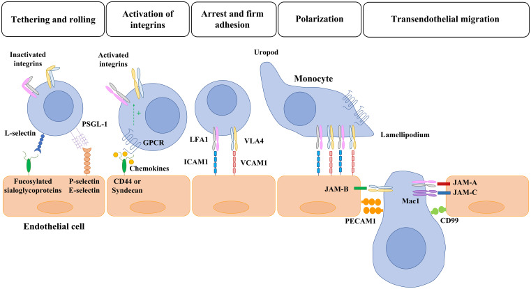
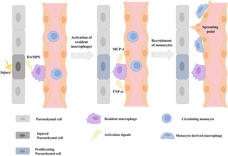
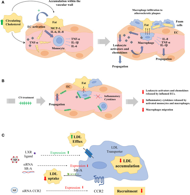
Monocytes are circulating leukocytes of innate immunity derived from the bone marrow that interact with endothelial cells under physiological or pathophysiological conditions to orchestrate inflammation, angiogenesis, or tissue remodeling. Monocytes are attracted by chemokines and specific receptors to precise areas in vessels or tissues and transdifferentiate into macrophages with tissue damage or infection. Adherent monocytes and infiltrated monocyte-derived macrophages locally release a myriad of cytokines, vasoactive agents, matrix metalloproteinases, and growth factors to induce vascular and tissue remodeling or for propagation of inflammatory responses. Infiltrated macrophages cooperate with tissue-resident macrophages during all the phases of tissue injury, repair, and regeneration. Substances released by infiltrated and resident macrophages serve not only to coordinate vessel and tissue growth but cellular interactions as well by attracting more circulating monocytes (e.g. MCP-1) and stimulating nearby endothelial cells (e.g. TNF-α) to expose monocyte adhesion molecules. Prolonged tissue accumulation and activation of infiltrated monocytes may result in alterations in extracellular matrix turnover, tissue functions, and vascular leakage. In this review, we highlight the link between interactions of infiltrating monocytes and endothelial cells to regulate vascular and tissue remodeling with a special focus on how these interactions contribute to pathophysiological conditions such as cardiovascular and chronic liver diseases.

Keywords: angiogenesis; cardiovascular diseases; endothelial cell; liver diseases.; macrophage; monocyte; tumor.


Conflict of interest statement

The authors declare that the research was conducted in the absence of any commercial or financial relationships that could be construed as a potential conflict of interest.

Figures

Figure 1
Figure 1
Overview of monocyte-endothelial cell interaction and transmigration. Monocyte migration to inflammatory sites is a multistep process with many molecules involved. First, selectins mediate the initial tethering and rolling of monocytes along the cytokine-activated endothelial cells. Then, the monocyte interacts with chemokines that are bound to transmembrane heparan sulphate proteoglycans (CD44 and sydecan) expressed in the endothelium. The activation of GPCR leads to the activation of integrins and the consequent monocyte arrest mediated by the interaction of LFA1 and VLA4 with ICAM1 and VCAM, respectively. Once a monocyte establishes firm adhesion to the vascular endothelium, it undergoes a morphological change known as polarization, in which chemokine receptors and activated integrins redistribute to the leading edge. After polarization, monocytes migrate toward interendothelial junctions and then transmigrate into the underlying tissues. The members of the JAM family expressed by endothelial cells (JAM-A, JAM-B, JAM-C) interact with the activated integrins of monocytes (LFA1, VLA4, Mac1) and allow the transmigration through tight junctions. Lastly, PECAM-1 (CD31) and CD99 hemophilic engagement and endothelial retraction lead to monocyte extravasation. PSGL: P-selectin glycoprotein ligand-1; GPCR: G-protein-coupled receptors; LFA1: lymphocyte function-associated antigen 1; VLA4: very late antigen 4; ICAM1: intercellular adhesion molecule 1 (ICAM1/CD54); VCAM: vascular cell-adhesion molecule 1; Mac1: macrophage receptor 1; PECAM: platelet/endothelial cell-adhesion molecule 1.
Figure 2
Figure 2
Monocyte-endothelial cell interactions and vascular sprouting occurring after hepatectomy. (A) Images of vessels from liver sections identified by staining for von Willebrand factor (VWF, in red) and recruited monocytes by staining for CD14 (in green). Nuclei were stained by DAPI (in blue). Initial contacts of recruited monocytes (in yellow) take place in portal areas. (B) Amplification of multiphoton images (vessels in green and yellow, macrophages in red), visualized vascular buds (white circles) surrounded by spread recruited macrophages 16 hours after hepatectomy. Images reprinted with permission from “Melgar-Lesmes, P.; et al. Monocyte-endothelial cell interactions in the regulation of vascular sprouting and liver regeneration in mouse. J Hepatol. 2015; 63 4):917-25. doi: 10.1016/j.jhep.2015.05.011”.
Figure 3
Figure 3
Schematic illustration showing that signals from injured parenchymal cells stimulate the release of cytokines and chemokines from resident macrophages and the induction of monocyte adhesion molecules on the endothelium to stimulate vascular sprouting. DAMPS: Danger-associated molecular patterns; TNF-α: tumor necrosis factor alpha; MCP-1: Monocyte chemoattractant protein-1. Scheme modified and adapted from “Melgar-Lesmes, P.; et al. Monocyte-endothelial cell interactions in the regulation of vascular sprouting and liver regeneration in mouse. J Hepatol. 2015 Oct;63 (4):917-25. doi: 10.1016/j.jhep.2015.05.011”.
Figure 4
Figure 4
Monocyte-endothelial cell interactions locally release sprouting factors. (A) Hepatic staining of recruited monocytes (CD14, in red) enhancing the local expression of sprouting-related factors Wnt5a, Notch1, and Ang-1 (in green) in contact points of vessels (in yellow) in portal areas 16 hours post-hepatectomy. (B) Signals released by resident macrophages (KCs) recruit monocytes to selected areas driving the sprouting and angiogenic process. KCs deliver MCP-1 to blood stream and TNF-α towards nearby ECs to promote the expression of ICAM-1, which recruit monocytes and allow phosphorylation of the interendothelial VE-cadherin allowing monocyte migration throughout the vessel where they locally deliver sprouting factors (Notch1, Ang-1, and Wnt5a). Images reprinted with permission from “Melgar-Lesmes, P.; et al. Monocyte-endothelial cell interactions in the regulation of vascular sprouting and liver regeneration in mouse. J Hepatol. 2015;63 (4):917-25. doi: 10.1016/j.jhep.2015.05.011”.
Figure 5
Figure 5
(A) Hypercholesterolemia, high blood pressure, or disrupted flow patterns lead to LDL accumulation within the vascular wall. This accumulation activates endothelial cells (ECs), which recruit and activate monocytes by the secretion of chemokines and monocyte activators (MCP-1, IL-6, IL-8). Then, monocytes infiltrate atherosclerotic lesions, differentiating into macrophages and ultimately into foam cells. Macrophages and foam cells deliver pro-inflammatory mediators (TNF-α, IL-1β), which decisively participate in the propagation of the inflammatory response and plaque progression. (B) Exogenous administration of CS disrupts the release of leukocyte activators and chemokines from aortic ECs inflamed with TNF-α and interferes with the release of inflammatory cytokines in activated monocytes and macrophages, and with their migration. (C) Numerous nanoparticles (NPs) have been used to target and treat macrophages in experimental atherosclerosis. Nanoparticles have aimed at reducing low density lipoprotein (LDL) accumulation in macrophages either reducing the macrophage expression of LDL scavenger receptors (SRs) using siRNA or delivering the liver-x-receptor (LXR) ligand to increase the expression of cholesterol transporters. Other NPs have been designed to deliver siRNA against the expression of the chemokine receptor 2 (CCR2) and reduce monocyte recruitment to atherosclerotic plaques.

References

    1. Ginhoux F, Jung S. Monocytes and macrophages: developmental pathways and tissue homeostasis. Nat Rev Immunol (2014) 14:392–404. doi: 10.1038/nri3671 - DOI - PubMed
    1. Medrano-Bosch M, Moreno-Lanceta A, Melgar-Lesmes P. Nanoparticles to target and treat macrophages: the ockham’s concept? Pharmaceutics (2021) 13(9):1340. doi: 10.3390/pharmaceutics13091340 - DOI - PMC - PubMed
    1. He W, Kapate N, Shields C, Mitragotri S. Drug delivery to macrophages: a review of targeting drugs and drug carriers to macrophages for inflammatory diseases. Adv Drug Delivery Rev (2020) 165-166:15–40. doi: 10.1016/j.addr.2019.12.001 - DOI - PubMed
    1. Wang L, Lu Q, Gao W, Yu S. Recent advancement on development of drug-induced macrophage polarization in control of human diseases. Life Sci (2021) 284:119914. doi: 10.1016/j.lfs.2021.119914 - DOI - PubMed
    1. Hashimoto D, Chow A, Noizat C, Teo P, Beasley MB, Leboeuf M, et al. . Tissue-resident macrophages self-maintain locally throughout adult life with minimal contribution from circulating monocytes. Immunity (2013) 38:792–804. doi: 10.1016/j.immuni.2013.04.004 - DOI - PMC - PubMed

Publication types