

Ulvan/Silver nanoparticle hydrogel films for burn wound dressing
- PMID: 37483826
- PMCID: PMC10362238
- DOI: 10.1016/j.heliyon.2023.e18044
Ulvan/Silver nanoparticle hydrogel films for burn wound dressing
Abstract
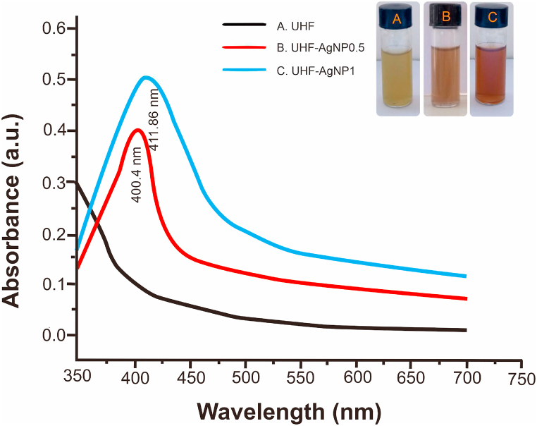
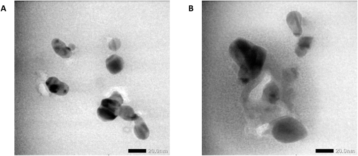
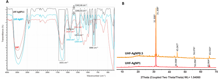
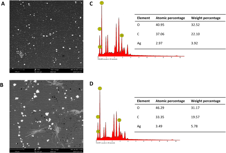
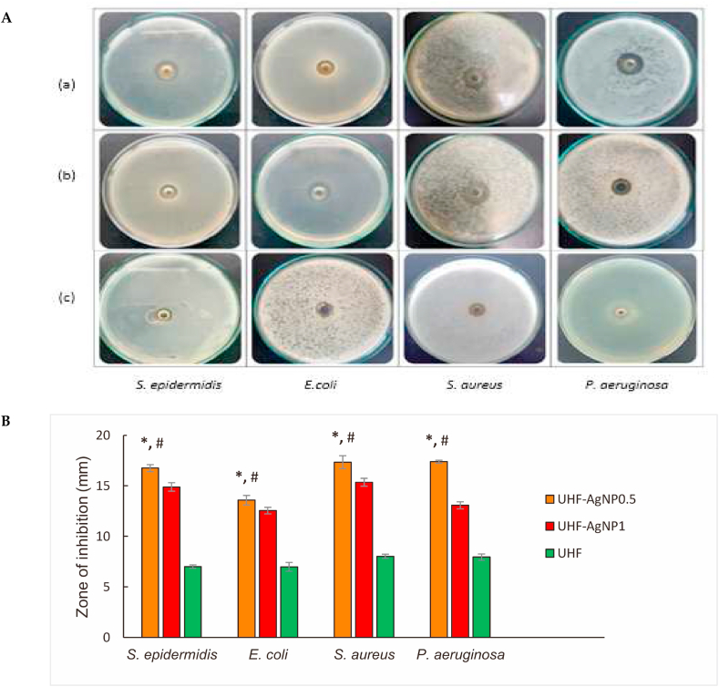
Ulvan is a polysaccharide from green algae that shows good hydrogel film dressing characteristics. Silver nanoparticles (AgNP) can be incorporated into the hydrogel film to improve antibacterial properties and provide a potential burn treatment. In this study, we developed a novel hydrogel film wound dressing composed of ulvan and silver nanoparticles. Two concentrations (0.5 mM and 1 mM) of silver nitrate were used to produce ulvan-silver nanoparticles hydrogel film (UHF-AgNP0.5 and UHF-AgNP1), respectively. The physicochemical characteristics of the hydrogel films were evaluated, including particle size, zeta potential, Fourier transform infrared (FTIR), X-ray diffractometry (XRD), scanning electron microscope and energy-dispersive X-ray (SEM-EDX). Furthermore, the in vitro antimicrobial activity, and second-degree burn wound healing test were evaluated. The UHF-AgNP0.5 showed the highest antimicrobial activity compared to UHF-AgNP1 and UHF film. Meanwhile, an in vivo study using Wistar rats induced second-degree burns showed that UHF-AgNP0.5 significantly accelerated the healing process by regulating the inflammatory process, increasing re-epithelialization, and improving the vascularization process. Ulvan-silver nanoparticle hydrogel films have the ability to accelerate the healing of second-degree burns and are potential candidates for wound dressings.
Keywords: Antimicrobial; Burn healing; Hydrogel film; Silver nanoparticles; Ulvan.
© 2023 The Authors. Published by Elsevier Ltd.
Conflict of interest statement
The authors declare that they have no known competing financial interests or personal relationships that could have appeared to influence the work reported in this paper.
Figures

References
-
- World Health Organization. Burns . 2018. Fact Sheet.https://www.who.int/news-room/fact-sheets/detail/burns Available online:
-
- Hop M.J., Polinder S., Van Der Vlies C.H., Middelkoop E., Van Baar M.E. Costs of burn care: a systematic review. Wound Repair Regen. 2014;22:436–450. - PubMed
-
- Tsai S.Y., Lio C.F., Yao W.C., Liu C.P., Shih S.C., Wang T.Y.T., Leong K.H., Sun F.J., Kuo C.F. Cost-drivers of medical expenses in burn care management. Burns. 2020;46:817–824. - PubMed
-
- Stashak T.S., Farstvedt E., Othic A. Update on wound dressings: indications and best use. Clin. Tech. Equine Pract. 2004;3:148–163.
LinkOut - more resources
Full Text Sources
