. 2022 Dec;1(12):1174-1186.
doi: 10.1038/s44161-022-00175-w. Epub 2022 Dec 12.

Loss of soluble guanylyl cyclase in platelets contributes to atherosclerotic plaque formation and vascular inflammation

Affiliations

Loss of soluble guanylyl cyclase in platelets contributes to atherosclerotic plaque formation and vascular inflammation

Carina Mauersberger et al. Nat Cardiovasc Res. 2022 Dec.

Abstract

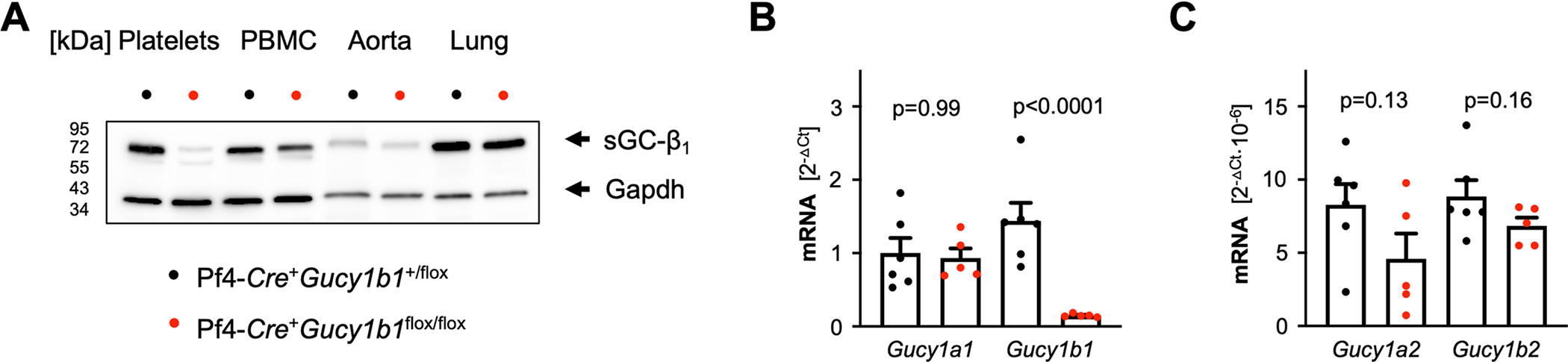
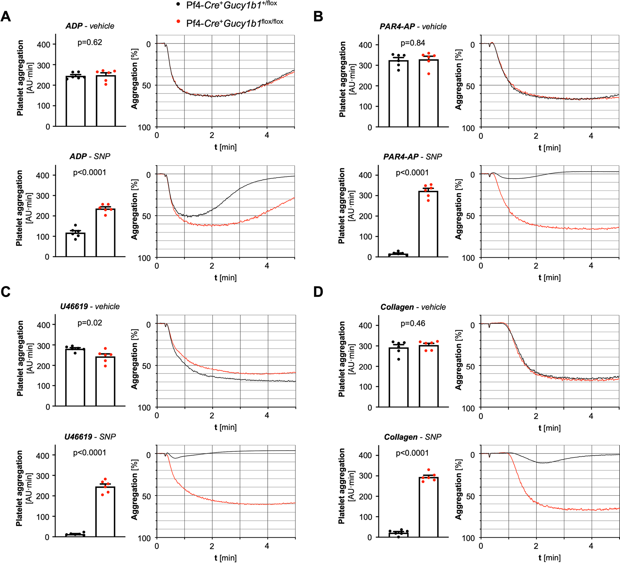
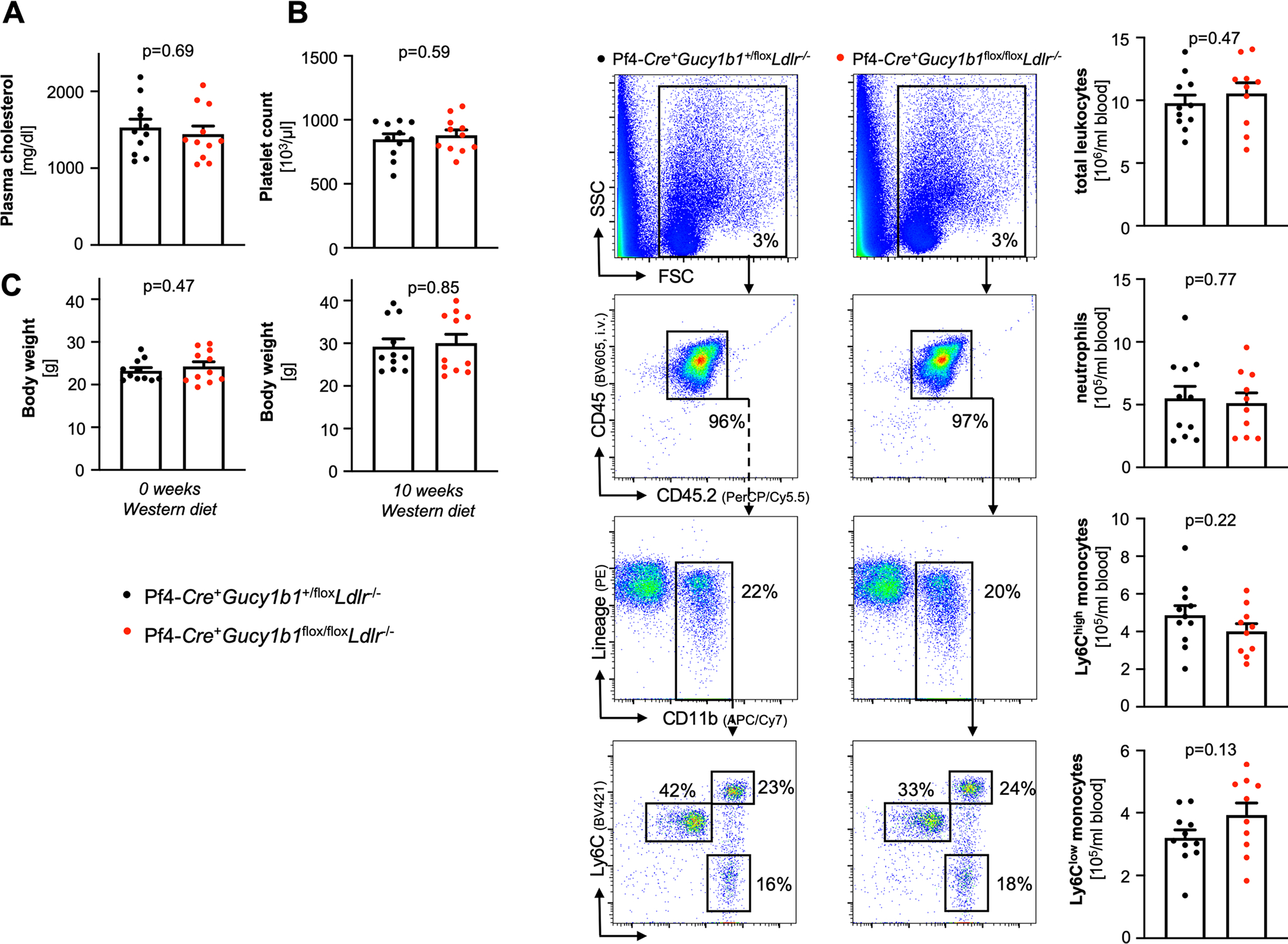
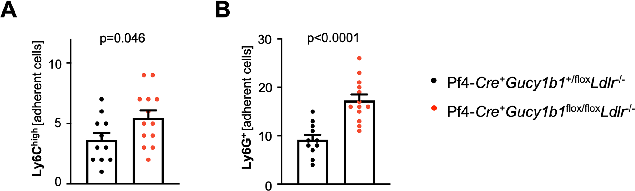
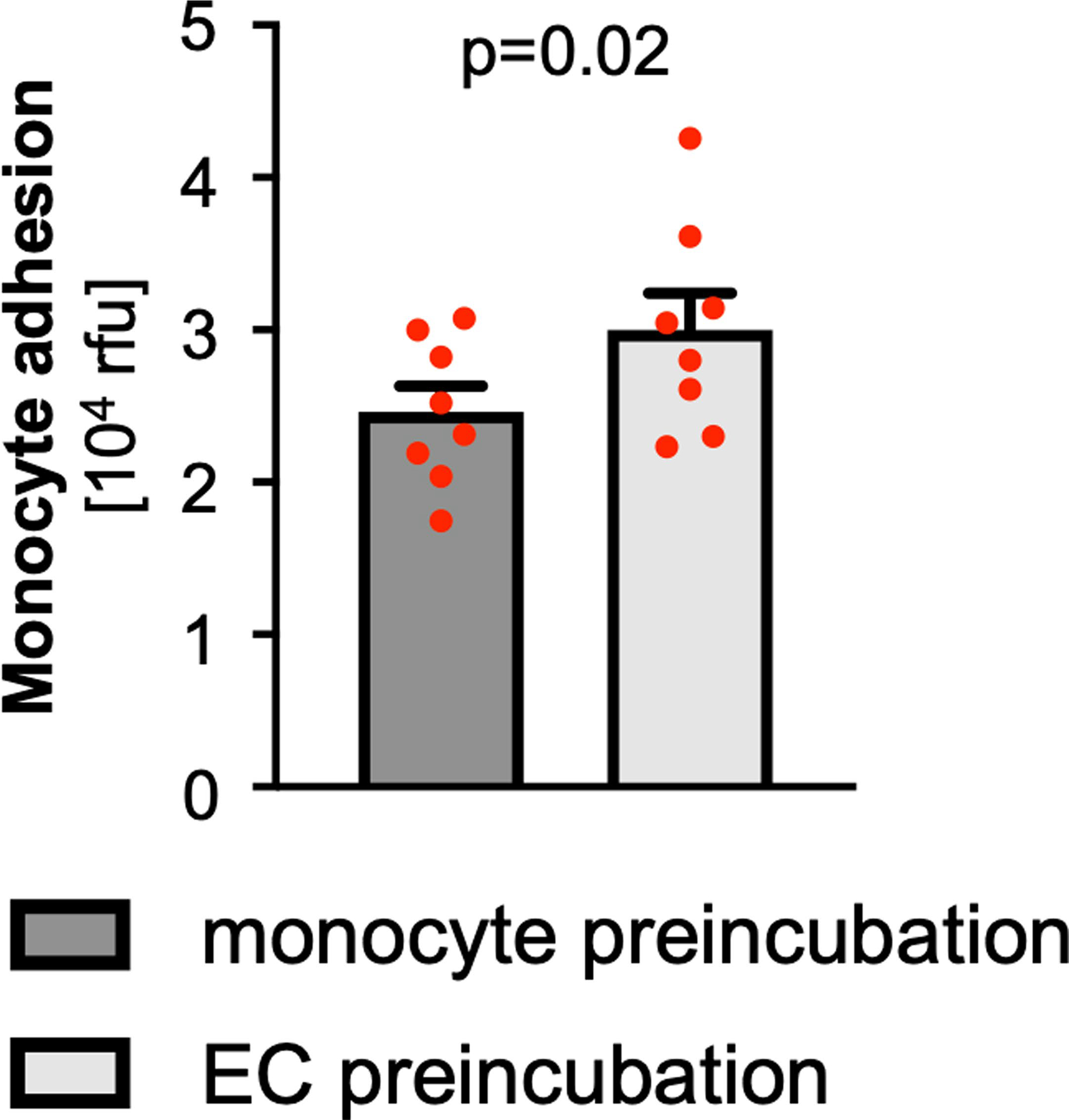
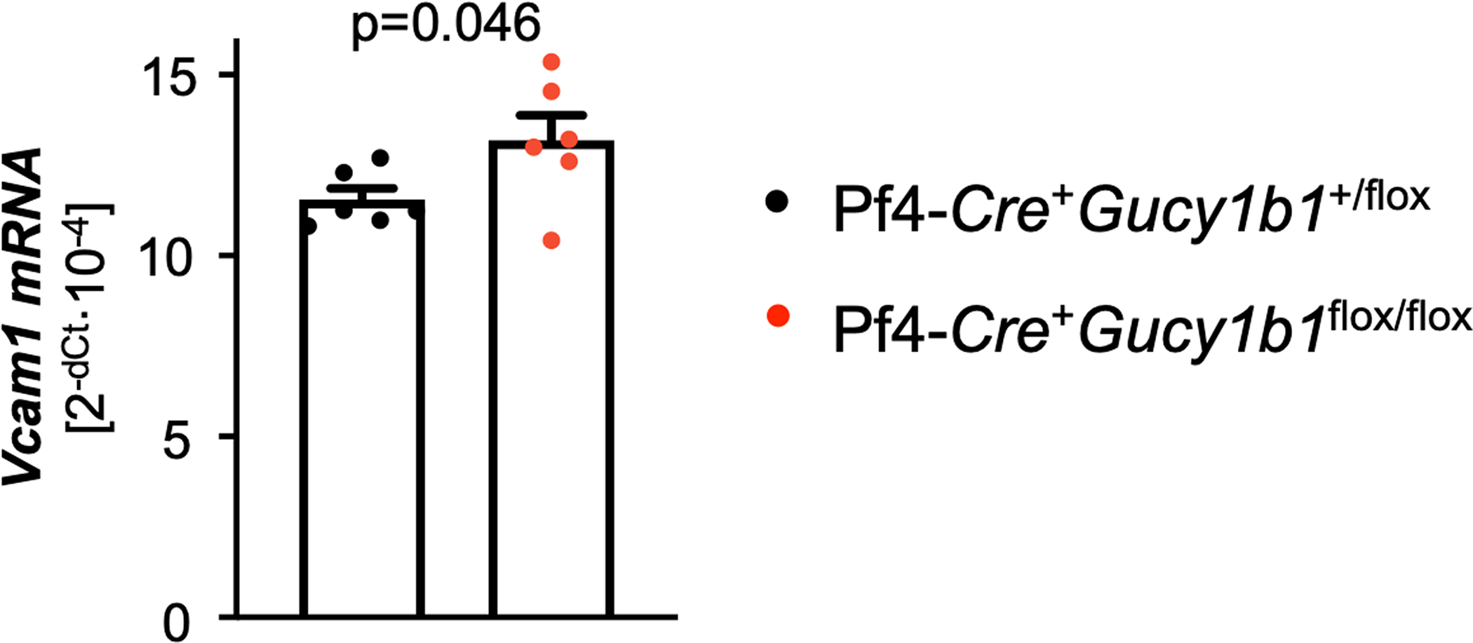
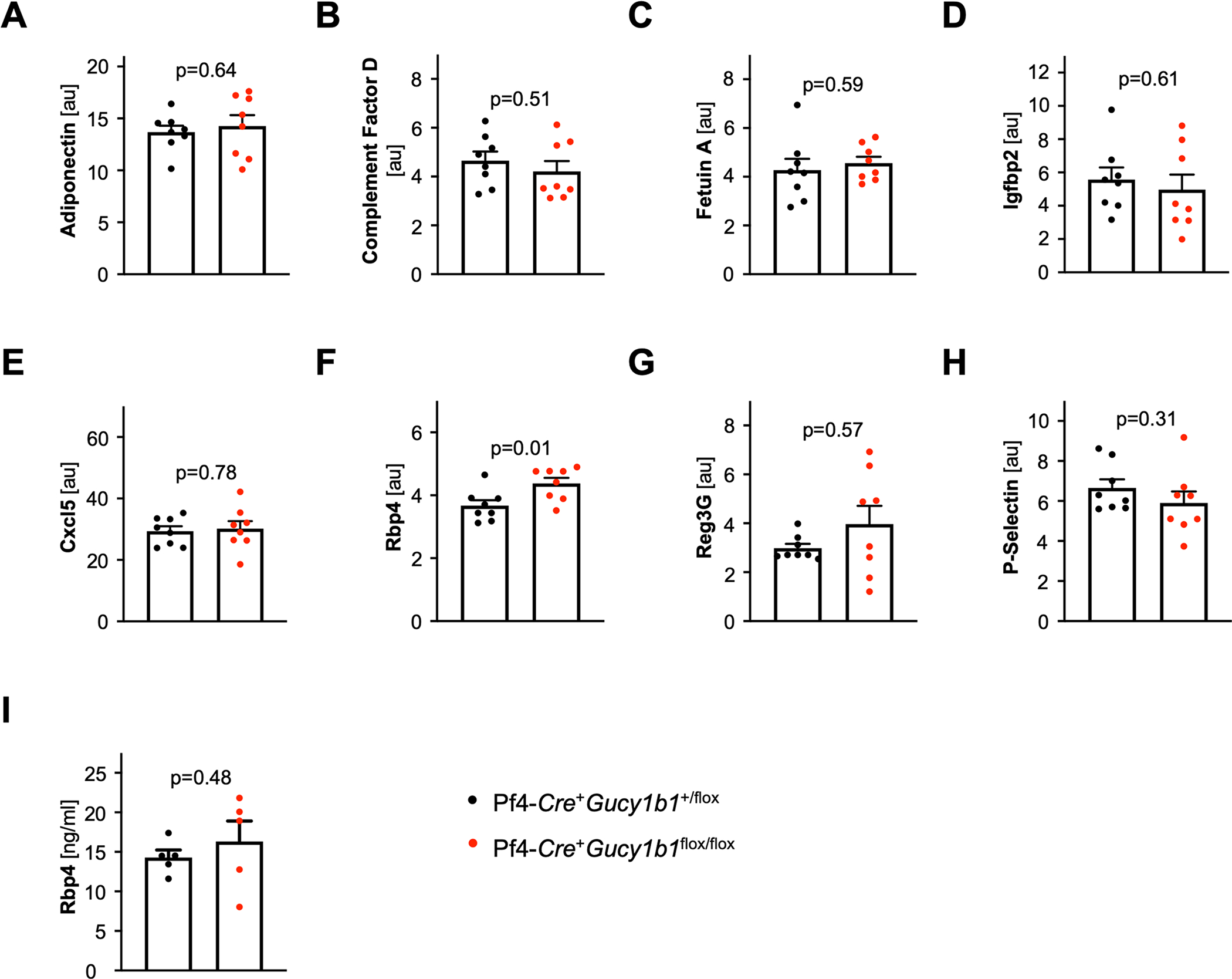
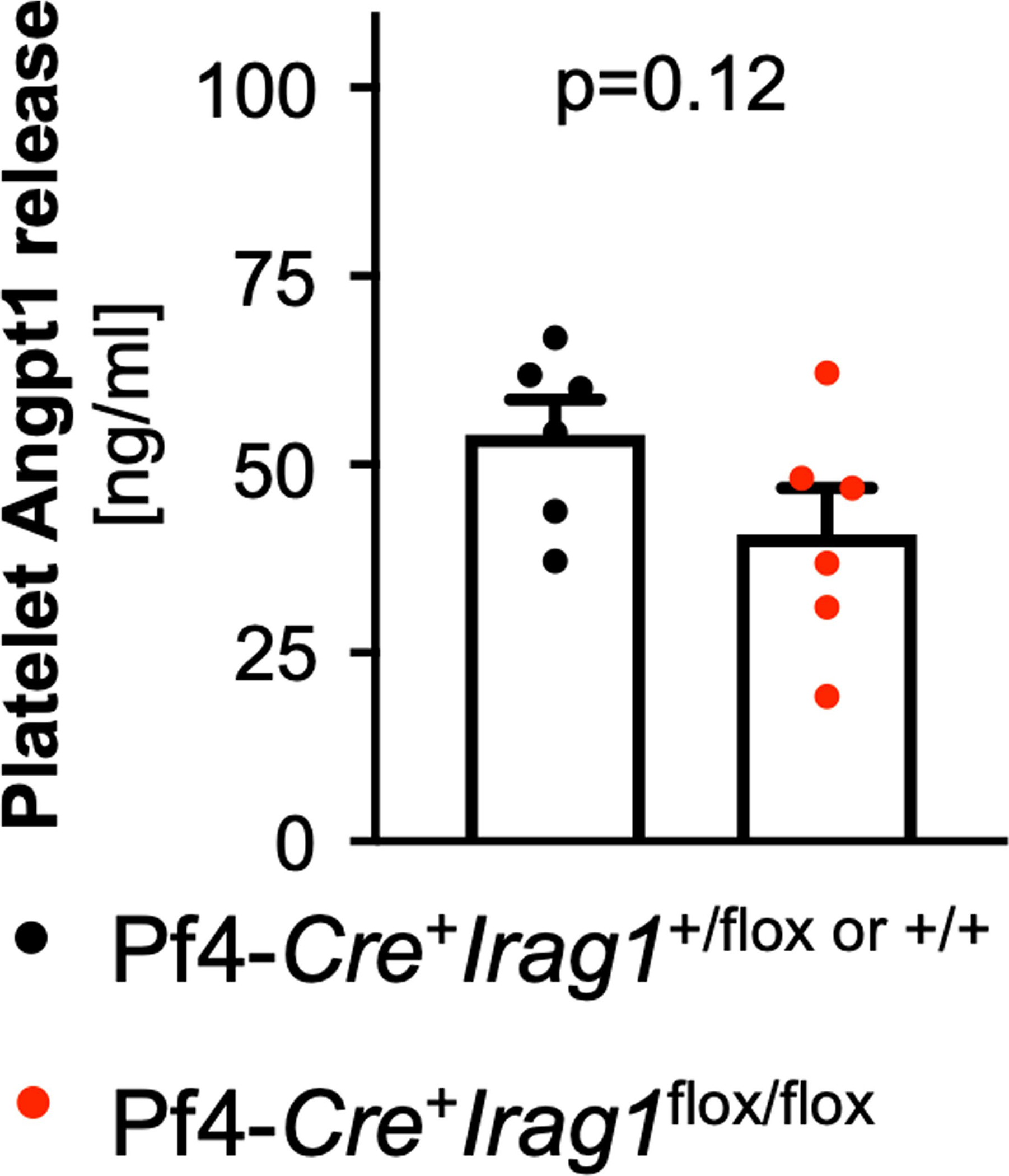
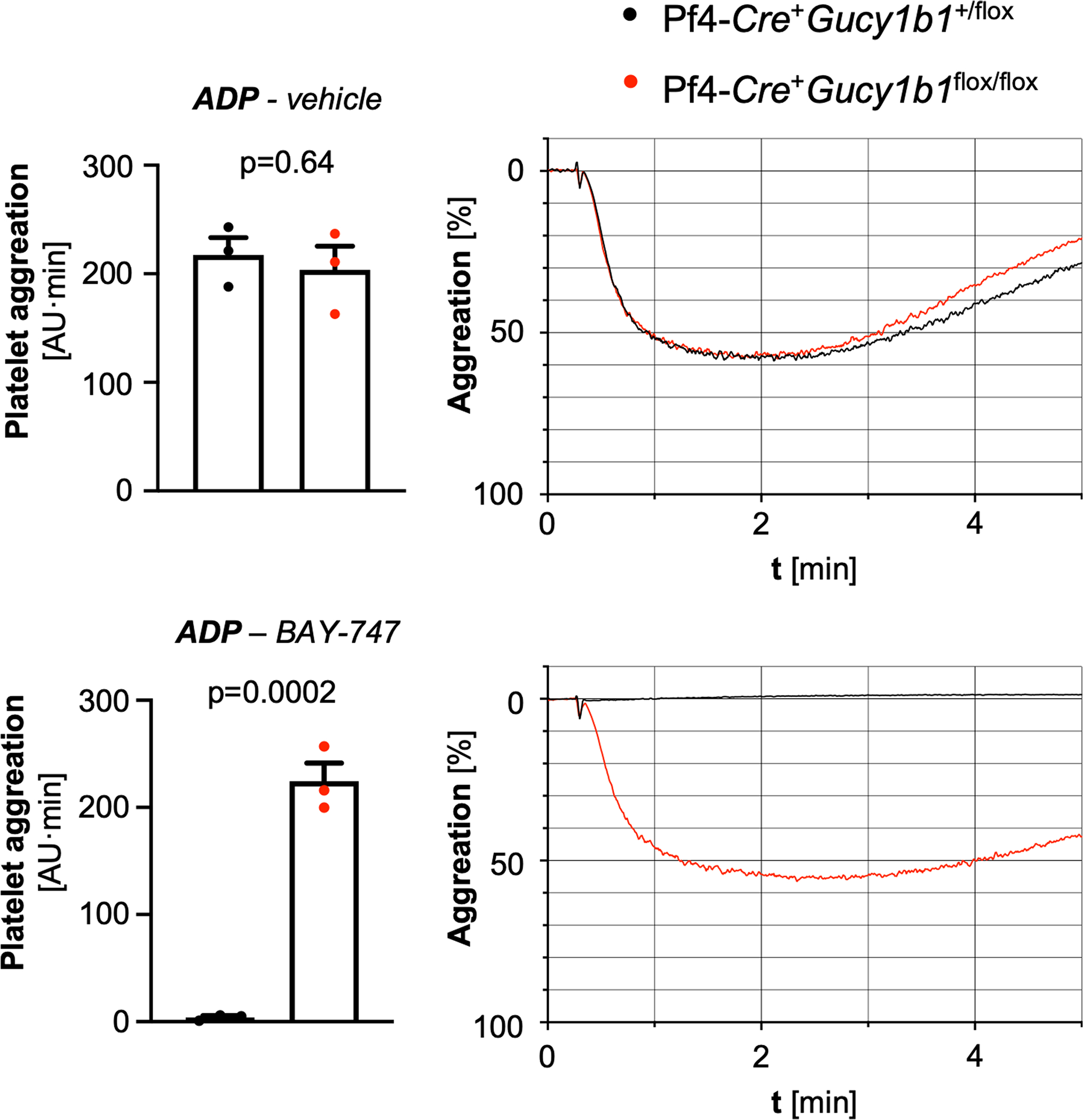
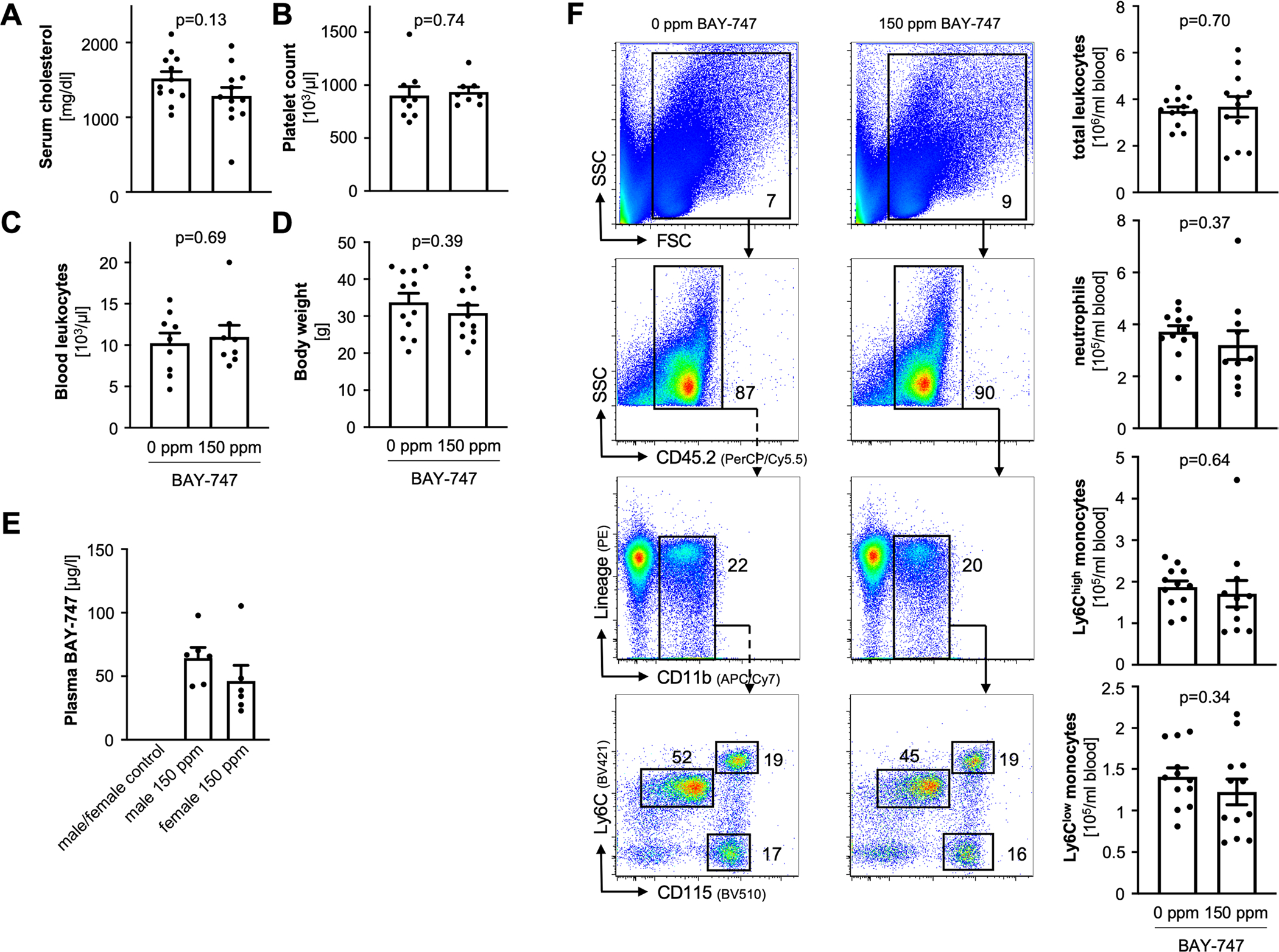
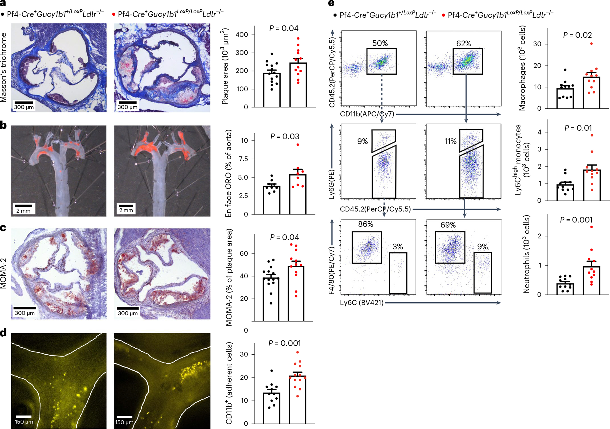
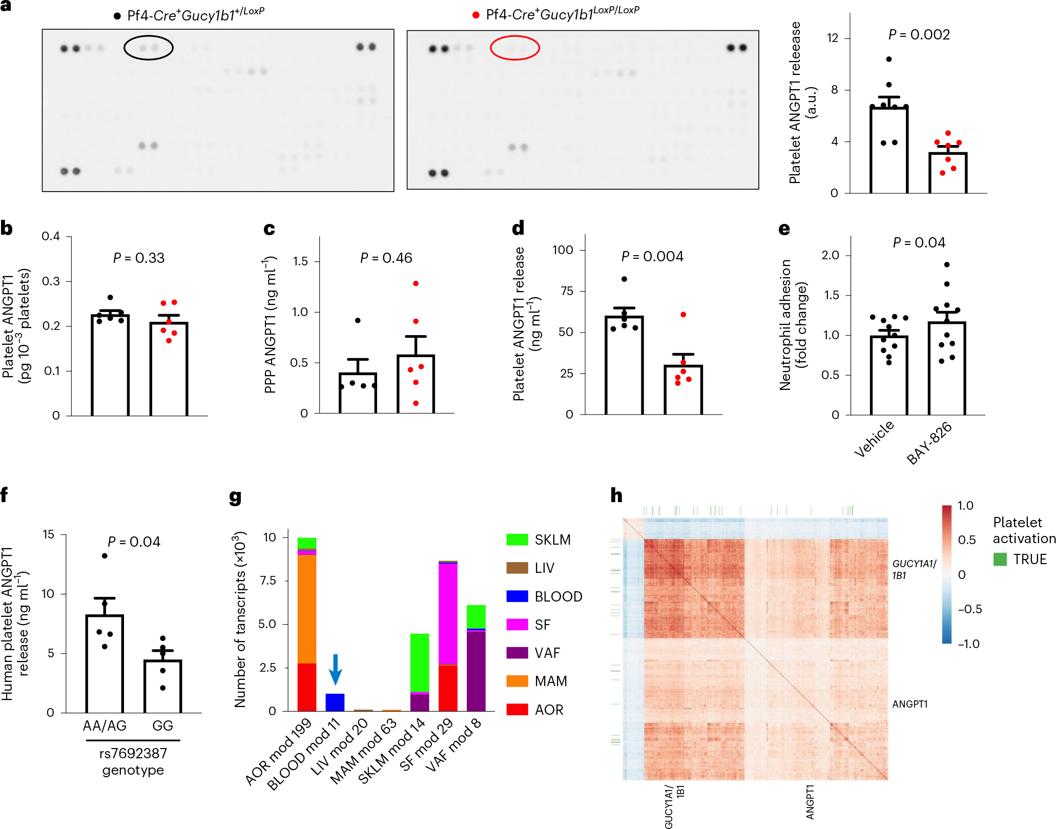
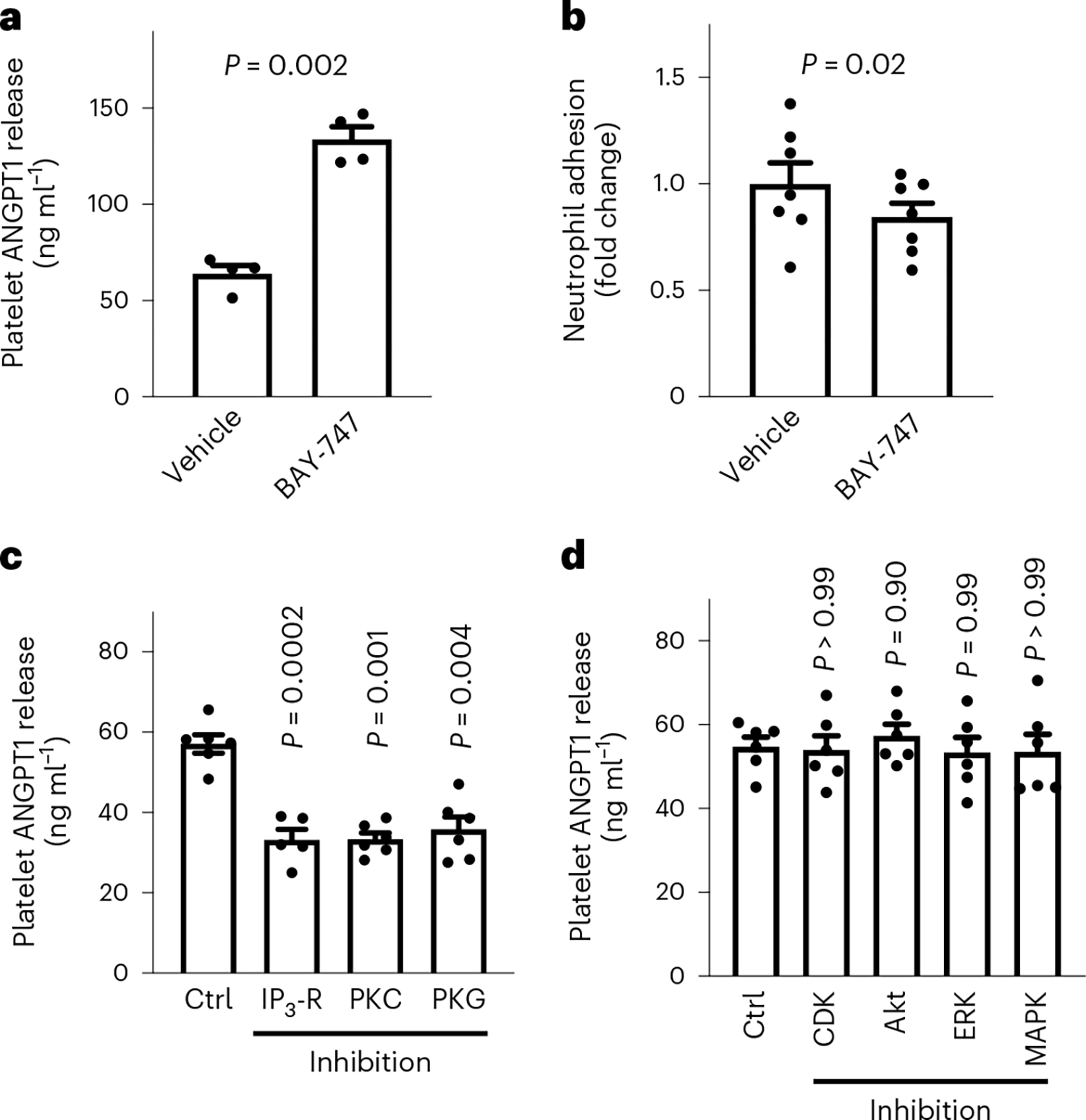
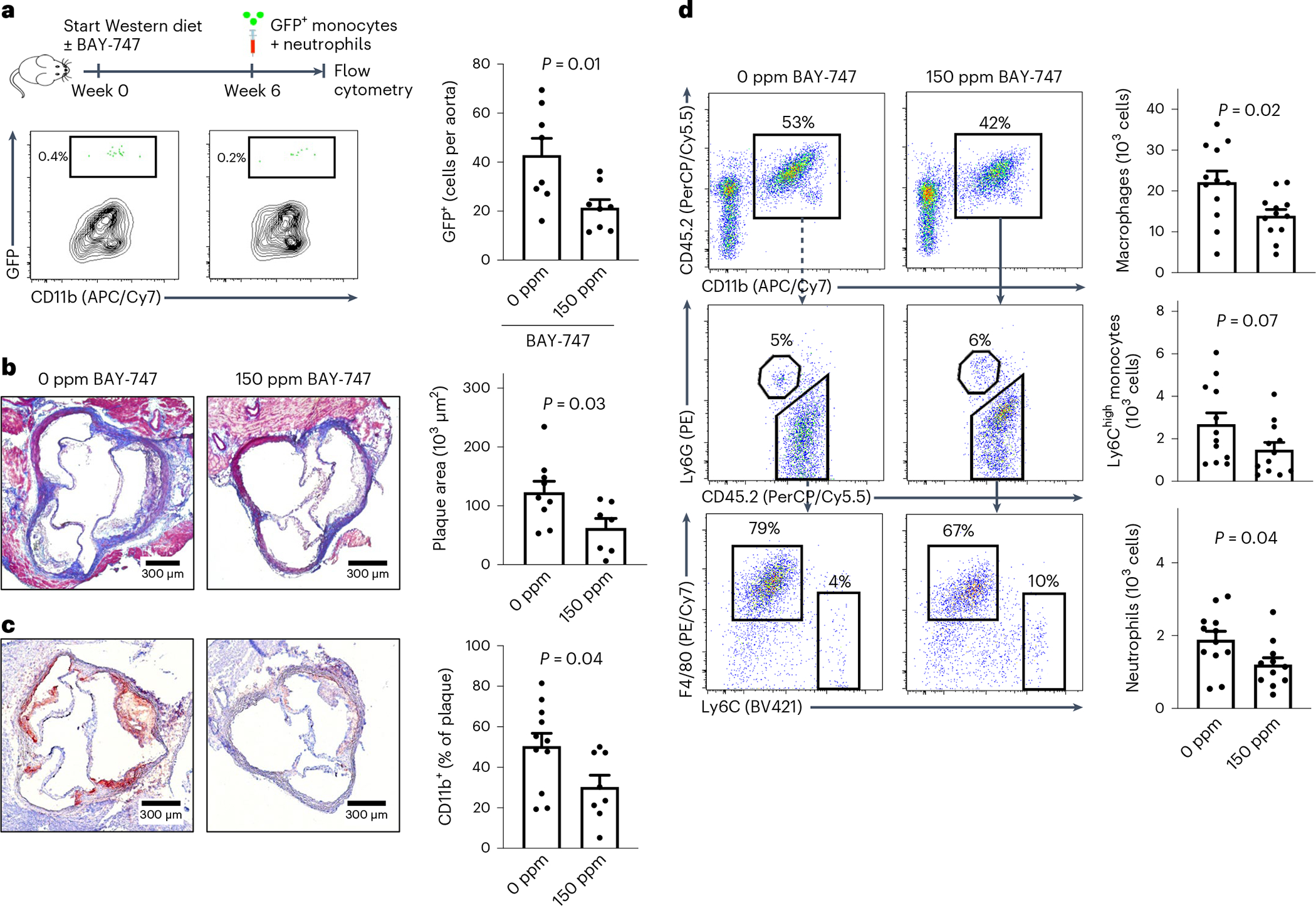
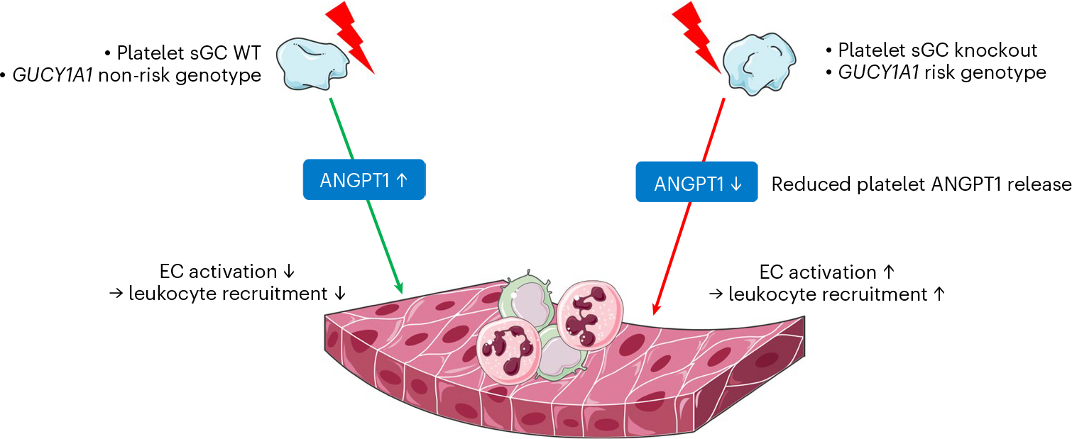
Variants in genes encoding the soluble guanylyl cyclase (sGC) in platelets are associated with coronary artery disease (CAD) risk. Here, by using histology, flow cytometry and intravital microscopy, we show that functional loss of sGC in platelets of atherosclerosis-prone Ldlr-/- mice contributes to atherosclerotic plaque formation, particularly via increasing in vivo leukocyte adhesion to atherosclerotic lesions. In vitro experiments revealed that supernatant from activated platelets lacking sGC promotes leukocyte adhesion to endothelial cells (ECs) by activating ECs. Profiling of platelet-released cytokines indicated that reduced platelet angiopoietin-1 release by sGC-depleted platelets, which was validated in isolated human platelets from carriers of GUCY1A1 risk alleles, enhances leukocyte adhesion to ECs. I mp or ta ntly, p ha rm ac ol ogical sGC stimulation increased platelet angiopoietin-1 release in vitro and reduced leukocyte recruitment and atherosclerotic plaque formation in atherosclerosis-prone Ldlr-/- mice. Therefore, pharmacological sGC stimulation might represent a potential therapeutic strategy to prevent and treat CAD.


Conflict of interest statement

Competing interests H.S. has received personal fees from MSD Sharp & Dohme, Amgen, Bayer Vital GmbH, Boehringer Ingelheim, Daiichi Sankyo, Novartis, Servier, Brahms, Bristol Myers Squibb, Medtronic, Sanofi Aventis, Synlab, Pfizer and Vifor T as well as grants and personal fees from AstraZeneca that are unrelated to the submitted work. H.S. and T.K. are named inventors on a patent application for the prevention of restenosis after angioplasty and stent implantation (patent applicants: Klinikum rechts der Isar, German Heart Centre Munich; inventors: T. Kessler, A. Kastrati, H. Schunkert; application no. PCT/EP2021/053116; status: pending), which is unrelated to the submitted work. T.K. received lecture fees from Bayer HealthCare Pharmaceuticals. L.D., F.W. and P.S. are full-time employees of Bayer HealthCare Pharmaceuticals. The other authors declare no competing interests.

Figures

Extended Data Fig. 1 |
Extended Data Fig. 1 |. sGC-Expression in Pf4-Cre+Gucy1b1LoxP/LoxP compared to Pf4-Cre+Gucy1b1+/LoxP mice.
a. Expression of sGC-β1 in platelets, peripheral blood mononuclear cells (PBMC), aorta, and lung. Representative of three independently performed Western blots on different samples. B+C. Gucy transcript expression analysis in megakaryocytes of Pf4-Cre+Gucy1b1+/LoxP and Pf4-Cre+Gucy1b1LoxP/LoxP mice. b. Gucy1a1 and Gucy1b1. C. Gucy1a2 and Gucy1b2. Each symbol represents one independent animal (n = 6 for control group, n = 5 for Pf4-Cre+Gucy1b1LoxP/LoxP). Two-sided unpaired t-test. Data are mean and s.e.m.
Extended Data Fig. 2 |
Extended Data Fig. 2 |. Platelet aggregation in control and platelet sGC knockout (Pf4-Cre+Gucy1b1LoxP/LoxP) mice.
Platelet aggregation in control and platelet sGC knockout (Pf4-Cre+Gucy1b1LoxP/LoxP) mice after stimulation with adenosine diphosphate (ADP, a) the platelet-activated receptor 4 agonist PAR4-AP (b), the thromboxane analog U46619 (c), and collagen (d). Each experiment was performed in the presence of DMSO (vehicle) or the nitric oxide donor sodium nitroprusside (SNP). Each symbol represents one independent animal (n = 6). Aggregation tracings represent the mean values of investigated animals per genotype. Two-sided unpaired t-test (except A, vehicle and D, vehicle: two-sided Mann-Whitney U-test). Data are mean and s.e.m.
Extended Data Fig. 3 |
Extended Data Fig. 3 |. Pf4-Cre+Gucy1b1LoxP/LoxPLdlr−/− compared to Pf4-Cre+Gucy1b1+/LoxPLdlr−/− mice after Western diet (see Fig. 1e).
a. Serum cholesterol levels (n = 11). b. Platelet count (n = 11). c. Body weight (n = 11) at baseline (left) and after ten weeks (right). d. Blood leukocyte numbers and subsets (n = 11 for controls, n = 10 for Pf4-Cre+Gucy1b1LoxP/LoxPLdlr−/−). Each symbol represents one independent animal. Two-sided unpaired t-test (except C 10 weeks: two-sided Mann-Whitney U-test). One outlier was removed in the analysis of D according to the ROUT method. Data are mean and s.e.m.
Extended Data Fig. 4 |
Extended Data Fig. 4 |. Leukocyte adhesion to atherosclerotic plaques of Pf4-Cre+Gucy1b1LoxP/LoxPLdlr−/− (n = 13) compared to Pf4-Cre+Gucy1b1+/LoxPLdlr−/− mice (n = 11) after Western diet assessed by fluorescence intravital microscopy.
a. Ly6Chigh monocytes. b. Neutrophils. Each symbol represents one independent animal. Two-sided unpaired t-test. Data are mean and s.e.m.
Extended Data Fig. 5 |
Extended Data Fig. 5 |. Quantification of monocyte adhesion after preincubation of either EC or monocytes with supernatant of activated platelets from Pf4-Cre + Gucy1b1LoxP/LoxP mice.
Each symbol represents one paired sample (derived from n = 8 independent animals). Two-sided paired t-test. Data are mean and s.e.m. Abbreviations: EC, endothelial cells; rfu, relative fluorescence units.
Extended Data Fig. 6 |
Extended Data Fig. 6 |. Vcam1 expression of wild-type endothelial cells after incubation with supernatant of activated Pf4-Cre+Gucy1b1+/LoxP or Pf4-Cre+Gucy1b1LoxP/LoxP platelets.
Each symbol represents one independent animal (n = 6). Data are mean and s.e.m. Two-sided paired t-test.
Extended Data Fig. 7 |
Extended Data Fig. 7 |. Cytokine profiling.
a-h. Cytokine profiling assay results for cytokines detected at a mean relative intensity ≥1 (in addition to Angiopoietin-1). I. Replication attempt for RBP4 release using ELISA. Each symbol represents one independent animal (A-H: n = 8; I: n = 5). Two-sided unpaired t-test (except B and G: Two-sided Mann-Whitney U-test). Data are mean and s.e.m. Abbreviations: au, arbitrary units; CXCL5, C-X-C Motif Chemokine Ligand 5; IGFPB2, Insulin Like Growth Factor Binding Protein 2; RBP4, Retinol Binding Protein 4; REG3G, Regenerating islet-derived protein 3 gamma.
Extended Data Fig. 8 |
Extended Data Fig. 8 |. ngiopoietin-1 (ANGPT1) release by control or Pf4-Cre+Irag1LoxP/LoxP platelets after activation by shaking.
A Each symbol represents one independent animal (n = 6). Two-sided unpaired t-test. Data are mean and s.e.m.
Extended Data Fig. 9 |
Extended Data Fig. 9 |. Adenosine diphosphate-induced platelet aggregation in Pf4-Cre+Gucy1b1+/LoxP and Pf4-Cre+Gucy1b1LoxP/LoxP platelets after pretreatment with vehicle or BAY-747.
Each symbol represents one independent animal (n = 3). Two-sided unpaired t-test. Data are mean and s.e.m. Abbreviation: ADP, adenosine diphosphate.
Extended Data Fig. 10 |
Extended Data Fig. 10 |. Characteristics of Ldlr−/− mice receiving 0 ppm compared to Ldlr−/− mice receiving 150 ppm BAY-747 after ten weeks of Western diet.
a. Serum cholesterol levels (n = 12). b. Platelet count (n = 9 in 0 ppm, n = 8 in 150 ppm group). c. Blood leukocytes (n = 9 in 0 ppm, n = 8 in 150 ppm group). d. Body weight after (n = 12). e. Plasma concentrations of the soluble guanylyl cyclase stimulator BAY-747 in treated mice. f. Blood leukocyte numbers and subsets. Each symbol represents one independent animal (n = 12). Two-sided unpaired t-test. Two outliers were removed in the analysis of F: neutrophils (150 ppm group; n = 10) and Ly6Chigh monocytes (150 ppm group; n = 11) according to the ROUT method. Data are mean and s.e.m.
Fig. 1 |
Fig. 1 |. Atherosclerotic plaque formation and vascular inflammation in mice lacking platelet sGC.
ac, Atherosclerotic plaque formation as assessed by aortic root histology (a), aortic en face ORO staining (b), and monocyte and macrophage content (c) in 12 (b: 8) Pf4-Cre+Gucy1b1LoxP/LoxPLdlr−/− mice compared to 14 (b: 9) Pf4-Cre+Gucy1b1+/LoxPLdlr−/− mice that were fed a Western diet for 10 weeks. Two-sided unpaired t-test. d, Leukocyte adhesion to atherosclerotic plaques in n = 13 Pf4-Cre+Gucy1b1LoxP/LoxPLdlr−/− mice compared to n = 11 Pf4-Cr e+Gucy1b1+/LoxPLdlr−/− mice that were fed a Western diet for 6 weeks to induce atherosclerotic plaque formation. Two-sided unpaired t-test. e, Quantification of vascular inflammation by flow cytometry analysis of aortic cell suspensions of Pf4-Cre+Gucy1b1LoxP/LoxPLdlr−/− mice compared to Pf4-Cre+Gucy1b1+/LoxPLdlr−/− mice (n = 11 per group). Two-sided Mann–Whitney U-test. Each symbol represents one independent animal. Data are the mean ± s.e.m.
Fig. 2 |
Fig. 2 |. Influence of platelet sGC on leukocyte adhesion in vitro.
a,b, WT monocyte (a) and neutrophil (b) adhesion to WT ECs after incubation with supernatant of activated platelets isolated from either Pf4-Cre+Gucy1b1LoxP/LoxP or Pf4-Cre+Gucy1b1+/LoxP mice. Each symbol represents 1 independent animal (n = 8 per group). Two-sided unpaired t-test. Data are the mean ± s.e.m. c, Quantification of neutrophil adhesion after preincubation of either ECs or neutrophils with supernatant of activated platelets from Pf4-Cre+Gucy1b1LoxP/LoxP mice in comparison to non-preincubation conditions. Each symbol represents 1 paired sample (each derived from n = 8 independent animals). Repeated measures one-way ANOVA with Tukey test for multiple testing. Data are the mean ± s.e.m.
Fig. 3 |
Fig. 3 |. Platelet sGC influences the release of ANGPT1.
a, Left, identification of ANGPT1 (encircled) as differentially released protein from activated Pf4-Cre+Gucy1b1+/LoxP and Pf4-Cre+Gucy1b1LoxP/LoxP platelets. Right, quantification of ANGPT1 signal in 8 Pf4-Cre+Gucy1b1+/LoxP and 7 Pf4-Cre+Gucy1b1LoxP/LoxP mice. Two-sided unpaired t-test. bd, Quantification of platelet ANGPT1 content (n = 6 independent animals) (b), PPP ANGPT1 (n = 5 and 6 independent animals, respectively) (c) and released ANGPT1 as determined in 6 independent animals per group by ELISA (d). Two-sided unpaired t-test. e, WT neutrophil adhesion to WT ECs after incubation with supernatant of activated WT platelets in the absence and presence of the Tie2 inhibitor BAY-826 (0.5 μM). Two-sided paired t-test on n = 11 sample pairs derived from independent animals. f, Platelet ANGPT1 release in five humans carrying the GUCY1A1 (rs7692387) non-risk (AA, AG genotype) allele and five homozygous carriers of the risk allele (GG genotype). Each symbol represents one individual. Two-sided unpaired t-test. Data are the mean ± s.e.m. g, STARNET coexpression modules containing ANGPT1 from multitissue RNA-seq sampling of approximately 600 patients with CAD. The arrow denotes coexpression module 11 from whole-blood samples (BLOOD). h, Heatmap of Pearson’s correlation coefficients of genes in coexpression module 11, showing positive correlation of ANGPT1, GUCY1A1 and GUCY1B1 along with enrichment for platelet activation genes (false discovery rate = 6.863 × 10⁻¹³, Enrichr, KEGG pathway). AOR, aorta; LIV, liver; MAM, mammary artery; SF, subcutaneous fat; SKLM, skeletal muscle; VAF, visceral fat.
Fig. 4 |
Fig. 4 |. ANGPT1 release from platelets is influenced by sGC via the PKC pathway.
a, ANGPT1 release by WT platelets incubated with 150 ppm BAY-747 or vehicle (n = 4 sample pairs from independent animals). b, WT neutrophil adhesion to WT ECs after incubation with supernatant from n = 7 activated WT platelets from independent animals that were preincubated with either vehicle or 150 ppm BAY-747. c, Inhibition of IP3-R (10 μM 2-APB), PKC (5 μM Ro 32–0432), PKG (10 μM KT-5823) and measurement of ANGPT1 release from platelets (n = 6). One outlier was removed from the PKG group (n = 5) according to the ROUT test. d, Inhibition of mitogen-activated protein kinase (MAPK) (10 μM VX-702), cyclin-dependent kinase (CDK) 2/5/9 (100 nM dinaciclib), extracellular signalregulated kinase (ERK) (10 μM ravoxertinib), and Akt kinase (1 μM MK-2206) and measurement of ANGPT1 release from platelets (n = 6). Each symbol represents paired samples derived from independent animals. a,b, Two-sided paired t-test. c,d, Mixed-effects analysis (ANOVA with Dunnett multiple comparison test). Data are the mean ± s.e.m.
Fig. 5 |
Fig. 5 |. Pharmacological sGC stimulation influences leukocyte recruitment, atherosclerotic plaque formation and vascular inflammation.
a, Adoptive transfer of GFP+ leukocytes: study scheme, flow cytometry plot (left) and quantification (right) of GFP+ cells (n = 8 independent animals). b, Aortic root atherosclerotic plaques in Ldlr−/− mice that were fed a Western diet for 10 weeks containing 0 (control group, n = 9) or 150 ppm BAY-747 (n = 7). c, CD11b+ area of aortic roots in mice from the control (n = 11) and treatment groups (n = 8). d, Quantification of vascular inflammation by flow cytometry analysis of aortic cell suspensions of mice in the control (n = 12) and treatment groups (n = 12). Each symbol represents one independent mouse. Two-sided unpaired t-test. Data are the mean ± s.e.m. One outlier was removed in d in the analysis of neutrophils (150 ppm; n = 11) according to the ROUT test.
Fig. 6 |
Fig. 6 |. In the event of platelet activation, for example, by shear stress, sGC counterbalances proinflammatory activation of ECs, for example, by release of ANGPT1.
If sGC levels are reduced, for example, in the mouse model used in this study or in platelets of homozygous carriers of the CAD-associated risk variant, less ANGPT1 is released. Subsequently enhanced EC activation and leukocyte recruitment contribute to atherosclerotic plaque formation. This figure contains modified image material available at Servier Medical Art under a Creative Commons Attribution 3.0 Unported License.

References

    1. Benjamin EJ et al. Heart disease and stroke statistics–2019 update: a report from the American Heart Association. Circulation 139, e56–e528 (2019). - PubMed
    1. Erdmann J, Kessler T, Venegas LM & Schunkert H A decade of genome-wide association studies for coronary artery disease: the challenges ahead. Cardiovasc. Res. 114, 1241–1257 (2018). - PubMed
    1. Dang TA, Schunkert H & Kessler T cGMP signaling in cardiovascular diseases: linking genotype and phenotype. J. Cardiovasc. Pharmacol. 75, 516–525 (2020). - PubMed
    1. Deloukas P et al. Large-scale association analysis identifies new risk loci for coronary artery disease. Nat. Genet. 45, 25–33 (2013). - PMC - PubMed
    1. Erdmann J et al. Dysfunctional nitric oxide signalling increases risk of myocardial infarction. Nature 504, 432–436 (2013). - PubMed