. 2023 Apr 23;5(8):100764.
doi: 10.1016/j.jhepr.2023.100764. eCollection 2023 Aug.

Identification and characterisation of a rare MTTP variant underlying hereditary non-alcoholic fatty liver disease

Affiliations

Identification and characterisation of a rare MTTP variant underlying hereditary non-alcoholic fatty liver disease

Jane I Grove et al. JHEP Rep. .

Abstract

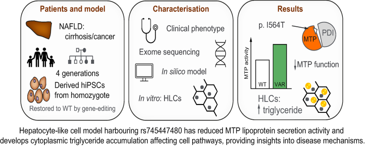
Background & aims: Non-alcoholic fatty liver disease (NAFLD) is a complex trait with an estimated prevalence of 25% globally. We aimed to identify the genetic variant underlying a four-generation family with progressive NAFLD leading to cirrhosis, decompensation, and development of hepatocellular carcinoma in the absence of common risk factors such as obesity and type 2 diabetes.

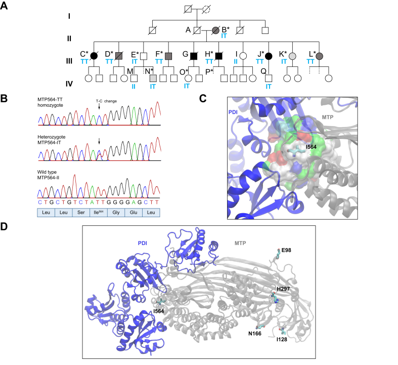
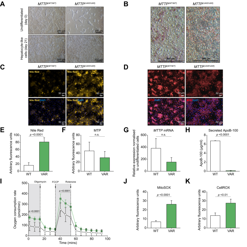
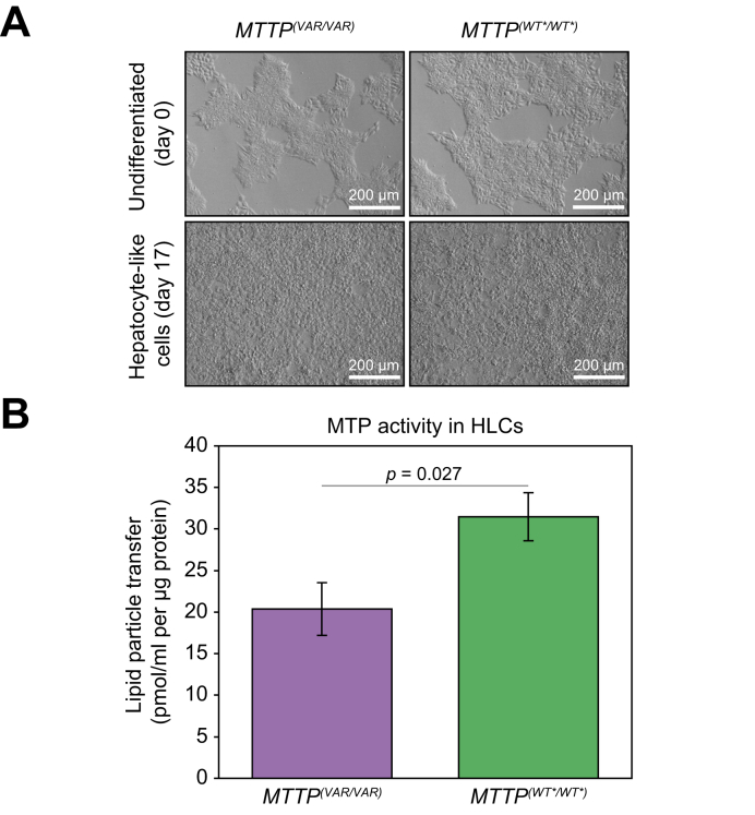
Methods: Exome sequencing and genome comparisons were used to identify the likely causal variant. We extensively characterised the clinical phenotype and post-prandial metabolic responses of family members with the identified novel variant in comparison with healthy non-carriers and wild-type patients with NAFLD. Variant-expressing hepatocyte-like cells (HLCs) were derived from human-induced pluripotent stem cells generated from homozygous donor skin fibroblasts and restored to wild-type using CRISPR-Cas9. The phenotype was assessed using imaging, targeted RNA analysis, and molecular expression arrays.

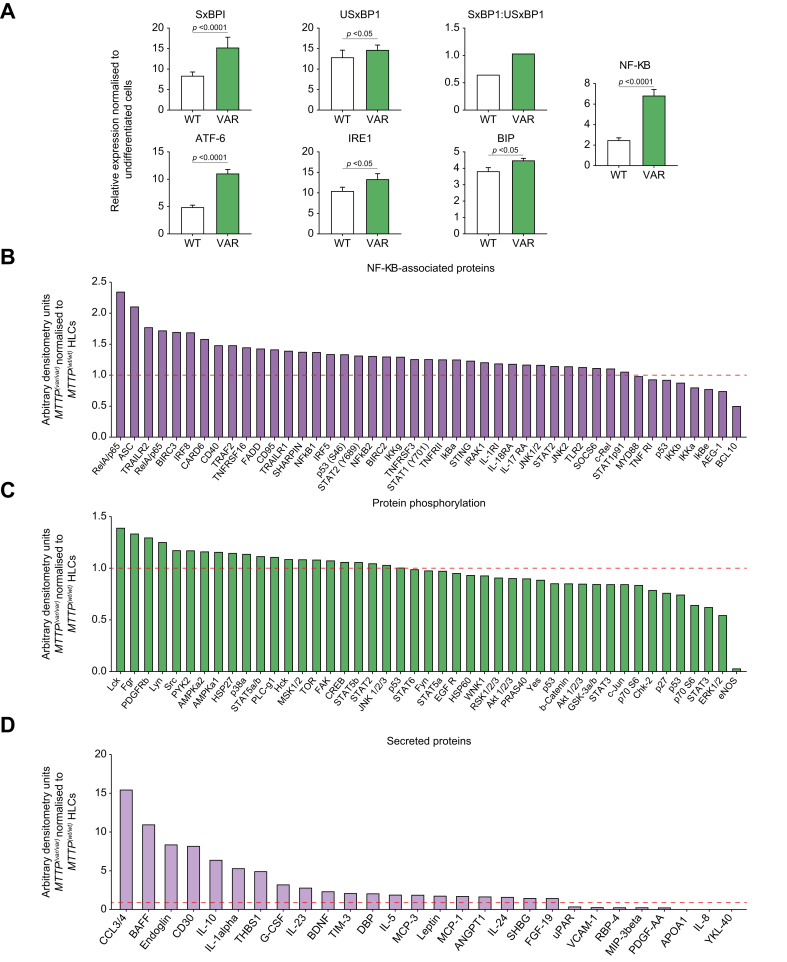
Results: We identified a rare causal variant c.1691T>C p.I564T (rs745447480) in MTTP, encoding microsomal triglyceride transfer protein (MTP), associated with progressive NAFLD, unrelated to metabolic syndrome and without characteristic features of abetalipoproteinaemia. HLCs derived from a homozygote donor had significantly lower MTP activity and lower lipoprotein ApoB secretion than wild-type cells, while having similar levels of MTP mRNA and protein. Cytoplasmic triglyceride accumulation in HLCs triggered endoplasmic reticulum stress, secretion of pro-inflammatory mediators, and production of reactive oxygen species.

Conclusions: We have identified and characterised a rare causal variant in MTTP, and homozygosity for MTTP p.I564T is associated with progressive NAFLD without any other manifestations of abetalipoproteinaemia. Our findings provide insights into mechanisms driving progressive NAFLD.

Impact and implications: A rare genetic variant in the gene MTTP has been identified as responsible for the development of severe non-alcoholic fatty liver disease in a four-generation family with no typical disease risk factors. A cell line culture created harbouring this variant gene was characterised to understand how this genetic variation leads to a defect in liver cells, which results in accumulation of fat and processes that promote disease. This is now a useful model for studying the disease pathways and to discover new ways to treat common types of fatty liver disease.

Keywords: Abetalipoproteinaemia; Lipoprotein ApoB; Microsomal triglyceride transfer protein; hiPSC-derived hepatocytes.


Conflict of interest statement

GPA has served as a consultant and an advisory board member for Pfizer Inc, Inventiva Pharma, GlaxoSmithKline, and KaNDy Therapeutics; he has been a consultant to Servier, Clinipace, Albireo Pharma, BenevolentAI Bio, DNDi, BerGenBio ASA, Median Technologies, FRACTYL, Amryt Pharma, and AstraZeneca; and has given presentations on behalf of Roche Diagnostics and Medscape. IN is employed by Gilead Sciences Ltd. (since August 2019). All other authors declare no conflict of interests. Please refer to the accompanying ICMJE disclosure forms for further details.

Figures

None
Graphical abstract
Fig. 1
Fig. 1
Identification of a pathogenic variant in a large family with non-alcoholic fatty liver disease. (A) Pedigree. Clinical features are described in Table 1. Diagnosis is indicated by shading: black, hepatocellular carcinoma; dark grey, cirrhosis; light grey, non-alcoholic steatohepatitis. Dashed lines indicate no investigations. ∗Exome sequenced. Blue letters indicate residue 564 in MTP. (B) Variant rs745447480 sequencing. (C) Local environment of I564 on MTP–PDI interface (hydrophobic pocket [white], polar [green], and charged [red/blue] residues). (D) 564T variant and common non-synonymous variants (side chains: C = cyan; O = red; N = blue) in a model derived from PDB ID:617S, a heterodimer of PDI (blue), and MTTP gene product (MTP) (grey). MTP, microsomal triglyceride transfer protein; PDI, protein disulfide isomerase.
Fig. 2
Fig. 2
Meal-response study to investigate metabolism in family members and matched controls. (A) Study design. (B) ApoB levels. Participants are grouped according to age and sex matching to family members F, J, K, Q, and M (Table S1). MTP residue 564 is indicated (TT/TI/II). PNPLA3 or TM6SF2 in parentheses indicates individuals homozygous for variant rs738409 or rs58542926, respectively. (C) Serum ApoB-100 (mean levels ± standard deviation). 1Milk and cornflakes. 280–175 min between breakfast start and pre-lunch sample. 3From participants J and 1 used to derive cell lines. ApoB, apolipoprotein B; ApoB-100, apolipoprotein B-100; HV, healthy volunteers; MTP, microsomal triglyceride transfer protein; NAFLD, non-alcoholic fatty liver disease; SNP, single-nucleotide polymorphism.
Fig. 3
Fig. 3
Triglyceride levels in study participants. Total serum triglycerides: (A) Participant F and matched controls and (B) participant J and matched controls. VLDL-triglyceride: (C) Participant F and matched controls and (D) participant J and matched controls. Chylomicron-triglyceride: (E) Participant F and matched controls and (F) participant J and matched controls. MTTP genotypes are shown for family members. PNPLA3 p.I148M and TM6SF2 p.E167 K genotypes are indicated in parentheses. HV, healthy volunteers; NAFLD, non-alcoholic fatty liver disease.
Fig. 4
Fig. 4
Characterisation of MTP-564T homozygote variant, MTTP(VAR/VAR), and wild-type HLCs. (A) Light microscopy image of terminally differentiated hiPSC-derived HLCs. (B) Oil Red O staining of HLCs (light microscopy). (C) Nile red staining of lipids ± DAPI staining (fluorescence microscopy). (D) MTP expression immunocytochemistry ± DAPI staining. Quantification of Nile red fluorescence (E) and MTP staining (F) in HLCs. (G) Expression of MTP determined by quantitaive PCR. (H) ApoB-100 secretion by HLCs (ELISA). (I) Basal and maximal mitochondrial respiratory rates in MTTP(WT/WT) (white circles) and MTTP(VAR/VAR) (green squares) HLCs. Quantification of cellular superoxide (J) and reactive oxygen species (K) from fluorescence microscopy. Mean ± SE. p <0.05 is significant (t test, paired, two-tailed). ApoB-100, apolipoprotein B-100; hiPSC, human induced pluripotent stem cell; HLC, hepatocyte-like cell; MTP, microsomal triglyceride transfer protein.
Fig. 5
Fig. 5
Phenotypic characterisation of MTTP(VAR/VAR) hiPSC-derived HLCs. (A) Expression of inflammation-related and ER stress-related genes. Mean ± SE. p <0.05 is significant (t test, paired, two-tailed) (B) Expression of NF-κB-associated intracellular signalling proteins in MTTP(VAR/VAR) HLCs, relative to expression in MTTP(WT/WT). (C) Phosphorylated proteins in MTTP(VAR/VAR) HLCs, normalised to MTTP(WT/WT). (D) Secreted proteins from MTTP(VAR/VAR) HLCs, normalised to MTTP(WT/WT). ATF6, activating transcription factor 6; BIP, binding immunoglobulin protein; ER, endoplasmic reticulum; hiPSC, human induced pluripotent stem cell; HLC, hepatocyte-like cell; IRE1, inositol requiring enzyme 1; SxBP1, spliced X-box binding protein-1; USxBP1, unspliced X-box binding protein-1.
Fig. 6
Fig. 6
Restoration of activities by gene editing of MTTPVAR/VAR 564-TT to 564-II. (A) Light microscopy showing terminally differentiated hiPSC-derived HLCs from MTTP(VAR/VAR) and gene-edited derivative MTTP(WT/WT). (B) MTP enzyme activity in HLCs. Mean ± SE; significance level p <0.05 (t test). hiPSC, human induced pluripotent stem cell; HLC, hepatocyte-like cell; MTP, microsomal triglyceride transfer protein.

References

    1. Younossi Z.M., Koenig A.B., Abdelatif D., Fazel Y., Henry L., Wymer M. Global epidemiology of nonalcoholic fatty liver disease-Meta-analytic assessment of prevalence, incidence, and outcomes. Hepatology. 2016;64:73–84. - PubMed
    1. Krawczyk M., Liebe R., Lammert F. Toward genetic prediction of nonalcoholic fatty liver disease trajectories: PNPLA3 and beyond. Gastroenterology. 2020;158:1865–1880.e1861. - PubMed
    1. Trépo E., Valenti L. Update on NAFLD genetics: from new variants to the clinic. J Hepatol. 2020;72:1196–1209. - PubMed
    1. Tan J., Zhang J., Zhao Z., Zhang J., Dong M., Ma X., et al. The association between SNPs rs1800591 and rs3816873 of the MTTP gene and nonalcoholic fatty liver disease: a meta-analysis. Saudi J Gastroenterol. 2020;26:171–178. - PMC - PubMed
    1. Haas M.E., Pirruccello J.P., Friedman S.N., Wang M., Emdin C.A., Ajmera V.H., et al. Machine learning enables new insights into genetic contributions to liver fat accumulation. Cell Genom. 2021;1 - PMC - PubMed

LinkOut - more resources